. 2009 Jun 15;439(1-2):63-70.
doi: 10.1016/j.gene.2009.03.016. Epub 2009 Mar 31.

Interferon regulatory factors (IRFs) repress transcription of the chicken ovalbumin gene

Affiliations

Interferon regulatory factors (IRFs) repress transcription of the chicken ovalbumin gene

Dawne C Dougherty et al. Gene. .

Abstract

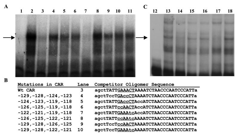
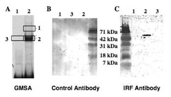
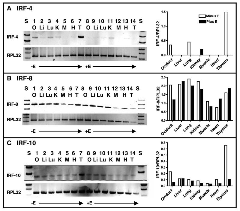
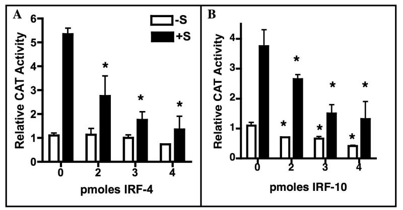
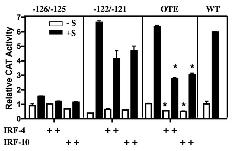
Although the ovalbumin (Ov) gene has served as a model to study tissue-specific, steroid hormone-induced gene expression in vertebrates for decades, the mechanisms responsible for regulating this gene remain elusive. Ov is repressed in non-oviduct tissue and in estrogen-deprived oviduct by a strong repressor site located from -130 to -100 and designated CAR for COUP-TF adjacent repressor. The goal of this study was to identify the CAR binding protein(s). A transcription factor database search revealed that a putative interferon-stimulated response element (ISRE), which binds interferon regulatory factors (IRFs), is located in this region. Gel mobility shift assays demonstrated that the protein(s) binding to the CAR site is recognized by an IRF antibody and that mutations in the ISRE abolish that binding. In hopes of identifying the IRF(s) responsible for the tissue-specific regulation of Ov, mRNA levels for IRFs-4, -8, and -10 were measured in seven tissues from chicks treated with or without estrogen. PCR experiments showed that both IRF-8 and -10 are expressed in all chick tissues tested whereas IRF-4 has a much more limited expression pattern. Transfection experiments with OvCAT (chloramphenicol acetyltransferase) reporter constructs demonstrated that both IRF-4 and IRF-10 are capable of repressing the Ov gene even in the presence of steroid hormones and that nucleotides in the ISRE are required for repression. These experiments indicate that the repressor activity associated with the CAR site is mediated by IRF family members and suggest that IRF members also repress Ov in non-oviduct tissues.


Figures

Fig. 1
Fig. 1
Regulatory elements in the Ov promoter. A. Map of the two major regulatory units in Ov, the SDRE (-892 to -793) and the NRE (-308 to -100). Genomic footprinting and/or mutagenesis indicate that protein or protein complexes bind to all of the sites that are numbered. Designated names are listed above the box depicting Ov, and real names are given in the box. The circles represent various transcription factors or factor complexes. Any circles with the same pattern represent the same factors or members of the same transcription factor family. B. The sequences in the regions of the OTE and CAR regulatory sites are shown. Sequences common to both of these are underlined. The ISRE consensus sequence and a putative unknown transcription factor binding site sequence are aligned with the common sites.
Fig. 2
Fig. 2
The OTE and CAR sites bind a similar protein(s). GMSAs were performed using the conditions described in Section 2.2. A. GMSA with the labeled CAR oligomer (-130 to -100). B. GMSA with the labeled OTE oligomer (-198 to -170). Lane 1: probe alone. Lanes 2-8 contain 5 μg of laying hen oviduct nuclear protein extract. Lanes 3 and 4 contain 10X and 50X molar unlabeled OTE oligomer, respectively. Lanes 5 and 6 contain 10X and 50X molar unlabeled CAR oligomer, respectively. Lanes 7 and 8 contain 10X and 50X molar unlabeled nonspecific competitor, respectively.
Fig. 3
Fig. 3
Oligomers with critical nucleotides mutated in the ISRE attenuate competition with the CAR oligomer. A. GMSAs were performed using the conditions described in Section 2.2 and . Lanes 1 to 11 contain the labeled CAR oligomer (-130 to -100). Lanes 2 to 11 contain 5 μg of 3 day estrogen-withdrawn oviduct nuclear protein. Lane 3 contains 25X molar unlabeled wt CAR oligomer. Lanes 4 to 10 contain 25X molar unlabeled mutated CAR oligomers that are described in Panel B. Lane 11 contains 25X molar unlabeled non-specific DNA. The arrow designates the shifted DNA-protein complex that is specific. B. The CAR competitor oligomer sequences (mutated nucleotides are shown in lower case) in the order used in the experiment. The positions of the mutated bases relative to the transcription start site are given. Comparable results were found with 50X molar excess competitors. C. GMSAs were performed using the conditions described in Section 2.2. Lanes 12 to 18 contain the labeled CAR oligomer. Lanes 13 to 18 contain 5 μg of nuclear extract protein from sexually immature chickens that had never been treated with estrogen. Lane 14 also contains 25X molar unlabeled wt CAR oligomer. Lane 15 contains 25X molar unlabeled mutated CAR oligomer. Lane 16 contains 25X molar unlabeled non-specific DNA. Lane 17 contains 25X molar unlabeled wt consensus IRF oligomer. Lane 18 contains 25X molar unlabeled mutated IRF consensus oligomer. The arrow designates the specific shifted DNA-protein complex. The GMSAs in A and C differ somewhat in their binding pattern because the hormonal treatment of the chicks differed. Also, the pattern in Fig.3A differs from that of Fig. 2 because the protein extracts used in 3A were not fresh. The upper band seen in Fig. 2 is labile.
Fig. 4
Fig. 4
The CAR complex contains a protein that cross reacts with an IRF-4 antibody. A. GMSA with the wt CAR oligogomer (-130 to -100) with (lane 2) and without (lane 1) nuclear protein from estrogen-withdrawn chicks. B. Gel regions as indicated by the boxes from a gel comparable to that in A were run on an SDS/PAGE gel and subjected to western blotting with an irrelevant (δEF1) antibody. C. Areas 1, 2 and 3 were excised from the gel in A and analyzed by western blotting using an IRF-4 antibody (Santa Cruz). This antibody also recognizes chicken IRFs-8, and -10 (data not shown). Area 3 was used as a control.
Fig. 5
Fig. 5
Tissue distribution of IRF-4, -8, and -10 mRNAs in estrogen-withdrawn and estrogen-stimulated chick tissues. Panel A, left side: RNA was isolated from 3 day estrogen-withdrawn (lanes 1 to 7) or chronically estrogen-stimulated (lanes 8 to 14) chicken tissues. RT-PCR reactions for IRF-4 and RPL32 were run as described in Section 2.3. Lanes 1 and 8: oviduct, Lanes 2 and 9: liver, Lanes 3 and 10: lung, Lanes 4 and 11: kidney, Lanes 5 and 12: muscle, Lanes 6 and 13: heart, and Lanes 7 and 14: thymus. Right side: IRF-4 mRNA was quantified relative to that of RPL32 (ribosomal protein L32) for each tissue. For quantification, the background was removed for each lane. When no bars are present, the ratio was too low to graph. This is representative of an experiment done three times. Panel B: As in Panel A except that IRF-8 mRNA was quantified. Panel C: As in Panel A except that IRF-10 mRNA was quantified. Note that the y-axes in histograms differ among the IRF mRNAs.
Fig. 6
Fig. 6
Overexpression of IRF-4 or IRF-10 represses Ov. Primary oviduct cells were cultured for 24 hrs without and with estrogen and corticosterone (+S). The pOvCAT-900 reporter vector was cotransfected with increasing amounts of an IRF-4 (panel A) or IRF-10 (panel B) expression plasmid. Total expression plasmid DNA was held constant at 4 pmol using the empty vector. Data are presented as CAT activity relative to pOvCAT-900 in the absence of steroid hormones (-S). Statistical significance was determined using one way ANOVA with Tukey’s Multiple Comparison post test. Stars indicate samples that are significantly different from pOvCAT with the corresponding hormonal treatment in the absence of added IRF (0 pmoles IRF). For IRF-4 +S, p<0.021; for IRF-10 -S, p<0.05; for IRF-10 +S, p<0.01
Fig. 7
Fig. 7
Mutation of critical nucleotides in the ISRE located in the CAR site reveal roles for both positive and negative regulation of Ov. Primary oviduct cells were cultured and transfected as described in the legend to Fig. 6. The pOvCAT reporter vectors with mutations in the indicated sites were cotransfected with IRF-4 or IRF-10 expression plasmid. Mutations were made at -126 and -125, at -122 and -121, or at -193 to -191 in the OTE ISRE as indicated in Section 2.4. Statistical significance was determines using one way ANOVA with Tukey’s Multiple Comparison post test. Stars indicate samples that are significantly different from the corresponding vector and hormonal treatment in the absence of added IRF. For OTE -S, p<0.05; for OTE +S, p<0.01

Similar articles

Cited by

References

    1. Bellard M, Dretzen G, Belard F, Oudet P, Chambon P. Disruption of the typical chromatin structure in a 2500 base-pair region at the 5′-end of the actively transcribed ovalbumin gene. EMBO J. 1982;1:223–230. - PMC - PubMed
    1. Chamberlain EM, Sanders MM. Identification of the novel player ™EF1 in estrogen transcriptional cascades. Mol. Cell. Biol. 1999;19:3600–3606. - PMC - PubMed
    1. Dadoune JP, Pawlak A, Alfonsi MF, Siffroi JP. Identification of transcripts by macroarrays, RT-PCR and in situ hybridization in human ejaculate spermatozoa. Mol. Human Reprod. 2005;11:133–140. - PubMed
    1. Dean DM, Jones PM, Sanders MM. Alterations in chromatin structure are implicated in the activation of the steroid hormone response unit in the ovalbumin gene. DNA Cell Biol. 2001;20:27–39. - PubMed
    1. Dean DM, Jones PS, Sanders MM. Regulation of the chick ovalbumin gene by estrogen and corticosterone requires a novel DNA element that binds a labile protein, Chirp-1. Mol. Cell. Biol. 1996;16:2015–2024. - PMC - PubMed

Publication types

MeSH terms

LinkOut - more resources