. 2009 Apr 1;23(7):849-61.
doi: 10.1101/gad.1748409.

GCN5 is a required cofactor for a ubiquitin ligase that targets NF-kappaB/RelA

Affiliations

GCN5 is a required cofactor for a ubiquitin ligase that targets NF-kappaB/RelA

Xicheng Mao et al. Genes Dev. .

Abstract

The transcription factor NF-kappaB is a critical regulator of inflammatory and cell survival signals. Proteasomal degradation of NF-kappaB subunits plays an important role in the termination of NF-kappaB activity, and at least one of the identified ubiquitin ligases is a multimeric complex containing Copper Metabolism Murr1 Domain 1 (COMMD1) and Cul2. We report here that GCN5, a histone acetyltransferase, associates with COMMD1 and other components of the ligase, promotes RelA ubiquitination, and represses kappaB-dependent transcription. In this role, the acetyltransferase activity of GCN5 is not required. Interestingly, GCN5 binds more avidly to RelA after phosphorylation on Ser 468, an event that is dependent on IKK activity. Consistent with this, we find that both GCN5 and the IkappaB Kinase (IKK) complex promote RelA degradation. Collectively, the data indicate that GCN5 participates in the ubiquitination process as an accessory factor for a ubiquitin ligase, where it provides a novel link between phosphorylation and ubiquitination.


Figures
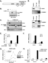
Figure 1.
Figure 1.
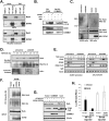
GCN5 binds to COMMD1 and inhibits NF-κB-mediated transcription. (A) Identification of GCN5 as a COMMD1-associated factor. Schematic representation of GCN5 with the boundaries of its HAT domain and Bromo domain (BD). The C-terminal peptide of GCN5 (amino acids 817–828) identified by LC/MS-MS is shown. The HAT activity and Bromo domain function are dependent on residues E575 and Y814, respectively. (B) COMMD1 binds GCN5 through its COMM domain. GCN5 was coexpressed with full-length COMMD1 (FL), its N terminus (N), or its COMM domain (CD) fused with GST. Subsequently, COMMD1 was precipitated and the recovered material was immunoblotted for GCN5. (C) GCN5 interacts with other COMMD proteins. HEK293 cells were cotransfected with GCN5 and the indicated COMMD proteins fused to GST, which were subsequently precipitated from cell lysates using GSH Sepharose beads. The presence of coprecipitated GCN5 was determined by immunoblotting. (D) Coprecipitation of endogenous GCN5 and COMMD1. Cell extracts were subjected to immnoprecipitation for COMMD1 (top panel) or GCN5 (bottom panel). Immunoblotting for GCN5 and COMMD1 was performed as indicated (ns, nonspecific band; input is 0.5% of the IP material). (EG) GCN5 represses NF-κB-dependent gene expression. HEK293 cells were transiently transfected with siRNA against GCN5 (E) or a GCN5 expression vector. U2OS cells were stably transduced with a lentivirus expressing shRNA against GCN5 (F). After TNF stimulation, the indicated transcript levels were measured by qRT–PCR. (H) The effect of GCN5 is dependent on RelA. HEK293 cells were transiently transfected with siRNA against GCN5, and additionally, cells received control siRNA transfection (open bars) or siRNA transfection against RelA (black bars). The effect on TNF transcript levels was assessed by qRT–PCR.
Figure 2.
Figure 2.
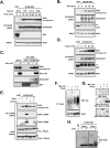
GCN5 interacts with RelA and promotes its ubiquitination. (A) Coprecipitation of endogenous RelA and GCN5. WCLs or nuclear extracts were prepared from unstimulated and TNF-stimulated HEK293 cells. GCN5 was immunoprecipitated, and the recovered material was immunoblotted for RelA (input is 0.8% of the IP material). (B) The GCN5/COMMD1 complex interacts with endogenous RelA. GCN5 fused with GST and COMMD1 fused to a biotinylation tag were expressed in cells. GCN5 was purified through a GSH affinity column (PD: GCN5); GCN5 was eluted from the column and COMMD1 was precipitated from this fraction using streptavidin-agarose beads (Re-PD: COMMD1). Western blots for endogenous RelA, GCN5, and COMMD1 are presented. (C) GCN5 promotes the ubiquitination of endogenous RelA. Nuclear extracts from U2OS GCN5-deficient cells (left panels) or HEK293 cells transfected with GCN5 (right panels) were prepared. A denatured immunoprecipitation for RelA was subsequently performed and the presence of ubiquitinated RelA was determined by immunoblotting for ubiquitin. The deficiency and overexpression of GCN5 were determined by Western blotting for GCN5. (D) GCN5 deficiency increases nuclear RelA level. The GCN5-deficient U2OS cell line or the corresponding control line were treated with TNF for 10 min, followed by cycloheximide (CHX). At the indicated time points, cells were harvested and nuclear extracts were prepared and immunobloted for RelA and RNA Pol II (as a loading control). (Bottom panel) IκB-α levels in cytosolic extracts were determined by Western blotting to monitor the effect of TNF and CHX. (E) GCN5 deficiency prolongs RelA binding to NF-κB-responsive promoters. The same GCN5-deficient and control U2OS cell lines were treated with TNF as before. At the indicated time points, cells were lysed and used for ChIP. Occupancy of RelA, GCN5, and RNA Pol II on the ICAM1 and IL8 gene promoters was analyzed. (F) GCN5 promotes RelA ubiquitination despite inactivating mutations in its HAT or Bromo domains. GCN5 wild-type, E575Q (HAT-deficient), or Y814A (Bromo domain-deficient) were cotransfected with HA-RelA and His6-tagged ubiquitin, and the levels of ubiquitinated RelA were determined as before. (G) GCN5 promotes the degradation of RelA independently of its HAT activity. HEK293 cells were transfected with RelA along with GCN5 wild-type or E575Q mutant, in combination with COMMD1 or Cul2 as indicated. Cells were lysed and the expression of RelA was determined by Western blot. (H) The HAT activity of GCN5 is dispensable for repression of κB-dependent transcription. The effects of GCN5 E575Q on an NF-κB-responsive reporter (3κB-luc) or a Smad-responsive reporter (SBE-JONK) were examined by luciferase assay.
Figure 3.
Figure 3.
GCN5 interacts with the COMMD1-containing ligase. (A) GCN5 precipitates ubiquitin ligase activity. GCN5–GST or GST precipitated from transfected HEK293 cells, or recombinant (r) GST, or GCN5–GST prepared in E. coli were added to an in vitro ubiquitination reaction. In addition, rGST or rGCN5–GST were mixed with an HEK293 cell lysate, incubated at 4°C for 2 h, and then extensively washed prior to being used as a source for ubiquitin ligase activity. Formation of polyubiquitin chains in the reaction was determined by SDS-PAGE and immunoblotting for ubiquitin. (B) GCN5 was copurified as a Cul2-associated factor. Lysates were prepared from HEK293 cells overexpressing a GST-Cul2 fusion protein (Input) and were applied to a GSH-Sepharose affinity column, from which Cul2-containing complexes were subsequently eluted and concentrated by filtration (Eluate). The input, the flow-through material not bound to the column (FT), and eluate were subjected to immunoblotting for Cul2, COMMD1, Elongin C (EloC), GCN5, and TAF6. Silver stain of the eluate is shown in the right panel with the bands corresponding to Cul2, and the immunoblotted subunits noted by small arrows. Nonspecific bands are indicated by an asterisk (*). (C) GCN5 binds to Cul2, the main scaffold protein of the COMMD1-containing ligase. HEK293 cells were transfected with GST or GST–GCN5, which were precipitated from cell lysates. The presence of coprecipitated endogenous Cul2 was determined by Western blot. The position of the 97-kDa molecular weight marker is indicated. (D) The C terminus of GCN5 binds to COMMD1 and Cul2. Cells were transfected with GCN5 full-length (FL) fused to GST or the indicated truncation mutants along with Flag-Cul2 or COMMD1-Flag. GCN5 was precipitated by GSH Sepharose beads, and the presence of Cul2 or COMMD1 in the precipitated material was determined by Western blot analysis. (E) The C terminus of GCN5 containing the HAT and Bromo domains precipitates E3 ligase activity. GCN5 full-length (FL) fused to GST or truncation mutants spanning its N terminus (N-term, amino acids 1–491) or C terminus (HAT/Bromo, amino acids 492–837) were expressed in mammalian cells. These proteins were precipitated from cell lysates, added to in vitro ubiquitination reactions, and polyubiquitin chain formation was determined as before.
Figure 4.
Figure 4.
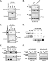
RelA phosphorylation enhances GCN5-RelA interactions. (A) Various IKK-activating stimuli promote GCN5–RelA binding. HEK293 cells were transfected with GCN5–GST or GST and subsequently treated with TNF, IL-1β, or Flagellin prior to lysis and GSH precipitation. The presence of coprecipitated endogenous RelA was determined by immunoblotting. (B) TNF treatment prior to lysis promotes RelA–GCN5 binding in vitro. HEK293 cells were treated with TNF or Calyculin A, and the obtained lysates were mixed with bacterially made rGST or rGCN5–GST, followed by precipitation. The presence of endogenous RelA in the recovered material was determined by immunoblotting. (C) IκB does not impair the GCN5–RelA interaction. HEK293 cells were transfected with IκB-α superdominant (SD), and the ability of recombinant GCN5 to precipitate endogenous RelA post-lysis was examined as in B. (D) The phosphatase inhibitor Calyculin A promotes RelA–GCN5 binding in vitro. HEK293 cells were treated with Calyculin A and the obtained lysates were mixed with rGST or rGCN5–GST, followed by precipitation. The presence of endogenous RelA in the recovered material was determined by immunoblotting. (E) Dephosphorylation of RelA abrogates its induced binding to GCN5. HEK293 cells were treated with Calyculin A and subsequently lysed in a buffer without phosphatase inhibitors. The lysate was incubated with λ-protein phosphatase (λ-PPase) as indicated, and in vitro binding of RelA to recombinant GCN5 was performed as in D. The recovered material was immunoblotted to detect the coprecipitation of endogenous RelA and phosphorylated RelA. (F) Phosphorylation promotes RelA ubiquitination. Wild-type MEFs were treated with MG-132 (30 min) and Calyculin A as indicated. RelA was subsequently immunoprecipitated from cell lysates and the presence of ubiquitinated RelA was determined by immunoblotting for ubiquitin. (G) Phosphorylated RelA is labile and is stabilized by proteasomal blockade. Wild-type MEFs were treated with Calyculin A and MG-132 as indicated. Phosphorylated RelA levels were determined by Western blot using two phospho-specific antibodies. (H) GCN5-associated ligase activity ubiquitinates RelA in vitro. HEK293 cells transfected with GCN5–GST or GST were treated without or with Calyculin A as shown. Material precipitated by GSH beads was applied to an in vitro ubquitination reaction as in Figure 3A. Coprecipitated RelA and its ubiquitination were shown by immunoblotting.
Figure 5.
Figure 5.
The GCN5–RelA interaction requires the C terminus of RelA and is enhanced by phosphorylation at Ser 468. (A) GCN5 interacts with the transactivation domain of RelA. Recombinant RelA fragments fused to GST were purified from E. coli and their ability to precipitate radiolabeled in vitro translated GCN5 was examined by autoradiography. Coomasie staining demonstrates the amount of recombinant protein used. (B) The TAD of RelA is responsible for phosphorylation-inducible binding. The same experiment as in B was performed, but this time using lysates from cells transfected with RelA–TAD (306–551). (Bottom panel) In addition, λ-PPase treatment of the lysate was also performed prior to binding. (C) GCN5 interacts with TA1, encompassed by the last 21 amino acids of RelA. The indicated recombinant RelA fragments fused to GST were purified from HEK293 cells, and their ability to precipitate radiolabeled in vitro translated GCN5 was examined by autoradiography. Coomasie staining demonstrates the amount of recombinant protein used. (D) A peptide from TA1 is capable of disrupting GCN5–RelA binding in vitro. Recombinant RelA fragments fused to GST were purified from E. coli, and their ability to precipitate radiolabeled in vitro translated GCN5 was examined as in A. In addition, peptides spanning the indicated amino acid residues in RelA were added to the in vitro binding reaction as potential competitive inhibitors. (E) The phosphorylation residue that mediates RelA–GCN5 inducible binding is not contained in the last 21 amino acids of RelA. The same experiment as in Figure 4E, but this time using two mutations in the TAD: deletion of the last 21 amino acids (306–530), Δ21; or mutation of the last six serines in RelA (S535, 536, 543, 547, 550, 551A), 6S/A. (F) Ser 468 phosphorylation is required for RelA–GCN5-inducible binding. The same experiment as in B and E, but using either Calyculin A treatment (left) of TNF stimulation (right). Wild-type RelA or a point mutant at Ser 468 are compared.
Figure 6.
Figure 6.
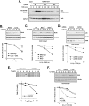
IKK and GCN5 promote RelA degradation. (A) IKK is required for TNF-induced RelA–GCN5 binding. Wild-type (WT), Ikkα −/−, or Ikkβ −/− MEFs were stimulated with TNF and subsequently lysed. This material was applied to rGCN5–GST for in vitro binding as before. The coprecipitation of endogenous RelA was determined by immunoblotting. (B) IL1-β stimulation decreases RelA stability. Cycloheximide (CHX) was used to determine the stability of RelA in wild-type MEFs that were untreated or stimulated with IL1-β. RelA levels were determined by immunoblotting and densitometry analysis. (C) Ikk deficiency stabilizes RelA in IL1-β treated cells. Similar to B, but the effects of IL1-β treatment were compared between wild type, Ikkα −/−, or Ikkβ −/− MEFs. (D) GCN5 deficiency prevents IL1-β-promoted degradation. HEK293 cells were transfected with the indicated siRNA and subsequently treated with IL1-β. The stability of RelA after cycloheximide (CHX) treatment was examined by immunoblotting and densitometry analysis. (E) GCN5 deficiency stabilizes RelA in U2OS cells. GCN5-deficient U2OS cells and the corresponding control cells were metabolically labeled by 35S-methionine and 35S-cysteine. At the indicated time points after labeling, cells were lysed for a denatured immunoprecipitation for RelA. The recovered material was examined by autoradiography after SDS-PAGE separation. (F) RelA 1–530 and RelA S468A are more stable than full-length (FL) RelA. The indicated human RelA constructs were introduced into rela-deficient MEFs through lentiviral infection and selection. Protein stability was examined as before.

Similar articles

Cited by

References

    1. Arenzana-Seisdedos F., Turpin P., Rodriguez M., Thomas D., Hay R.T., Virelizier J.L., Dargemont C. Nuclear localization of IκBα promotes active transport of NF−κB from the nucleus to the cytoplasm. J. Cell Sci. 1997;110:369–378. - PubMed
    1. Baeuerle P.A., Baltimore D. I κ B: A specific inhibitor of the NF-κ B transcription factor. Science. 1988;242:540–546. - PubMed
    1. Bu P., Evrard Y.A., Lozano G., Dent S.Y. Loss of Gcn5 acetyltransferase activity leads to neural tube closure defects and exencephaly in mouse embryos. Mol. Cell. Biol. 2007;27:3405–3416. - PMC - PubMed
    1. Burstein E., Ganesh L., Dick R.D., van De Sluis B., Wilkinson J.C., Klomp L.W., Wijmenga C., Brewer G.J., Nabel G.J., Duckett C.S. A novel role for XIAP in copper homeostasis through regulation of MURR1. EMBO J. 2004;23:244–254. - PMC - PubMed
    1. Burstein E., Hoberg J.E., Wilkinson A.S., Rumble J.M., Csomos R.A., Komarck C.M., Maine G.N., Wilkinson J.C., Mayo M.W., Duckett C.S. COMMD proteins: A novel family of structural and functional homologs of MURR1. J. Biol. Chem. 2005;280:22222–22232. - PubMed

Publication types