. 2009 Apr 1;29(13):4023-34.
doi: 10.1523/JNEUROSCI.4566-08.2009.

Fast adaptation and Ca2+ sensitivity of the mechanotransducer require myosin-XVa in inner but not outer cochlear hair cells

Affiliations

Fast adaptation and Ca2+ sensitivity of the mechanotransducer require myosin-XVa in inner but not outer cochlear hair cells

Ruben Stepanyan et al. J Neurosci. .

Abstract

In inner ear hair cells, activation of mechanotransduction channels is followed by extremely rapid deactivation that depends on the influx of Ca(2+) through these channels. Although the molecular mechanisms of this "fast" adaptation are largely unknown, the predominant models assume Ca(2+) sensitivity as an intrinsic property of yet unidentified mechanotransduction channels. Here, we examined mechanotransduction in the hair cells of young postnatal shaker 2 mice (Myo15(sh2/sh2)). These mice have no functional myosin-XVa, which is critical for normal growth of mechanosensory stereocilia of hair cells. Although stereocilia of both inner and outer hair cells of Myo15(sh2/sh2) mice lack myosin-XVa and are abnormally short, these cells have dramatically different hair bundle morphology. Myo15(sh2/sh2) outer hair cells retain a staircase arrangement of the abnormally short stereocilia and prominent tip links. Myo15(sh2/sh2) inner hair cells do not have obliquely oriented tip links, and their mechanosensitivity is mediated exclusively by "top-to-top" links between equally short stereocilia. In both inner and outer hair cells of Myo15(sh2/sh2) mice, we found mechanotransduction responses with a normal "wild-type" amplitude and speed of activation. Surprisingly, only outer hair cells exhibit fast adaptation and sensitivity to extracellular Ca(2+). In Myo15(sh2/sh2) inner hair cells, fast adaptation is disrupted and the transduction current is insensitive to extracellular Ca(2+). We conclude that the Ca(2+) sensitivity of the mechanotransduction channels and the fast adaptation require a structural environment that is dependent on myosin-XVa and is disrupted in Myo15(sh2/sh2) inner hair cells, but not in Myo15(sh2/sh2) outer hair cells.


Figures

Figure 1.
Figure 1.
Recordings of MET responses in cochlear hair cells. A, Cultured organ of Corti explant with a piezo-driven probe (left) and a patch pipette (right). A third pipette (top left) was used to deliver test solutions to the hair bundle. The image of the probe was projected to a pair of photodiodes (dashed rectangles) that monitor displacement stimuli. The specimen was visualized on an inverted microscope. Scale bar, 5 μm. B, In-scale schematic drawing of a 5 μm probe deflecting stereocilia of Myo15 +/sh2 (top) and Myo15 sh2/sh2 (bottom) IHCs. Stereocilia of Myo15 sh2/sh2 IHCs are deflected by the probe located at the top of the bundle.
Figure 2.
Figure 2.
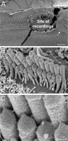
Postexperimental examination of specimens. A, The site of patch-clamp recordings is easy to identify even in the presence of the SEM preparation artifact (a fracture across the recording site). B, C, An IHC that is indicated by an arrow in A is shown at high (B) and very high (C) magnifications. Our oldest Myo15 +/sh2 preparation (P4 + 5 d in vitro) is shown to illustrate preservation of IHC morphology in vitro. Scale bars: A, 20 μm; B, 0.5 μm; C, 200 nm.
Figure 3.
Figure 3.
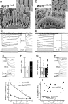
Loss of fast adaptation in Myo15 sh2/sh2 IHCs. A, B, SEM images of Myo15 +/sh2 (A) and Myo15 sh2/sh2 (B) IHCs from approximately the same mid-cochlear location. Insets show stereocilia links of these cells at higher magnification (and different viewing angle in B). Scale bars: 1 μm (main panels); 200 nm (insets). C, D, MET responses (top traces) evoked by the graded deflections of stereocilia in the same specimens that were imaged in A and B. Horizontal movement of the piezo-driven probe was monitored by a photodiode technique (bottom traces). Responses to negative deflections are shown in black color. Right traces show the beginning of MET responses on a faster time scale to reveal fast adaptation. Adaptation at the small bundle deflection of 150 nm (τad) is fitted with single exponential decay (dashed lines). E, H, The same MET records on an ultrafast time scale to reveal time constants of MET activation and probe movement. Holding potential was −90 mV. Age of the cells: (A, C, E) P2 + 2 d in vitro; (B, D, H) P3 + 1 d in vitro. F, Average maximum MET responses. G, Time constants of MET activation (τact): average and row data. Gray area indicates the range of time constants of the stimuli in the same experiments. I, Percentage changes of the MET current at the end of 25 ms stimulation step (extent of adaptation) as a function of stimulus intensity. J, The relationship between maximum MET current and time constant of adaptation at the small bundle deflection of 150 nm. Solid line shows least squares fit to data points from Myo15 +/sh2 IHCs, r = −0.91, p < 0.0001. The same records contribute to F, G, I, and J. Number of cells: n = 17 (Myo15 +/sh2), n = 18 (Myo15 sh2/sh2). All averaged data are shown as mean ± SE. Asterisks indicate statistical significance: *p < 0.05, **p < 0.01, ***p < 0.0001 (t test of independent samples).
Figure 4.
Figure 4.
Slow adaptation in Myo15 sh2/sh2 IHCs. A, B, SEM examination (A) and MET responses (B) in an experiment with one of the most prominent adaptation in Myo15 sh2/sh2 IHCs. Inset in A shows top-to-top stereocilia links; the specimen is viewed from an ∼30° angle. Age of the specimen was P3 + 2 d in vitro. Scale bars: A, 1 μm; A, inset, 100 nm. C, Scatter plot of the time constants of adaptation in Myo15 sh2/sh2 IHCs and slow adaptation in Myo15 +/sh2 IHCs versus maximum MET current. The same set of experiments as in Figure 3. Time constants of slow adaptation in Myo15 +/sh2 IHCs were determined for MET responses to 300–600 nm deflections by fitting these responses to double exponential function.
Figure 5.
Figure 5.
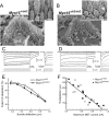
Apparently normal adaptation in Myo15 sh2/sh2 OHCs. A, B, SEM images of Myo15 +/sh2 (A) and Myo15 sh2/sh2 (B) OHCs from approximately the same mid-cochlear location. Insets show stereocilia links at higher magnification. Presumable tip links in Myo15 sh2/sh2 OHCs are indicated by arrowheads. Scale bars: 0.5 μm (main panels) and 200 nm (insets). C, D, MET responses (top traces) evoked by the graded deflections of stereocilia in the same specimens that were imaged in A and B. Movement of the piezo-driven probe was calibrated by video recording and command voltage was converted to displacement units. Right traces show the beginning of MET responses on a faster time scale. Fast adaptation to small stimuli is fitted with single exponential decays (dashed lines). Holding potential was −85 mV. Age of the cells: (A, C) P2 + 1 d in vitro; (B, D) P2 + 2 d in vitro. E, Percentage changes of the MET current at the end of 25 ms stimulation step (extent of adaptation) as a function of stimulus intensity. Data are shown as mean ± SE F, The relationship between maximum MET current and the time constant of fast adaptation to small bundle deflection of 150 nm. Solid line shows least squares fit to all points, r = −0.88, p < 0.0001. The same records contribute to E and F. Number of cells: n = 8 (Myo15 +/sh2), n = 12 (Myo15 sh2/sh2).
Figure 6.
Figure 6.
Disrupted Ca2+ sensitivity of the MET machinery in Myo15 sh2/sh2 IHCs. A, B, MET responses in Myo15 +/sh2 (A) and Myo15 sh2/sh2 (B) IHCs evoked by the graded deflections of stereocilia in an extracellular solution containing 1.5 mm Ca2+ (top traces) and in low Ca2+ solution containing 20 μm Ca2+ (middle traces). Bottom traces show command voltage converted to displacement units. Right panels show the same records on a faster time scale. Scale bars of the MET current apply to both top and middle traces. C, E, Changes of the whole-cell current produced by low extracellular Ca2+ in the same IHCs. Dashed lines indicate the breaks, during which we examined MET responses in low Ca2+ environment. Age of the cells: (A, C) P3 + 5 d in vitro; (B, E) P3 + 4 d in vitro. D, Resting whole-cell current at holding potential of −60 mV (I rest) and the changes of this current (ΔI) evoked by the application of low Ca2+ extracellular medium to the stereocilia bundles of Myo15 +/sh2 (open bars) and Myo15 sh2/sh2 (closed bars) IHCs. F, Maximum amplitude of MET responses during and after application of low Ca2+ extracellular solution as a percentage of maximal amplitude of MET responses before application. MET responses were recorded at holding potential of −90 mV. Averaged data in D and F are shown as mean ± SE. Number of cells: n = 6 (IHCs, Myo15 +/sh2), n = 8 (IHCs, Myo15 sh2/sh2), n = 3 (OHCs, Myo15 sh2/sh2). Asterisks indicate statistical significance: *p < 0.05, **p < 0.01 (t test of independent samples in D and paired t test in F).
Figure 7.
Figure 7.
Mechanotransduction in Myo15 sh2/sh2 IHCs is mediated by top-to-top stereocilia links. A–C, SEM images of the organ of Corti explants incubated in standard HBSS (A), Ca2+-free HBSS supplemented with 5 mm BAPTA (B), and in standard HBSS supplemented with 50 μg/ml subtilisin (C). Insets show magnified images of stereocilia links. All incubations were performed at room temperature. The duration of incubations: (A, B) 5 min; (C) 60 min. D, MET responses in a Myo15 sh2/sh2 IHC before (left) and after (right) application of Ca2+-free HBSS supplemented with 5 mm BAPTA for 1 min. Holding potential: −90 mV. E, Whole-cell current responses evoked by the application of 250 μm dihydrostreptomycin to the stereocilia bundle of Myo15 +/sh2 (top) and Myo15 sh2/sh2 (bottom) IHCs. Holding potential: −60 mV. F, Brief application of 10 μm FM1–43 to the cultured Myo15 sh2/sh2 organ of Corti explant (45 s at room temperature) results in accumulation of the dye in both OHCs and IHCs. Arrows at the right side of the panel point to three rows of OHCs and one row of IHCs. G, SEM image of a Myo15 sh2/sh2 IHC showing membrane tenting (arrows) at the attachments of top-to-top stereocilia links. The ages of the cells: (A–C) P3 + 3 d in vitro; (D) P3 + 1 d in vitro; (E) P3 + 4 d in vitro; (F) P4 + 4 d in vitro; (G) P1 + 4 d in vitro. Scale bars: A–C, 1 μm; F, 50 μm; G, 200 nm.
Figure 8.
Figure 8.
Average relationships (mean ± SE) between peak transduction current and probe displacement in Myo15 +/sh2 (open circles, n = 17) and Myo15 sh2/sh2 (closed circles, n = 18) IHCs. Asterisks indicate statistical significance of the difference (p < 0.05, t test of independent samples). The same set of experiments as in Figures 3 F,G,I,J and 4C.
Figure 9.
Figure 9.
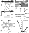
Abnormal directional sensitivity of Myo15 sh2/sh2 IHCs. A, MET currents evoked by negative displacements of stereocilia with a piezo-driven stiff probe in Myo15 sh2/sh2 (top) and Myo15 +/sh2 (middle) IHCs. Bottom traces show command voltage converted to displacement units. Holding potential: −90 mV. B, MET responses evoked by consecutive negative and positive deflections of the same bundle by a fluid-jet. Top image illustrates a fluid-jet pipette (left), a Myo15 sh2/sh2 IHC bundle (middle), and a patch pipette (right). Traces below show the records obtained in this experiment (from top to bottom): MET responses, movement of the stereocilia bundle determined by off-line frame-by-frame analysis of the video record (Frolenkov et al., 1997), and the command pressure that was applied to the fluid-jet pipette. C, In an experiment when the probe was strongly adhered to the hair bundle, both positive and negative ramp-like deflections of stereocilia evoked MET responses in a Myo15 sh2/sh2 IHC. Top traces show MET responses at different holding potentials (right), which are indicated after correction for the voltage drop across series resistance. Note that the MET responses reverse at the holding potential close to zero as expected for nonselective cation channels. Bottom trace shows the stimulus. D, Current-displacement relationship derived from MET responses at + 71 mV holding potential in (C). To illustrate sampling frequency, the data are presented as a scatter plot of individual measurements during ramp deflections of stereocilia. The ages of the cells: (A) P3 + 4 d in vitro and P3 + 3 d in vitro; (B) P3 + 4 d in vitro; (C, D) P4 + 4 d in vitro. Insets in A, D show schematically the direction of the stimuli.
Figure 10.
Figure 10.
Some potential mechanisms that could result in the loss of Ca2+ sensitivity and fast adaptation of the mechanotransducer in Myo15 sh2/sh2 IHCs. A, An accessory element may convey Ca2+ sensitivity to the MET channel. During Ca2+ binding, this accessory element may force conformational changes of the MET channel into the state with a lower single channel conductance. In Myo15 sh2/sh2 IHCs, this element may somehow become dissociated from the MET channel. For example, it may be localized at the tip of a shorter stereocilium. The MET channels (open circles) may be localized at either or both ends of the tip link (Denk et al., 1995). B, According to the tension release model of fast adaptation, Ca2+ ions enter the cell and bind to the nearby tension release element that releases the tension applied to the MET channel (Bozovic and Hudspeth, 2003; Martin et al., 2003). If the tension release element is situated at the tip of a stereocilium, it may become separated from the MET channel in Myo15 sh2/sh2 IHCs, resulting in loss of fast adaptation. C, Because myosin-Ic molecules are thought to be assembled in series along the length of a stereocilium (Gillespie and Cyr, 2004), the overall displacement produced by this assembly might also be directed along the stereocilium. If this tension release displacement is oriented along the stereocilium, it would decrease the tension of an obliquely oriented tip link in a normal Myo15 +/sh2 IHC but would not be effective in changing the tension of a perpendicularly oriented top-to-top stereocilia link in a mutant Myo15 sh2/sh2 IHC.

Similar articles

Cited by

References

    1. Adamek N, Coluccio LM, Geeves MA. Calcium sensitivity of the cross-bridge cycle of Myo1c, the adaptation motor in the inner ear. Proc Natl Acad Sci U S A. 2008;105:5710–5715. - PMC - PubMed
    1. Ahmed ZM, Goodyear R, Riazuddin S, Lagziel A, Legan PK, Behra M, Burgess SM, Lilley KS, Wilcox ER, Riazuddin S, Griffith AJ, Frolenkov GI, Belyantseva IA, Richardson GP, Friedman TB. The tip-link antigen, a protein associated with the transduction complex of sensory hair cells, is protocadherin-15. J Neurosci. 2006;26:7022–7034. - PMC - PubMed
    1. Assad JA, Corey DP. An active motor model for adaptation by vertebrate hair cells. J Neurosci. 1992;12:3291–3309. - PMC - PubMed
    1. Assad JA, Hacohen N, Corey DP. Voltage dependence of adaptation and active bundle movement in bullfrog saccular hair cells. Proc Natl Acad Sci U S A. 1989;86:2918–2922. - PMC - PubMed
    1. Assad JA, Shepherd GM, Corey DP. Tip-link integrity and mechanical transduction in vertebrate hair cells. Neuron. 1991;7:985–994. - PubMed

Publication types

MeSH terms

LinkOut - more resources