. 2009 May;20(10):2615-25.
doi: 10.1091/mbc.e08-12-1162. Epub 2009 Apr 1.

Mrpl36 is important for generation of assembly competent proteins during mitochondrial translation

Affiliations

Mrpl36 is important for generation of assembly competent proteins during mitochondrial translation

Martin Prestele et al. Mol Biol Cell. 2009 May.

Abstract

The complexes of the respiratory chain represent mosaics of nuclear and mitochondrially encoded components. The processes by which synthesis and assembly of the various subunits are coordinated remain largely elusive. During evolution, many proteins of the mitochondrial ribosome acquired additional domains pointing at specific properties or functions of the translation machinery in mitochondria. Here, we analyzed the function of Mrpl36, a protein associated with the large subunit of the mitochondrial ribosome. This protein, homologous to the ribosomal protein L31 from bacteria, contains a mitochondria-specific C-terminal domain that is not required for protein synthesis per se; however, its absence decreases stability of Mrpl36. Cells lacking this C-terminal domain can still synthesize proteins, but these translation products fail to be properly assembled into respiratory chain complexes and are rapidly degraded. Surprisingly, overexpression of Mrpl36 seems to even increase the efficiency of mitochondrial translation. Our data suggest that Mrpl36 plays a critical role during translation that determines the rate of respiratory chain assembly. This important function seems to be carried out by a stabilizing activity of Mrpl36 on the interaction between large and small ribosomal subunits, which could influence accuracy of protein synthesis.


Figures

Figure 1.
Figure 1.
Both domains of Mrpl36 are critical for respiratory growth. (A) Schematic representation of Mrpl36 and mrpl36ΔC. The predicted probabilities to form coiled-coil structures (Lupas, 1997) are expressed in percentage. MTS, mitochondrial targeting signal. (B) Deletion of the CE domain of Mrpl36 reduces growth on nonfermentable carbon sources. The indicated cells were grown in full medium containing galactose to log phase. Serial 10-fold dilutions were spotted on YP plates containing 2% glucose or 2% glycerol, and plates were incubated at 30°C for 2 and 4 d, respectively. (C) The L31 domain is sufficient to support respiratory growth. The indicated strains were grown on synthetic medium containing galactose to log phase. Serial 10-fold dilutions were spotted on synthetic media containing 2% glucose or 2% glycerol, and plates were incubated at 30°C for 3 and 6 d, respectively. (D) Steady-state levels of the different Mrpl36 forms. Lysates of the indicated cells grown on galactose-containing media were prepared and analyzed by Western blotting using antibodies against Tom70 (loading control), the HA-tag, and Mrpl36. Black arrow indicates Mrpl36ΔC, and white arrow indicates Mrpl36ΔC-HA. Asterisk (*), possible degradation product.
Figure 2.
Figure 2.
mrpl36ΔC cells exhibit reduced translation efficiency and stability of mitochondrially encoded proteins. (A) Cells of the indicated strains were grown in synthetic media containing 2% galactose. Cytosolic translation was inhibited by cycloheximide, and mitochondrial translation products were radiolabeled for the indicated times. Labeling was stopped by alkaline lysis and samples were separated by SDS-PAGE. Western blotting against Tom70 and Tim23 served as loading controls. (B) Mitochondrial translation products were labeled in vivo in the indicated strains for 15 min. Next, [35S]methionine and cycloheximide were removed by extensive washing. The cells were further incubated in complete synthetic medium with 2% galactose. After the indicated times, proteins were extracted by alkaline lysis and applied to SDS-PAGE and autoradiography. (C) Signals of the experiment presented in B were densitometrically analyzed. The start value (0 min) was set to 1. Solid line, wild type; dashed line, mrpl36ΔC. (D) Stability of Cox3/Atp6 in wild-type and mrpl36ΔC cells as quantified from four independent experiments. The initial signal (0 min) was set to 1 and compared with the remaining signal after 150 min. A Student's t test was used to reveal significance.
Figure 3.
Figure 3.
Steady-state levels of mitochondrially encoded proteins are reduced in mrpl36ΔC cells. (A) Mitochondrially encoded proteins fail to accumulate on media not requiring respiration. Indicated amounts of mitochondria prepared from cultures grown on lactate or galactose were separated by SDS-PAGE and analyzed by Western blotting. (B) Mitochondria prepared from the two strains grown on lactate medium were solubilized in 2.6% digitonin (left) or 1% dodecyl-maltoside (right) and separated by BN-PAGE. The gel was stained with Coomassie (left) or blotted onto PVDF membrane (right) and analyzed by Western blotting. The positions of the supercomplexes are indicated. V2, ATPase dimer; III2/IV2, dimeric bc1 complexes associated with a dimer of COX complexes; III2/IV, dimer of bc1 complexes associated with a monomeric COX complex; V, ATPase monomer. Arrow (left) indicates a band containing nonassembled nuclear encoded subunits of the respiratory chain. (C) The enzyme activities depicted were measured with the mitochondria isolated from lactate grown cultures. Data were normalized to malate dehydrogenase and activities of the wild-type mitochondria were set to 1. (D) Overexpression of Mrpl36ΔC-HA allows the accumulation of mitochondrially encoded proteins under growth conditions not requiring respiration. Lysates of cells grown with galactose as carbon source were prepared and analyzed with Western blotting against Tom70 (loading control), Cox2, and aconitase (Aco1).
Figure 4.
Figure 4.
Mitochondrial translation generates unproductive proteins in mrpl36ΔC cells. (A) Labeling profiles of mitochondrial translation in cells with wild-type and cox2::ARG8m mitochondrial genomes. (B) Cells of the indicated strains were grown in synthetic media containing 2% galactose. Cytosolic translation was inhibited by cycloheximide. Mitochondrial translation products were radiolabeled for the indicated times, after which labeling was stopped by alkaline lysis. (C) Steady-state levels of Arg8. Proteins of isolated mitochondria were separated by SDS-PAGE and probed with antibodies against Arg8. Western blotting against Tim23 and Mrp20 was used as loading control. (D) Cells of the indicated strains were spotted in 10-fold dilutions onto media with or without arginine and incubated for 3 d at 30°C.
Figure 5.
Figure 5.
Interaction of ribosomal subunits is destabilized in mrpl36ΔC mitochondria. Translation products of wild-type (A) or mrpl36ΔC (B) mitochondria were labeled with [35S]methionine. (C) Labeling of translation products in wild-type mitochondria was stopped after 20 min by the addition of puromycin. Next, mitochondria were reisolated, lysed with 1% dodecyl-maltoside, and the cleared lysate subjected to velocity centrifugation on a linear sucrose gradient. The gradient was fractionated and analyzed by autoradiography and Western blotting. Quantifications of the signals for Mrp20 and Mrp51 are presented.
Figure 6.
Figure 6.
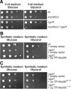
MRPL36 interacts genetically with GUF1. (A) The indicated strains were grown on galactose-containing medium to log phase. Serial 10-fold dilutions were spotted on YP plates containing 2% glucose or 2% glycerol, and plates were incubated at 30°C for 2 and 4 d, respectively. (B) The indicated cells were transformed with 2μ plasmids either containing or not containing MRPL36 under the control of the TPI promoter. The cells were grown on galactose-containing synthetic medium to log phase. Serial 10-fold dilutions were spotted on synthetic media without leucine containing either 2% glucose or 2% glycerol and incubated at 30°C for 3 and 6 d, respectively. (C) Mrpl36ΔC or the CE domain of Mrpl36 cannot suppress the Δguf1 phenotype. Δguf1 cells containing the indicated plasmids were analyzed as described in B.
Figure 7.
Figure 7.
Overexpressed Mrpl36 accumulates in a soluble pool in the matrix, from where the protein can dynamically integrate into the ribosome. (A) Mrpl36 is stable as a soluble matrix protein. Indicated mitochondria were lysed with Triton X-100. One-half of the obtained lysate was kept and served as a 100%-total (T); the other half was fractionated by centrifugation through a sucrose cushion. The resulting fractions, supernatant (S) and the pellet (P), together with the totals, were separated by SDS-PAGE and analyzed by Western blotting. (B) Imported Mrpl36 can dynamically integrate into preexisting ribosomes. Radiolabeled precursor of Mrpl36 was incubated with energized mitochondria of the indicated strains. After 30 min, mitochondria were reisolated and either exposed to hypotonic buffers to rupture the outer membrane (Swelling) or to iso-osmotic buffers. Mitochondria were treated with proteinase K (PK) to remove nonimported material; Western blotting against Tom70 was used to assess successful removal of exposed domains. A portion of the import reaction was lysed and separated by centrifugation through a sucrose cushion into a soluble and a ribosomal fraction. (C) Imported Mrpl3 cannot integrate into the ribosome. The experiment was conducted as described in B by using radiolabeled precursor of Mrpl3.
Figure 8.
Figure 8.
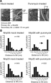
Mrpl36 dynamically interacts with the ribosome in vivo. (A) Wild-type cells were grown in lactate containing media in the absence (left) or presence (right) of puromycin and fixed with glutaraldehyde and formaldehyde. Next, mitochondrial ribosomal proteins were localized by postembedding immunogold labeling of cryosectioned cells. The distance of each gold particle relative to the inner membrane was measured. (B) Statistical analysis of the distribution of gold particles in mitochondria from cells grown in the absence (left) or presence of puromycin (right). Particles were grouped according to their distance to the inner membrane. n indicates the number of gold particles analyzed.
Figure 9.
Figure 9.
Hypothetical model for the role of Mrpl36 in mitochondrial protein synthesis. Under normal conditions (left), Mrpl36 binds close to the interface of large and small ribosomal subunit. This contact leads to a tight interaction of both subunits and hence allows efficient protein synthesis. In ribosomes from the mrpl36ΔC strain (right), this contact is destabilized, resulting in the production of defective polypeptides. During translation, Mrpl36 dynamically interacts with the ribosome at an as-yet unidentified step.

Similar articles

Cited by

References

    1. Ackerman S. H., Tzagoloff A. Function, structure, and biogenesis of mitochondrial ATP synthase. Prog. Nucleic Acid Res. Mol. Biol. 2005;80:95–133. - PubMed
    1. Agirrezabala X., Lei J., Brunelle J. L., Ortiz-Meoz R. F., Green R., Frank J. Visualization of the hybrid state of tRNA binding promoted by spontaneous ratcheting of the ribosome. Mol. Cell. 2008;32:190–197. - PMC - PubMed
    1. Arnold I., Pfeiffer K., Neupert W., Stuart R. A., Schagger H. Yeast mitochondrial F1F0-ATP synthase exists as a dimer: identification of three dimer-specific subunits. EMBO J. 1998;17:7170–7178. - PMC - PubMed
    1. Barrientos A., Zambrano A., Tzagoloff A. Mss51p and Cox14p jointly regulate mitochondrial Cox1p expression in Saccharomyces cerevisiae. EMBO J. 2004;23:3472–3482. - PMC - PubMed
    1. Bauerschmitt H., Funes S., Herrmann J. M. The membrane-bound GTPase Guf1 promotes mitochondrial protein synthesis under suboptimal conditions. J. Biol. Chem. 2008;283:17139–17146. - PubMed

Publication types

MeSH terms

Substances