. 2009 Jun;20(11):2661-72.
doi: 10.1091/mbc.e09-01-0094. Epub 2009 Apr 1.

LULL1 retargets TorsinA to the nuclear envelope revealing an activity that is impaired by the DYT1 dystonia mutation

Affiliations

LULL1 retargets TorsinA to the nuclear envelope revealing an activity that is impaired by the DYT1 dystonia mutation

Abigail B Vander Heyden et al. Mol Biol Cell. 2009 Jun.

Abstract

TorsinA (TorA) is an AAA+ ATPase in the endoplasmic reticulum (ER) lumen that is mutated in early onset DYT1 dystonia. TorA is an essential protein in mice and is thought to function in the nuclear envelope (NE) despite localizing throughout the ER. Here, we report that transient interaction of TorA with the ER membrane protein LULL1 targets TorA to the NE. FRAP and Blue Native PAGE indicate that TorA is a stable, slowly diffusing oligomer in either the absence or presence of LULL1. Increasing LULL1 expression redistributes both wild-type and disease-mutant TorA to the NE, while decreasing LULL1 with shRNAs eliminates intrinsic enrichment of disease-mutant TorA in the NE. When concentrated in the NE, TorA displaces the nuclear membrane proteins Sun2, nesprin-2G, and nesprin-3 while leaving nuclear pores and Sun1 unchanged. Wild-type TorA also induces changes in NE membrane structure. Because SUN proteins interact with nesprins to connect nucleus and cytoskeleton, these effects suggest a new role for TorA in modulating complexes that traverse the NE. Importantly, once concentrated in the NE, disease-mutant TorA displaces Sun2 with reduced efficiency and does not change NE membrane structure. Together, our data suggest that LULL1 regulates the distribution and activity of TorA within the ER and NE lumen and reveal functional defects in the mutant protein responsible for DYT1 dystonia.


Figures
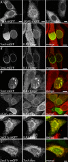
Figure 1.
Figure 1.
LULL1 promotes relocalization of TorA to the NE. (A) Distribution of TorA-mGFP, ΔGAG-TorA-mGFP, and LULL1-myc expressed individually in U2OS cells. (B) Distribution of coexpressed TorA-mGFP and LULL1-myc, shown in maximum intensity projections (B and B″) and a confocal slice (B′). (C) Distribution of coexpressed ΔGAG-TorA-mGFP and LULL1-myc. (D) Distribution of coexpressed Sec61γ-mGFP and LULL1-myc. (E) Distribution of coexpressed Sec61γ-mGFP and TorA-myc. (A–C and E) are all maximum intensity projections of confocal z-series. (D) An epifluorescence image. Scale bars, 10 μm.
Figure 2.
Figure 2.
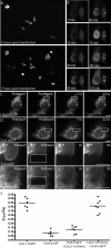
Live imaging and FRAP analysis of LULL1-directed TorA relocalization to the NE. (A) Selected images from time-lapse observation of TorA-mGFP in cells expressing unlabeled LULL1-myc. (B) Selected images from zoomed-in portion of a field similar to A. TorA-mGFP fills the NE over an average time of 45 min (±26 min, n = 52 cells). (C) Time series showing FRAP of LULL1-mGFP in U2OS cells. (D) Comparable time series showing FRAP of TorA-mGFP in U2OS cells. (E) Time series showing FRAP of TorA-mGFP in a cell coexpressing LULL1-mCherry. (E′) Corresponding time series showing FRAP of LULL1-mCherry in a cell expressing TorA-mGFP. (F) Diffusion coefficient values for LULL1 and TorA–mGFP, determined as described in Siggia et al. (2000) and Snapp et al. (2003a) from recovery curves such as those shown in Supplemental Figure S3. LULL1's diffusion coefficient averages ∼0.4 μm2/s in U2OS cells and in cells coexpressing TorA-mGFP, whereas TorA's diffusion coefficient ranges between 0.09 and 0.13 μm2/s in both U2OS and LULL1-mCherry–expressing cells. For comparison, luminal ER-GFP has a Deff of ∼10 μm2/s (Snapp et al., 2003a), an ER-localized transmembrane protein has a Deff of ∼0.4 μm2/s (Snapp et al., 2003a), and a polysome-associated translocon has a Deff of ∼0.05 μm2/s (Nikonov et al., 2002). Scale bars, 10 μm.
Figure 3.
Figure 3.
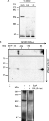
BN-PAGE separation of TorA oligomers. (A) BN-PAGE separation of untagged TorA expressed in U2OS cells. TorA is detectable as a species of approximately hexameric size in 0.25% wt/vol DDM; this species is decreased in abundance in the presence of higher concentrations of DDM. (B) 2D PAGE of untagged TorA expressed in U2OS cells. TorA was solubilized in 0.5% wt/vol digitonin, run on first dimension BN-PAGE and then separated in a second dimension by SDS-PAGE. (C) BN-PAGE separation of untagged TorA expressed alone or with LULL1-myc, solubilized in 1% wt/vol digitonin.
Figure 4.
Figure 4.
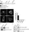
Structural requirements for LULL1-directed TorA redistribution. (A) Schematic of TorA structure. (B) Representative epifluorescence images of TorA-mGFP containing indicated mutation in LULL1-myc (not shown) expressing U2OS cells. Scale bars, 10 μm. (C) Quantitation of redistribution. n > 150 cells for each mutant. (D) Immunoblots of equal fractions of aqueous and hydrophobic phases from a Triton X-114 phase-partitioning assay show that wild-type TorA partitions with the hydrophobic phase, but shifts to the aqueous phase after deleting the protein's N-terminal hydrophobic domain (amino acids 26–43). (E) E171Q-TorA-mGFP immunoprecipitates efficiently with LULL1-myc, and deletion of the N-terminal hydrophobic domain (amino acids 26–43) does not abolish binding.
Figure 5.
Figure 5.
Removing LULL1 by RNAi reverses enrichment of ΔGAG-TorA in NE. (A) Immunoblot of lysates from cells transduced with the indicated shRNA probed for LULL1. (B and C) Representative confocal images of ΔGAG-TorA-mGFP and ER-RFP coexpressed in control U2OS cells (B) versus in U2OS cells transduced with shRNA no. 2088 (C). (D) Relative fluorescence intensity of ER-RFP, wt-TorA-mGFP, and ΔGAG-TorA-mGFP in NE versus ER. ΔGAG-TorA-mGFP has a significantly higher NE–ER ratio in U2OS cells than in LULL1 knockdown cells (p = 0.003). The NE–ER ratio of ΔGAG-TorA-mGFP is not significantly different from that of ER-RFP in LULL1 knockdown cells (p = 0.19). n >21 cells for each condition. Scale bars, 10 μm.
Figure 6.
Figure 6.
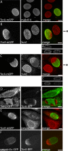
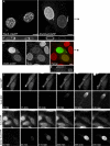
Redistributed TorA displaces LINC complex components from the NE. (A) Nuclear pore components stained with mAb414 in cells expressing LULL1-myc (not shown) and TorA-mGFP, epifluorescence image. (B) Sun1 in cells expressing LULL1-myc (not shown) and TorA-mGFP. Shown are maximum intensity projections of a confocal z-series. (B′) XZ orthogonal view: top, TorA-mGFP; bottom, Sun1. Approximate position of xz slice is marked by asterisk in B. (C) Sun2 immunostaining in the same cells. Sixty percent of cells containing TorA-mGFP in the NE lack a nuclear rim of Sun2, compared with 4% of untransfected cells (n >110 for each). (C′) XZ orthogonal view: top, TorA-mGFP; bottom, Sun2. Approximate position of xz slice is marked by asterisk in C. (D) Sun2-GFP and TorA-TagRFP in cells also expressing LULL1-myc, projected z-series. (E) Nesprin2-Giant and TorA-mGFP in cells also expressing LULL1-myc (not shown), imaged by epifluorescence microscopy. Fifty-six percent of cells containing TorA-mGFP in the NE lack NE-localized nesprin2-Giant, compared with 1% of untransfected cells (n >35 for each). (F) Transfected nesprin3α-GFP in cells also expressing TorA-TagRFP and LULL1-myc (not shown), projected z-series. Scale bars, 10 μm.
Figure 7.
Figure 7.
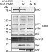
Sun2 protein levels are decreased as a consequence of LULL1-directed NE localization of TorA. Western blot analysis of cells coexpressing LULL1-myc and TorA-mGFP for 12 or 22 h and sorted by GFP fluorescence indicates that Sun2 protein levels decrease, whereas NPCs (mAb 414) and a representative ER protein (Sec61β) are not affected.
Figure 8.
Figure 8.
Comparison of wild-type and ΔGAG-TorA-mGFP effects on the NE. (A) Comparison of NE in cells expressing LULL1-myc (not shown) with redistributed wild-type TorA-mGFP (left) and ΔGAG-TorA-mGFP (right). NE membrane distortions occur in 68% of cells with NE-localized wild-type TorA (n = 136) and in 7% of cells with NE-localized ΔGAG-TorA (n = 74). Shown are maximum intensity projections of confocal z-series, with orthogonal xz views in A′. Approximate position of xz orthogonal slice marked by asterisk in A. (B) Effect of LULL1-redistributed ΔGAG-TorA-mGFP on Sun2. Projected z-series, with orthogonal views of ΔGAG-TorA (top) and Sun2 (bottom) in B′. Approximate position of xz slice is marked by asterisk in B. (C) Time-lapse imaging of Sun2-GFP and TorA-TagRFP in LULL1-myc–expressing cells. (D) Time-lapse imaging of Sun2-GFP and ΔGAG-TorA-TagRFP in LULL1-myc–expressing cells. The median delay between the onset of TorA-TagRFP redistribution and initiation of Sun2-GFP loss was <15 min (between consecutive frames) for wild type (±20 min, n = 32 cells) and 420 min for ΔGAG-TorA (±378 min, n = 33 cells). Scale bars, 10 μm.
Figure 9.
Figure 9.
Model of LULL1-dependent TorA function at the NE. LULL1 interacts with the TorA hexamer in the peripheral ER, which promotes an activating change in TorA that requires catalytic residues and association with the membrane. TorA then moves into the NE, where it displaces components of LINC complexes, including Sun2, nesprin2-Giant, and nesprin3. This gives TorA the ability to alter contacts between the nuclear lamina (or nuclear contents), the nuclear envelope membranes, and the cytoskeleton.

Similar articles

Cited by

References

    1. Breakefield X. O., Blood A. J., Li Y., Hallett M., Hanson P. I., Standaert D. G. The pathophysiological basis of dystonias. Nat. Rev. Neurosci. 2008;9:222–234. - PubMed
    1. Callan A. C., Bunning S., Jones O. T., High S., Swanton E. Biosynthesis of the dystonia-associated AAA+ ATPase torsinA at the endoplasmic reticulum. Biochem. J. 2007;401:607–612. - PMC - PubMed
    1. Chen I. H., Huber M., Guan T., Bubeck A., Gerace L. Nuclear envelope transmembrane proteins (NETs) that are up-regulated during myogenesis. BMC Cell Biol. 2006;7:38. - PMC - PubMed
    1. Crisp M., Burke B. The nuclear envelope as an integrator of nuclear and cytoplasmic architecture. FEBS Lett. 2008;582:2023–2032. - PubMed
    1. Crisp M., Liu Q., Roux K., Rattner J. B., Shanahan C., Burke B., Stahl P. D., Hodzic D. Coupling of the nucleus and cytoplasm: role of the LINC complex. J. Cell Biol. 2006;172:41–53. - PMC - PubMed

Publication types

MeSH terms

LinkOut - more resources