. 2009 Mar 20;136(6):1056-72.
doi: 10.1016/j.cell.2008.12.040.

A role of DNA-PK for the metabolic gene regulation in response to insulin

Affiliations

A role of DNA-PK for the metabolic gene regulation in response to insulin

Roger H F Wong et al. Cell. .

Abstract

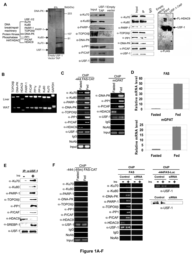
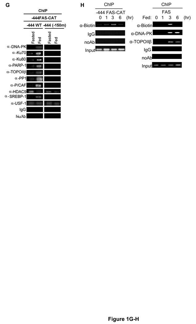
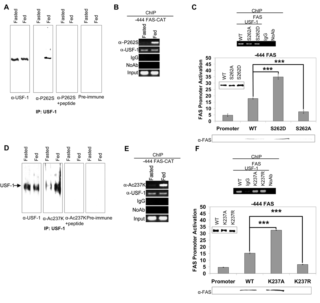
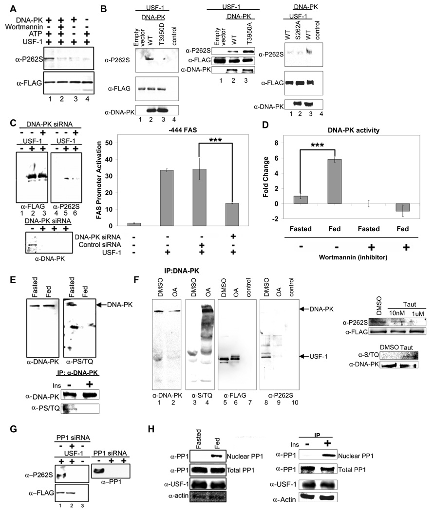
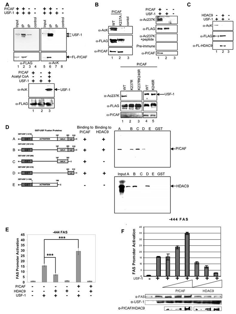
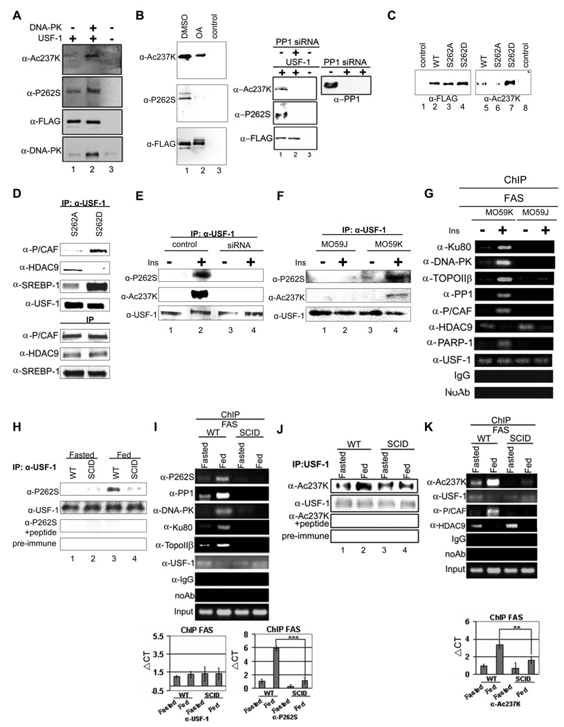
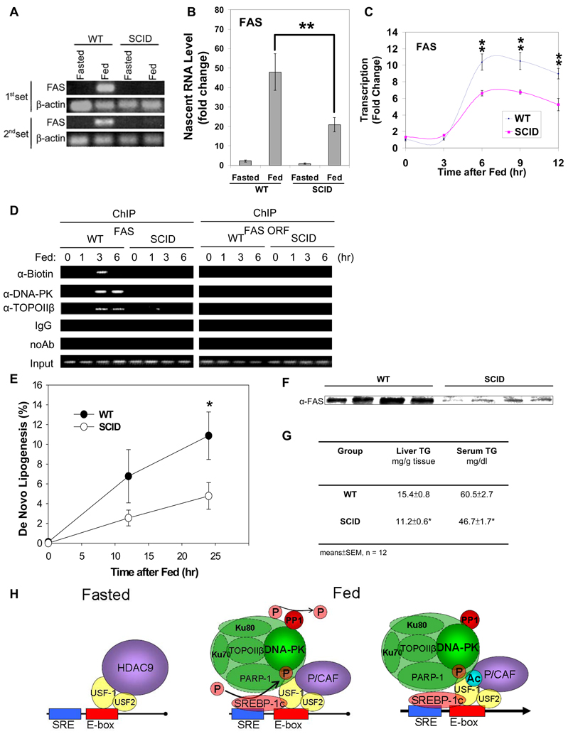
Fatty acid synthase (FAS) is a central enzyme in lipogenesis and transcriptionally activated in response to feeding and insulin signaling. The transcription factor USF is required for the activation of FAS transcription, and we show here that USF phosphorylation by DNA-PK, which is dephosphorylated by PP1 in response to feeding, triggers a switch-like mechanism. Under fasting conditions, USF-1 is deacetylated by HDAC9, causing promoter inactivation. In contrast, feeding induces the recruitment of DNA-PK to USF-1 and its phosphorylation, which then allows recruitment of P/CAF, resulting in USF-1 acetylation and FAS promoter activation. DNA break/repair components associated with USF induce transient DNA breaks during FAS activation. In DNA-PK-deficient SCID mice, feeding-induced USF-1 phosphorylation/acetylation, DNA breaks, and FAS activation leading to lipogenesis are impaired, resulting in decreased triglyceride levels. Our study demonstrates that a kinase central to the DNA damage response mediates metabolic gene activation.


Figures

Figure 1
Figure 1. Purification of USF-1 interacting proteins
(A) The identities (left) of USF-1-associated polypeptides analyzed by MS are listed. Purified USF-1 interacting proteins were separated by SDS-PAGE for silver staining (2nd left). Immunoblotting of TAP eluates (middle) using antibodies against indicated proteins. Immunoprecipitated USF-1 (2nd left) from 293F cells with monoclonal anti-USF-1 antibodies was Western blotted with indicated antibodies. TAP eluates from 293F cells transfected with USF-1-TAP and HDAC9-FLAG were immunoblotted with indicated antibodies (right). (B) RNA from mouse liver and adipose tissue were used for RT-PCR. (C) ChIP for association of USF-1 interacting proteins to the −444 FAS-CAT promoter (left) or the mGPAT promoter (right) in livers from fasted or fed FAS-CAT transgenic mice (left). (D) FAS and mGPAT expression in liver determined by RT-qPCR. (E) IP of FLAG-tagged USF-1 from HepG2 cells treated with or without 100 nM insulin for 30 min. Immunoprecipitated USF-1 was immunoblotted with indicated antibodies. (F) ChIP for association of USF-1 interacting proteins to the −444(−65m) FAS-CAT (left) promoter in liver or the FAS promoter in HepG2 cells (right) transfected with control or USF-1 siRNA. USF-1 protein levels were analyzed by immunoblotting (bottom right). (G) ChIP for binding of USF-1 interacting proteins to the −444 FAS-CAT (left) and −444(−150m) FAS-CAT (right) promoter regions in mouse liver. (H) ChIP analysis for biotin incorporation into 3'-ends of DNA breaks and DNA-PK and TopoIIβ binding to the FAS-CAT (left) or the endogenous FAS promoter (right) in livers from fasted or fed FAS-CAT transgenic or wild type mice.
Figure 2
Figure 2. Feeding-induced S262 phosphorylation and K237 acetylation of USF-1
(A) USF-1 immunoprecipitates using monoclonal anti-USF-1 antibodies was Western blotted with polyclonal anti-USF-1 or anti-P-USF-1 antibodies. Immunoblotting with anti-P-USF-1 in the presence of peptide or with pre-immune serum are shown as controls. (B) ChIP for indicated proteins binding to the −444 FAS-CAT promoter in mouse liver. (C) ChIP using anti-FLAG antibodies (top) for WT USF-1 and S262 USF-1 mutant association to the FAS promoter in 293FT cells. The FAS promoter activity (bottom) in cells transfected with −444 FAS-Luc and WT USF-1 or S262 USF-1 mutants was monitored. Immunoblotting for protein levels of WT, S262 USF-1 mutants (insert) and FAS are shown. (D) Immunoprecipitated USF-1 was Western blotted with indicated antibodies. (E) ChIP for binding of indicated proteins to the −444 FAS-CAT promoter in mouse liver. (F) ChIP (top) for association of WT USF-1 and K237 USF-1 mutant to the FAS promoter in 293F cells. The promoter activity of the −444 FAS-Luc (bottom) in cells transfected with WT USF-1 or K237 USF-1 mutants was measured.
Figure 3
Figure 3. Feeding dependent S262 phosphorylation of USF-1 is mediated by DNA-PK that is dephosphorylated/activated in feeding
(A) Bacterially expressed USF-1 was incubated with DNA-PK in the presence or absence of ATP or wortmannin (2 uM). (B) IP of cells overexpressing WT USF-1 and DNA-PK, T3950D DNA-PK (kinase dead) (left), T3950A DNA-PK or empty vector (middle) or overexpressing S262A USF-1 mutant with or without DNA-PK (right), using FLAG antibodies. DNA-PK protein levels were analyzed by immnoblotting. (C) IP (left) of cells cotransfected with USF-1, control siRNA or DNA-PK siRNA. The promoter activity of −444 FAS-Luc in cells transfected with WT USF-1 or siRNA was measured (right). (D) DNA-PK activity in liver nuclear extracts pretreated with or without wortmannin (2uM) was assayed using[ γ32P] ATP and biotinylated p53 peptide as substrate. (E) IP of liver nuclear extracts (top) or nuclear extracts from HepG2 cells (bottom) using anti-DNA-PK antibodies. (F) Total and phosphorylated DNA-PK levels were analyzed by westernblotting. IP of USF-1-FLAG from cells overexpressing USF-1 treated with either control DMSO or OA at 1uM (left) and Taut at indicated concentrations for 2 hrs (right). DNA-PK phosphorylation was detected from cells treated with Taut at 1uM for 2hrs. (G) IP of cells cotransfected with USF-1 and control or PP1 siRNA with anti-USF-1 antibodies. PP1 protein levels were analyzed by Western blotting as shown. (H) IP of nuclear extracts or total lysates from livers (left) or HepG2 cells (right) with anti-PP1 antibodies. USF-1 and β-actin protein levels were analyzed by Western blotting.
Figure 4
Figure 4. Acetylation of K237 of USF-1 by P/CAF and deacetylation by HDAC9
(A) IP of cells overexpressing USF-1 and P/CAF with FLAG antibodies (top). Bacterially expressed USF-1 was in vitro acetylated with P/CAF (bottom). (B) IP of WT USF-1 or K237A USF-1 from cells transfected with P/CAF using FLAG antibodies (top left). P/CAF protein levels were analyzed by immunoblotting. IP of cells transfected with USF-1 and P/CAF (top right) or cells transfected with P/CAF along with WT USF-1 or USF-1 mutants (bottom). (C) IP of cells overexpressing USF-1 and P/CAF with HDAC9 using FLAG antibodies. (D) Bacterially expressed USF-1-GST was incubated with in vitro translated 35S-labeled P/CAF and HDAC9 before subjecting to GST-pull down. Purified USF-1 was subjected to autoradiography for labeled proteins. GST alone was used as a control. (E) The promoter activity of the -444 FAS-Luc upon cotransfection of USF-1 along with P/CAF or HDAC9. (F) The promoter activity of −444 FAS-Luc upon cotransfection of USF-1 along with increasing amount of P/CAF or HDAC9 was measured. Total cell lysates were immunoblotted for indicated proteins.
Figure 5
Figure 5. Feeding/insulin induced phosphorylation and acetylation of USF-1 are greatly reduced in DNA-PK deficiency
(A) IP of FLAG-tagged USF-1 from cells transfected with DNA-PK using FLAG antibodies. Empty vector was used as control. (B) IP with FLAG antibodies of USF-1-FLAG from DMSO or OA treated cells overexpressing USF-1 (left) and from cells cotransfected with USF-1 and control or PP1 siRNA (right). (C) IP with FLAG antibodies of 293F cells transfected with FLAG-tagged WT USF-1 or S262 USF-1 mutants. Nuclear extracts from non-transfected cells were used as a control. (D) IP of USF-1-FLAG from HepG2 cells transfected with FLAG-tagged S262 USF-1 mutants. Immunoprecipitated USF-1 was immunoblotted with indicated antibodies (top). Total protein levels were also analyzed by immnnoblotting (bottom). (E) IP of nuclear extracts from HepG2 cells transfected with control or DNA-PK siRNA using monoclonal anti-USF-1 antibodies. (F) IP of nuclear extracts from M059J or M059K cells using anti-USF-1 antibodies. (G) ChIP for binding of indicated proteins to the FAS promoter in M059J or M059K cells. (H) IP of liver nuclear extracts from WT and SCID mice using monoclonal anti-USF-1 antibodies. (I) ChIP for indicated protein association to the FAS promoter in mouse liver. ChIP samples were analyzed by semi-quantitative PCR (top) or qPCR (bottom). (J) IP of liver nuclear extracts using anti-USF-1 antibodies. (K) ChIP for indicated protein association to the FAS promoter in liver.
Figure 6
Figure 6. Diminished FAS induction leading to blunted de novo lipogenesis and decreased triglyceride levels in liver and serum
(A) Nascent RNA from liver nuclei were used for RT-PCR. (B) Nascent RNA from liver nuclei were used for RT-qPCR. Fold induction normalized by β-actin was shown. (C) Run-on of biotin labeled nascent transcripts from liver nuclei at different time points upon feeding were analyzed by RT-qPCR. (D) ChIP for biotin incorporation and indicated protein binding to the FAS promoter in liver. (E) Newly synthesized fatty acids during a 4 hr-period of body water labeling with 2H2O in livers from 9-wk old WT and SCID mice during feeding were measured. Values are means ± SEM, n = 12. (F) Equal amounts of Liver extracts from 9-wk old WT and SCID mice after 24 hrs of feeding were immunoblotted for total FAS protein levels. (G) Hepatic and serum triglyceride levels were measured in 9-wk old fed WT and SCID mice. (H) Schematic representation of USF-1 and its interacting partners and their effects on lipogenic gene transcription in fasting/feeding.

Similar articles

Cited by

References

    1. Allen PB, Kwon YG, Nairn AC, Greengard P. Isolation and characterization of PNUTS, a putative protein phosphatase 1 nuclear targeting subunit. J. Biol. Chem. 1998;273:4089–4095. - PubMed
    1. Brady MJ, Saltiel AR. The role of protein phosphatase-1 in insulin action. Recent Prog. Horm. Res. 2001;56:157–173. - PubMed
    1. Casado M, Vallet VS, Kahn A, Vaulont S. Essential role in vivo of upstream stimulatory factors for a normal dietary response of the fatty acid synthase gene in the liver. J. Biol. Chem. 1999;274:2009–2013. - PubMed
    1. Collis SJ, DeWeese TL, Jeggo PA, Parker AR. The life and death of DNA-PK. Oncogene. 2005;24:949–961. - PubMed
    1. Corre S, Galibert MD. Upstream stimulating factors: highly versatile stress-responsive transcription factors. Pigment Cell Res. 2005;18:337–348. - PubMed

Publication types