

Selective expression of latency-associated peptide (LAP) and IL-1 receptor type I/II (CD121a/CD121b) on activated human FOXP3+ regulatory T cells allows for their purification from expansion cultures
- PMID: 19299332
- PMCID: PMC2686183
- DOI: 10.1182/blood-2009-01-199950
Selective expression of latency-associated peptide (LAP) and IL-1 receptor type I/II (CD121a/CD121b) on activated human FOXP3+ regulatory T cells allows for their purification from expansion cultures
Abstract
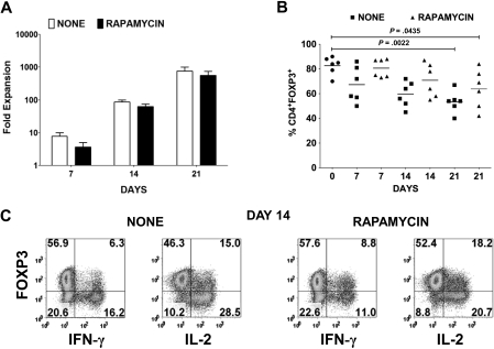
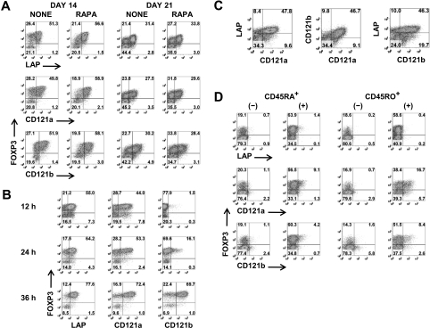
Although adoptive transfer of regulatory T cells (Foxp3(+) Tregs) has proven to be efficacious in the prevention and treatment of autoimmune diseases and graft-versus-host disease in rodents, a major obstacle for the use of Treg immunotherapy in humans is the difficulty of obtaining a highly purified preparation after ex vivo expansion. We have identified latency-associated peptide (LAP) and IL-1 receptor type I and II (CD121a/CD121b) as unique cell-surface markers that distinguish activated Tregs from activated FOXP3(-) and FOXP3(+) non-Tregs. We show that it is feasible to sort expanded FOXP3(+) Tregs from non-Tregs with the use of techniques for magnetic bead cell separation based on expression of these 3 markers. After separation, the final product contains greater than 90% fully functional FOXP3(+) Tregs. This novel protocol should facilitate the purification of Tregs for both cell-based therapies as well as detailed studies of human Treg function in health and disease.
Figures


Similar articles
-
Human LAP+GARP+FOXP3+ regulatory T cells attenuate xenogeneic graft versus host disease.Theranostics. 2019 Apr 12;9(8):2315-2324. doi: 10.7150/thno.30254. eCollection 2019. Theranostics. 2019. PMID: 31149046 Free PMC article.
-
FOXP3 promoter demethylation reveals the committed Treg population in humans.PLoS One. 2008 Feb 20;3(2):e1612. doi: 10.1371/journal.pone.0001612. PLoS One. 2008. PMID: 18286169 Free PMC article.
-
Combining FoxP3 and Helios with GARP/LAP markers can identify expanded Treg subsets in cancer patients.Oncotarget. 2016 Mar 22;7(12):14083-94. doi: 10.18632/oncotarget.7334. Oncotarget. 2016. PMID: 26885615 Free PMC article.
-
Identification of human regulatory T cells in the setting of T-cell activation and anti-CTLA-4 immunotherapy on the basis of expression of latency-associated peptide.Cancer Discov. 2012 Feb;2(2):122-30. doi: 10.1158/2159-8290.CD-11-0236. Epub 2011 Dec 27. Cancer Discov. 2012. PMID: 22585857
-
Heterogeneity in FoxP3- and GARP/LAP-Expressing T Regulatory Cells in an HLA Class II Transgenic Murine Model of Necrotizing Soft Tissue Infections by Group A Streptococcus.Infect Immun. 2018 Nov 20;86(12):e00432-18. doi: 10.1128/IAI.00432-18. Print 2018 Dec. Infect Immun. 2018. PMID: 30224551 Free PMC article.
Cited by
-
Cutting edge: progesterone directly upregulates vitamin d receptor gene expression for efficient regulation of T cells by calcitriol.J Immunol. 2015 Feb 1;194(3):883-6. doi: 10.4049/jimmunol.1401923. Epub 2014 Dec 29. J Immunol. 2015. PMID: 25548222 Free PMC article.
-
Extricating human tumour immune alterations from tissue inflammation.Nature. 2022 May;605(7911):728-735. doi: 10.1038/s41586-022-04718-w. Epub 2022 May 11. Nature. 2022. PMID: 35545675 Free PMC article.
-
Murine CD4 T cells produce a new form of TGF-β as measured by a newly developed TGF-β bioassay.PLoS One. 2011 Apr 11;6(4):e18365. doi: 10.1371/journal.pone.0018365. PLoS One. 2011. PMID: 21494571 Free PMC article.
-
Lrig1-expression confers suppressive function to CD4+ cells and is essential for averting autoimmunity via the Smad2/3/Foxp3 axis.Nat Commun. 2023 Sep 4;14(1):5382. doi: 10.1038/s41467-023-40986-4. Nat Commun. 2023. PMID: 37666819 Free PMC article.
-
Tuning inflammation and immunity by the negative regulators IL-1R2 and IL-1R8.Immunol Rev. 2018 Jan;281(1):233-247. doi: 10.1111/imr.12609. Immunol Rev. 2018. PMID: 29247989 Free PMC article. Review.
References
-
- Sakaguchi S, Ono M, Setoguchi R, et al. Foxp3+ CD25+ CD4+ natural regulatory T cells in dominant self-tolerance and autoimmune disease. Immunol Rev. 2006;212:8–27. - PubMed
-
- Valencia X, Yarboro C, Illei G, Lipsky PE. Deficient CD4+CD25high T regulatory cell function in patients with active systemic lupus erythematosus. J Immunol. 2007;178:2579–2588. - PubMed
-
- Lindley S, Dayan CM, Bishop A, Roep BO, Peakman M, Tree TI. Defective suppressor function in CD4(+)CD25(+) T-cells from patients with type 1 diabetes. Diabetes. 2005;54:92–99. - PubMed
-
- Hafler DA, Slavik JM, Anderson DE, O'Connor KC, De Jager P, Baecher-Allan C. Multiple sclerosis. Immunol Rev. 2005;204:208–231. - PubMed
Publication types
MeSH terms
Substances
Grants and funding
LinkOut - more resources
Full Text Sources
Other Literature Sources
Research Materials
