. 2009 Jun;83(11):5773-83.
doi: 10.1128/JVI.00103-09. Epub 2009 Mar 18.

Regulation of the catalytic activity of herpes simplex virus 1 protein kinase Us3 by autophosphorylation and its role in pathogenesis

Affiliations

Regulation of the catalytic activity of herpes simplex virus 1 protein kinase Us3 by autophosphorylation and its role in pathogenesis

Ken Sagou et al. J Virol. 2009 Jun.

Abstract

Us3 is a serine/threonine protein kinase encoded by herpes simplex virus 1 (HSV-1). We recently identified serine at Us3 position 147 (Ser-147) as a physiological phosphorylation site of Us3 (A. Kato, M. Tanaka, M. Yamamoto, R. Asai, T. Sata, Y. Nishiyama, and Y. Kawaguchi, J. Virol. 82:6172-6189, 2008). In the present study, we investigated the effects of phosphorylation of Us3 Ser-147 on regulation of Us3 catalytic activity in infected cells and on HSV-1 pathogenesis. Our results were as follows. (i) Only a small fraction of Us3 purified from infected cells was phosphorylated at Ser-147. (ii) Us3 phosphorylated at Ser-147 purified from infected cells had significantly higher kinase activity than Us3 not phosphorylated at Ser-147. (iii) Phosphorylation of Us3 Ser-147 in infected cells was dependent on Us3 kinase activity. (iv) Replacement of Us3 Ser-147 by alanine significantly reduced viral replication in the mouse cornea and the development of herpes stromal keratitis and periocular skin disease in mice. These results indicated that Us3 catalytic activity is tightly regulated by autophosphorylation of Ser-147 in infected cells and that regulation of Us3 activity by autophosphorylation appeared to play a critical role in viral replication in vivo and in HSV-1 pathogenesis.


Figures
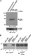
FIG. 1.
FIG. 1.
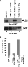
(A) Immunoblots of electrophoretically separated lysates from Vero cells infected with YK515 (Us3-S147A) (lane 1), YK517 (Us3-SA-repair) (lane 2), or wild-type HSV-1(F) (lanes 3 and 4). Infected cells were harvested at 18 h postinfection, solubilized, mock treated (lanes 1, 2, and 3) or treated with λ-PPase (lane 4), and immunoblotted with anti-Us3-S147P monoclonal antibody (upper panel) or anti-Us3 polyclonal antibody (lower panel). +, present; −, absent. (B) Immunoblots of electrophoretically separated lysates from Vero cells infected with wild-type HSV-1(F), YK515 (Us3-S147A), or YK517 (Us3-SA-repair). Infected cells were harvested at 18 h postinfection and immunoblotted with anti-Us3-S147P monoclonal antibody (left panel). The same membrane was reprobed with anti-Us3 polyclonal antibody (right panel). Molecular sizes are shown on the left of the panels. α, anti; IB, immunoblotting. Total Us3, Us3 protein detected by anti-Us3 polyclonal antibody.
FIG. 2.
FIG. 2.
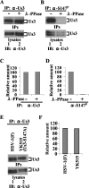
(A and B) Immunoblots of electrophoretically separated Us3 immunoprecipitates (upper panel) and lysates (lower panel) from Vero cells infected with HSV-1(F). Infected cells were harvested at 18 h postinfection, solubilized, and either mock treated (lanes 1) or treated with λ-PPase (lanes 2). Part of the treated cell lysates was analyzed by immunoblotting with anti-Us3 antibody (lower panels). The remainder of each lysate was immunoprecipitated with anti-Us3 (A) or anti-Us3-S147P antibody (B). The immunoprecipitates were separated in a denaturing gel and analyzed by immunoblotting with anti-Us3 antibody (upper panel). (C and D) Amount of Us3 protein immunoprecipitated with anti-Us3 (B, upper panel) and anti-Us3-S147P antibody (C, upper panel) relative to the amount of Us3 protein in the corresponding HSV-1(F)-infected cell lysates (A and B, lower panels). The data were normalized to the value for HSV-1(F)-infected cells without phosphatase treatment in panels A and B, lanes 1. (E) Immunoblots of electrophoretically separated Us3 immunoprecipitates (upper panel) and lysates (lower panel) from Vero cells infected with HSV-1(F) (lane 1) or YK515 (Us3-S147A) (lane 2). Infected Vero cells were harvested at 18 h postinfection and solubilized. Part of the cell lysates was immunoblotted with anti-Us3 polyclonal antibody (lower panel). The remainder of each lysate was immunoprecipitated with anti-Us3. The immunoprecipitates were separated in a denaturing gel and analyzed by immunoblotting with anti-Us3 antibody (upper panel). (F) Amount of Us3 protein immunoprecipitated with anti-Us3 antibody (E, upper panel) relative to the amount of Us3 protein in the corresponding HSV-1(F)- or YK515 (Us3-S147A)-infected cell lysates (E, lower panel). These data were normalized relative to the value for HSV-1(F)-infected cells in panel E, lane 1. IP, immunoprecipitation; IPs, immunoprecipitates; IB, immunoblotting; α, anti; +, present; −, absent.
FIG. 3.
FIG. 3.
(A) Immunoblots of electrophoretically separated lysates from Vero cells infected with wild-type HSV-1(F) (lane 1), YK511 (Us3-K220M) (lane 2), YK513 (Us3-KM-repair) (lane 3), or YK515 (Us3-S147A) (lane 4). Infected Vero cells were harvested at 18 h postinfection and analyzed by immunoblotting with anti-Us3-S147P antibody (upper panel) or anti-Us3 antibody (lower panel). (B) Amount of Us3-S147P protein detected with anti-Us3-S147P antibody (A, upper panel) relative to the amount of Us3 protein detected with anti-Us3 antibody (A, lower panel) in HSV-1(F)-infected cells. The data were normalized to the value for HSV-1(F)-infected cells in panel A, lane 1. α, anti; Total Us3, Us3 protein detected by anti-Us3 polyclonal antibody.
FIG. 4.
FIG. 4.
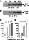
(A and B) Immunoblots of electrophoretically separated lysates from Vero cells infected with wild-type HSV-1(F). Infected Vero cells were harvested at the indicated times postinfection and analyzed by immunoblotting with anti-Us3-S147P antibody (A) or anti-Us3 antibody (B). (C and D) Amount of Us3-S147P protein shown in panel A (C) and total Us3 protein shown in panel B (D) as a function of time postinfection, relative to the amounts at 12 h postinfection. The data were normalized to the values at 12 h postinfection. α, anti; ND, not detected; Total Us3, Us3 protein detected by anti-Us3 polyclonal antibody.
FIG. 5.
FIG. 5.
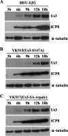
Immunoblots of electrophoretically separated lysates from Vero cells infected with wild-type HSV-1(F) (A), YK515 (Us3-S147A) (B), and YK517 (Us3-SA-repair) (C). Infected Vero cells were harvested at the indicated times postinfection and analyzed by immunoblotting with anti-Us3, anti-ICP8, and anti-α-tubulin antibodies.
FIG. 6.
FIG. 6.
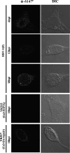
Digital confocal images showing the localization of Us3-S147P. Vero cells were infected with wild-type HSV-1(F), YK515 (Us3-S147A), or YK517 (Us3-SA-repair). Infected Vero cells were fixed at the indicated times postinfection, permeabilized, stained with anti-Us3-S147P antibody, and examined by confocal microscopy. hpi, hours postinfection; α, anti; DIC, digital interference contrast.
FIG. 7.
FIG. 7.
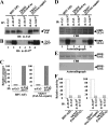
(A and B) Immunoblots of electrophoretically separated Us3 immunoprecipitates from Vero cells infected with wild-type HSV-1(F) (lanes 1 and 2), YK515 (Us3-S147A) (lanes 3 and 4), and YK517 (Us3-SA-repair) (lanes 5 and 6). Infected Vero cells were harvested at 18 h postinfection, solubilized, and immunoprecipitated with anti-Us3 (lanes 1, 3, and 5) or anti-Us3-S147P antibody (lanes 2, 4, and 6). The immunoprecipitates were then divided into four aliquots. Two immunoprecipitate aliquots were separated in a denaturing gel and analyzed by immunoblotting with anti-Us3 (A) and anti-Us3-S147P (B) antibodies. (C) Amount of Us3-S147P protein shown in panel B relative to the amount of total Us3 protein shown in panel A. In the left graph, the data were normalized to the relative amount for immunoprecipitates with anti-Us3-S147P antibody for cells infected with HSV-1(F) shown in panels A and B, lanes 2. In the right graph, the data were normalized to the relative amount for immunoprecipitates with anti-Us3-S147P antibody for cells infected with YK517 (Us3-SA-repair) shown in panels A and B, lanes 6. (D and E) For in vitro kinase assays of the other two immunoprecipitate aliquots, the immunoprecipitates described for panels A and B were incubated in kinase buffer containing [γ-32P]ATP and MBP-UL34 (D) or MBP-LacZ (E) and separated in denaturing gels. The gels were stained with Coomassie brilliant blue (CBB) (upper panels) and analyzed by autoradiography (lower panels). (F) Amount of radioactivity in 32P-labeled MBP-UL34 produced in a kinase reaction whose results are shown in panel D, lower panel, relative to the amount of total Us3 protein shown in panel A. In the left graph, the data were normalized to the relative value for the kinase activity of anti-Us3-S147P immunoprecipitates from cells infected with HSV-1(F) shown in panels D, lower panel, and A, lanes 2. In the right graph, the data were normalized to the relative value for the kinase activity of anti-Us3-S147P immunoprecipitates from cells infected with YK517 (Us3-SA-repair) shown in panels D, lower panel, and A, lanes 6. α, anti; IP, immunoprecipitation; IB, immunoblotting; Total Us3, Us3 protein detected by anti-Us3 polyclonal antibody.
FIG. 8.
FIG. 8.
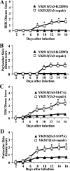
Effect of the Us3-S147A and Us3-K220M mutations on HSK and periocular skin disease in mice. (A and B) Six 5-week-old female ICR mice were infected with 1 × 10⁶ PFU YK511 (Us3-K220M) or YK513 (Us3-KM-repair) by corneal scarification and scored for HSK and periocular skin disease every other day for 16 days. The data shown are the averages and standard errors of the observations. A statistically significant difference between HSK and periocular skin disease scores in mice infected with YK511 (Us3-K220M) and those infected with YK513 (Us3-KM-repair) was noted (*, P < 0.01). (C and D) Two independent experiments were done, each using six 5-week-old female ICR mice infected with 1 × 10⁶ PFU YK515 (Us3-S147A) or YK517 (Us3-SA-repair) by corneal scarification and scored for HSK and periocular skin disease every other day for 16 days. The results from two independent experiments (each with six mice) were combined. The data shown are the averages and standard errors of the observations. A statistically significant difference between HSK and periocular skin disease scores in mice infected with YK515 (Us3-S147A) and those infected with YK517 (Us3-SA-repair) was noted (*, P < 0.01).
FIG. 9.
FIG. 9.
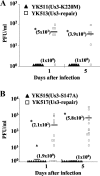
Effects of the Us3-S147A and Us3-K220M mutations on virus growth in the tear film of mice following corneal infection. In the experiment whose results are shown in Fig. 6, viral titers in the tear film of infected mice at 1 and 5 days postinfection were determined by standard plaque assays. Each data point represents the titer in the tear film of one mouse. The horizontal bars and figures in parentheses show the average for each group. A statistically significant difference in viral titers between mice infected with YK511 (Us3-K220M) and YK513 (Us3-KM-repair) (A) and between mice infected with YK515 (Us3-S147A) and YK517 (Us3-SA-repair) (B) was noted (*, P < 0.05).
FIG. 10.
FIG. 10.
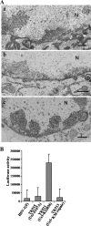
(A) Electron microscopy of Vero cells infected with wild-type HSV-1(F) (a), YK515 (Us3-S147A) (b), or R7041 (ΔUs3) (c) for 20 h. Scale bars, 500 nm; N, nucleus. (B) Caspase 3/7 activity of infected Vero cells after induction of apoptosis by osmotic shock. Vero cells were infected with wild-type HSV-1(F), YK511 (Us3-K220M), YK513 (Us3-KM-repair), or YK515 (Us3-S147A). At 12 h postinfection, the cells were exposed to sorbitol for 1 h, incubated for an additional 5 h, harvested, and assayed for caspase 3/7 activity using a Z-DEVD-aminoluciferin substrate (15). The values are the averages and standard deviations of the results of three independent experiments.

Similar articles

Cited by

References

    1. Benetti, L., and B. Roizman. 2004. Herpes simplex virus protein kinase US3 activates and functionally overlaps protein kinase A to block apoptosis. Proc. Natl. Acad. Sci. USA 1019411-9416. - PMC - PubMed
    1. Colbran, R. J. 2004. Targeting of calcium/calmodulin-dependent protein kinase II. Biochem. J. 3781-16. - PMC - PubMed
    1. Coller, K. E., and G. A. Smith. 2008. Two viral kinases are required for sustained long distance axon transport of a neuroinvasive herpesvirus. Traffic 91458-1470. - PMC - PubMed
    1. Daikoku, T., Y. Yamashita, T. Tsurumi, K. Maeno, and Y. Nishiyama. 1993. Purification and biochemical characterization of the protein kinase encoded by the US3 gene of herpes simplex virus type 2. Virology 197685-694. - PubMed
    1. Edelman, A. M., D. K. Blumenthal, and E. G. Krebs. 1987. Protein serine/threonine kinases. Annu. Rev. Biochem. 56567-613. - PubMed

Publication types

MeSH terms

LinkOut - more resources