. 2009 May 22;284(21):14245-57.
doi: 10.1074/jbc.M806902200. Epub 2009 Mar 16.

Oxidizable residues mediating protein stability and cytoprotective interaction of DJ-1 with apoptosis signal-regulating kinase 1

Affiliations

Oxidizable residues mediating protein stability and cytoprotective interaction of DJ-1 with apoptosis signal-regulating kinase 1

Jens Waak et al. J Biol Chem. .

Abstract

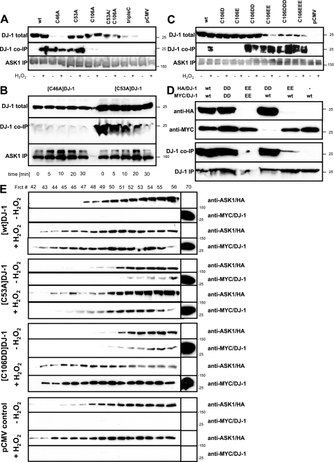
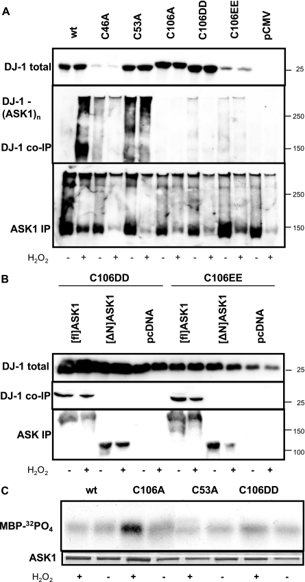
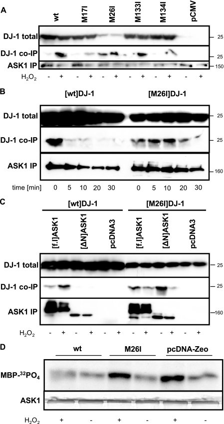
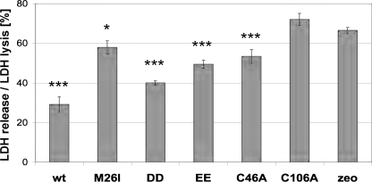
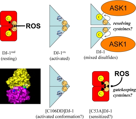
Parkinson disease (PD)-associated genomic deletions and the destabilizing L166P point mutation lead to loss of the cytoprotective DJ-1 protein. The effects of other PD-associated point mutations are less clear. Here we demonstrate that the M26I mutation reduces DJ-1 expression, particularly in a null background (knockout mouse embryonic fibroblasts). Thus, homozygous M26I mutation causes loss of DJ-1 protein. To determine the cellular consequences, we measured suppression of apoptosis signal-regulating kinase 1 (ASK1) and cytotoxicity for [M26I]DJ-1, and systematically all other DJ-1 methionine and cysteine mutants. C106A mutation of the central redox site specifically abolished binding to ASK1 and the cytoprotective activity of DJ-1. DJ-1 was apparently recruited into the ASK1 signalosome via Cys-106-linked mixed disulfides. The designed higher order oxidation mimicking [C106DD]DJ-1 non-covalently bound to ASK1 even in the absence of hydrogen peroxide and conferred partial cytoprotection. Interestingly, mutations of peripheral redox sites (C46A and C53A) and M26I also led to constitutive ASK1 binding. Cytoprotective [wt]DJ-1 bound to the ASK1 N terminus (which is known to bind another negative regulator, thioredoxin 1), whereas [M26I]DJ-1 bound to aberrant C-terminal site(s). Consequently, the peripheral cysteine mutants retained cytoprotective activity, whereas the PD-associated mutant [M26I]DJ-1 failed to suppress ASK1 activity and nuclear export of the death domain-associated protein Daxx and did not promote cytoprotection. Thus, cytoprotective binding of DJ-1 to ASK1 depends on the central redox-sensitive Cys-106 and may be modulated by peripheral cysteine residues. We suggest that impairments in oxidative conformation changes of DJ-1 might contribute to PD neurodegeneration.


Figures

FIGURE 1.
FIGURE 1.
Expression of DJ-1 cysteine and methionine mutants. A and B, DJ-1 cysteine mutants with C-terminal V5 tag were transiently transfected in HEK293T cells (A) and with N-terminal Myc tag or vector control in HEK293E cells (B). Western blots from whole cell lysates were concomitantly reacted with anti-V5 and the protein loading control anti-β-actin (A) and with anti-Myc and anti-α-tubulin (B), respectively. Molecular weight markers are indicated to the left. C, the indicated N-terminally Myc-tagged DJ-1 constructs (lanes 1–5) or vector control (lane 6) were transiently transfected in HEK293E cells. Western blots from whole cell lysates were reacted with anti-Myc (top panel). Blots were stripped and reprobed with anti-DJ-1 (middle panel), the upper arrowhead points to the transfected, tagged DJ-1, and the lower arrowhead to the endogenous DJ-1. Equal protein loading was confirmed by reprobing the blot with anti-α-tubulin (bottom panel). D, the indicated DJ-1 variants fused to N-terminal Myc tag (lanes 1–5), C-terminal V5 tag (lanes 7 and 8), or without epitope tag (lanes 10 and 11), and the respective vector controls (lanes 6, 9, and 12) were transiently transfected into DJ-1–/– MEF cells. DJ-1 on Western blots from whole cell lysates was detected with anti-Myc (lanes 1–6, top panel) or anti-V5 (lanes 7–9, top panel), and anti-DJ-1 (middle panels). Even loading was confirmed by reprobing with anti-α-tubulin (bottom panels). E, total RNA was extracted from DJ-1–/– MEF cells transiently transfected with Myc/[wt]DJ-1, Myc/[M26I]DJ-1, and pCMV vector plasmid. After reverse transcription, cDNA samples were amplified by PCR using primers specific for Myc/DJ-1 (upper panel) or β-actin (lower panel), and subjected to agarose gel electrophoresis followed by ethidium bromide staining. F, DJ-1–/– MEF cells were transiently transfected with the indicated DJ-1 constructs or vector controls. Reverse transcription-PCR analysis was performed as above, and the relative expression of DJ-1 mRNA of the various constructs was determined. There were no significant differences in mRNA expression of the reduced-protein mutants [M26I]DJ-1 and [C46A]DJ-1 compared with [wt]DJ-1, although the overall expression levels driven by pCDNA were lower than the other two mammalian expression vectors.
FIGURE 2.
FIGURE 2.
Influence of cysteine mutations on ASK1 binding and DJ-1 dimerization. AC, HEK293E cells were co-transfected with ASK1/HA together with the indicated N-terminal Myc-tagged DJ-1 cysteine mutant constructs, along with pCMV vector controls. After 36-h culturing, the cells were treated for 30 min without (–) or with (+) 1 mm H2O2 and lysed directly (A and C), or treated for 30 min with 1 mm H2O2 followed by replacement with fresh medium for the indicated times (B). Then cells were lysed and direct Western blots prepared for the determination of total DJ-1 steady-state expression levels (top panels) or ASK1 immunoprecipitations performed. Western blots were probed for co-immunoprecipitated DJ-1 (middle panels) and ASK1 to demonstrate even immunoprecipitation efficiency (bottom panels). D, to determine their dimerization potential, HA-tagged wt, C106DD, and C106EE mutant DJ-1 were transiently co-transfected with Myc-tagged DJ-1 variants. The more stable [C106DD]DJ-1 but not the less stable [C106EE]DJ-1 (upper two input panels) Myc-co-immunoprecipitated with HA-tagged DJ-1 (Myc-immunoprecipitates Western probed with anti-Myc and co-immunoprecipitates probed with anti-HA and the lower two panels, respectively). E, to determine the incorporation of DJ-1 into the native ASK1 complexes, HEK293E cells were transiently co-transfected with ASK1/HA and the indicated N-terminal Myc tagged DJ-1 cysteine mutant constructs or pCMV vector control. After 36-h culturing, the cells were treated for 30 min without or with 1 mm H2O2, as indicated. Cell lysates were subjected to Sephacryl S-500 gel filtration. Fractions were collected and resolved by SDS-PAGE followed by Western probing with anti-ASK1 to detect ASK1/HA and 9E10 anti-Myc to detect Myc/DJ-1, as indicated. Shown are the fractions 42–56 containing ASK1 complexes well above 1000 kDa, and the peak fractions containing the expected Myc/DJ-1 dimer. Molecular mass standards are indicated to the rightof all Western blots. The results shown are representative for two or three independent experiments.
FIGURE 3.
FIGURE 3.
H2O2-induced formation of ASK1-DJ-1-mixed disulfides. A and B, HEK293E cells were co-transfected with full-length ASK1/HA (A) or as indicated the [ΔN]ASK1/HA mutant (B) together with vector controls. After 36-h culturing, the cells were treated for 30 min without (–) or with (+) 1 mm H2O2. Then cells were lysed and direct Western blots prepared for the determination of total DJ-1 steady-state expression levels (top panels) or ASK1 immunoprecipitations performed. ASK1 immunocomplexes were resolved by non-reducing (A) and reducing (B) SDS-PAGE. Western blots were probed for co-immunoprecipitated DJ-1 (middle panels) and ASK1 to demonstrate even immunoprecipitation efficiency (bottom panels). C, DJ-1–/– MEF cells stably transfected with the indicated, untagged DJ-1 constructs were treated for 30 min with 500μm H2O2 (+) or left untreated (–). Endogenous ASK1 was immunoprecipitated for immune complex kinase assays. Equal amounts of ASK1 in the reaction mixes are demonstrated by Coomassie Blue staining (lower panel). ASK1 phosphotransferase activity is measured by autoradiography of 32PO4 incorporation into the substrate myelin basic protein (upper panel). The results shown are representative for two or three independent experiments.
FIGURE 4.
FIGURE 4.
Influence of DJ-1 methionine mutations on ASK1 binding and kinase activity. HEK293E cells were co-transfected with full-length ASK1/HA (A and B) or as indicated the [ΔN]ASK1/HA mutant and pcDNA3 vector control (C) together with the indicated N-terminal Myc-tagged methionine mutant constructs. After 36-h culturing, the cells were treated for 30 min without (–) or with (+) 1 mm H2O2 and lysed directly (A and C), or treated for 30 min with 1 mm H2O2 followed by replacement with fresh medium for the indicated times (B). Whole cell lysates were directly Western probed with anti-Myc (top panels) to assess the relative steady-state expression levels of the DJ-1 mutants. Anti-HA-agarose immunoprecipitates were Western blotted and sequentially probed with anti-DJ-1 (middle panels) and anti-ASK1 (bottom panels). D, DJ-1–/– MEF cells stably transfected with untagged [wt]DJ-1 and [M26I]DJ-1 or pcDNA-Zeo vector control were treated for 30min with 500 μm H2O2 (+) or left untreated (–). Endogenous ASK1 was immunoprecipitated for immune complex kinase assays. Equal amounts of ASK1 in the reaction mixes are demonstrated by Coomassie Blue staining (lower panel). ASK1 phosphotransferase activity is measured by autoradiography of 32PO4 incorporation into the substrate myelin basic protein (upper panel). The results shown are representative for two or three independent experiments.
FIGURE 5.
FIGURE 5.
C106A and M26I mutations reduce DJ-1 cytoprotective activity. DJ-1–/– MEFs were stably transfected with the indicated DJ-1 constructs or vector control. Cells were exposed for 16 h to 20 μm H2O2, and cytotoxicity was measured by LDH release. Error bars delineate standard deviation of triplicate samples; *, p < 0.05; ***, p < 0.001. These results are representative of two independent experiments.
FIGURE 6.
FIGURE 6.
Loss of suppression of Daxx translocation by the M26I DJ-1 mutation. DJ-1–/– MEF cells stably transfected with untagged [wt]DJ-1 and [M26I]DJ-1 or pcDNA-Zeo vector control were treated for 30 min with 500 μm H2O2 (+) or left untreated (–) after transient transfection with FLAG/Daxx. Nuclear export of FLAG/Daxx was scored by counting the percentage of cells with cytosolic anti-FLAG immunostaining. In the absence of H2O2 (panels 1–3), FLAG/Daxx shows distinct subnuclear localization and little cytosolic staining outside the nucleus (counterstained with Hoechst 33342). After H2O2 challenge, vector controls show significant increase in cytosolic FLAG/Daxx staining (panel 6), which was suppressed in DJ-1–/– MEFs stably re-transfected with [wt]DJ-1 (panel 4). In contrast, stable re-transfection with [M26I]DJ-1 failed to suppress nuclear export of FLAG/Daxx (panel 5). The graph shows mean values from three independent experiments ± S.E.; **, p < 0.02.
FIGURE 7.
FIGURE 7.
Schematic drawing of proposed DJ-1 activation states. The surface contour plot of the DJ-1 dimer (lower left) shows the cysteine side chains of each homomer (green residues belong to the yellow homomer, blue residues to the purple homomer). The Cys-53 side chains are prominently localized at the edge of the dimer interface, Cys-46 partially buried within the dimer interface. The Cys-106 residues are deeply buried within the DJ-1 each homomer, necessitating a channel for ROS to enter the central active site Cys-106 (Cc). A peripheral (Cp) redox center that appears to comprise Cys-53 and perhaps other oxidizable and/or structural residues (Cys-46 and Met-26) could gate the accessibility of reactive oxygen species to Cys-106 and activate the central site (Cc*). Peripheral redox center mutants might be sensitized due to deregulated gating of reactive oxygen species, allowing unquenched access to the active center. Alternatively, Cp might function as “resolving cysteines,” reducing transiently formed mixed disulfide bonds of Cys-106 with effector proteins, such as ASK1. Formation of Cc* might “open” the DJ-1 conformation, allowing access of the buried Cys-106 to cysteines within with N terminus of ASK1 to form mixed disulfides. Such an open conformation may also be induced by the C106DD mutation, which causes tight but non-covalent binding of DJ-1 to ASK1. See text for further details.

Similar articles

Cited by

References

    1. Bonifati, V., Rizzu, P., van Baren, M. J., Schaap, O., Breedveld, G. J., Krieger, E., Dekker, M. C. J., Squitieri, F., Ibanez, P., Joosse, M., van Dongen, J. W., Vanacore, N., van Swieten, J. C., Brice, A., Meco, G., van Duijn, C. M., Oostra, B. A., and Heutink, P. (2003) Science 299 256–259 - PubMed
    1. Macedo, M. G., Anar, B., Bronner, I. F., Cannella, M., Squitieri, F., Bonifati, V., Hoogeveen, A., Heutink, P., and Rizzu, P. (2003) Hum. Mol. Genet. 12 2807–2816 - PubMed
    1. Miller, D. W., Ahmad, R., Hague, S., Baptista, M. J., Canet-Avilés, R., McLendon, C., Carter, D. M., Zhu, P.-P., Stadler, J., Chandran, J., Klinefelter, G. R., Blackstone, C., and Cookson, M. R. (2003) J. Biol. Chem. 278 36588–36595 - PubMed
    1. Moore, D. J., Zhang, L., Dawson, T. M., and Dawson, V. L. (2003) J. Neurochem. 87 1558–1567 - PubMed
    1. Olzmann, J. A., Brown, K., Wilkinson, K. D., Rees, H. D., Huai, Q., Ke, H., Levey, A. I., Li, L., and Chin, L.-S. (2004) J. Biol. Chem. 279 8506–8515 - PubMed

Publication types

MeSH terms

LinkOut - more resources