. 2009 Mar 11:6:28.
doi: 10.1186/1742-4690-6-28.

A role for CD81 on the late steps of HIV-1 replication in a chronically infected T cell line

Affiliations

A role for CD81 on the late steps of HIV-1 replication in a chronically infected T cell line

Boyan Grigorov et al. Retrovirology. .

Abstract

Background: HIV-1 uses cellular co-factors for virion formation and release. The virus is able to incorporate into the viral particles host cellular proteins, such as tetraspanins which could serve to facilitate HIV-1 egress. Here, we investigated the implication of several tetraspanins on HIV-1 formation and release in chronically infected T-lymphoblastic cells, a model that permits the study of the late steps of HIV-1 replication.

Results: Our data revealed that HIV-1 Gag and Env structural proteins co-localized with tetraspanins in the form of clusters. Co-immunoprecipitation experiments showed that Gag proteins interact, directly or indirectly, with CD81, and less with CD82, in tetraspanin-enriched microdomains composed of CD81/CD82/CD63. In addition, when HIV-1 producing cells were treated with anti-CD81 antibodies, or upon CD81 silencing by RNA interference, HIV-1 release was significantly impaired, and its infectivity was modulated. Finally, CD81 downregulation resulted in Gag redistribution at the cell surface.

Conclusion: Our findings not only extend the notion that HIV-1 assembly can occur on tetraspanin-enriched microdomains in T cells, but also highlight a critical role for the tetraspanin CD81 on the late steps of HIV replication.


Figures
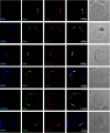
Figure 1
Figure 1
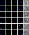
Localization of HIV-1 Gag and Env with tetraspanins at the cell surface of HIV-1 infected MOLT cells. MOLT/HIV-1 cells were fixed and the cell surface was stained directly with the anti-tetraspanin CD9, CD63, CD81 or CD82 antibodies, or with antibodies against CD45 or Lamp2. To reveal the viral proteins Gag and Env, the cells were co-stained with anti-MAp17 (Gag in green) and anti-SU gp120 (Env in red) antibodies. It can be observed that the tetraspanins are localized in microdomains close to or at the cell periphery. The percentage of Gag co-localization with the markers was calculated by image analysis and reported in the graph (Fig. 3).
Figure 2
Figure 2
Localization of HIV-1 Gag and Env with tetraspanins in permeabilized HIV-1 infected MOLT cells. MOLT/HIV-1 cells were fixed, permeabilized, and stained with the anti-tetraspanin CD9, CD63, CD81 or CD82 antibodies, or with antibodies against CD45 or Lamp2. To reveal the viral proteins Gag and Env, the cells were co-stained with anti-MAp17 (Gag in green) and anti-SU gp120 (Env in red) antibodies. The percentage of Gag co-localization with the markers was calculated by image analysis and reported in the graph (Fig. 3).
Figure 3
Figure 3
Localization of HIV-1 Gag and Env with tetraspanins in HIV-1 infected MOLT cells. The percentage of Gag co-localization with the tetraspanins or the CD45 or Lamp2 proteins was calculated by image analysis by the MetaMorph® Software and reported in the graph. Quantifications in non permeabilized MOLT/HIV-1 cells are indicated in black color, and in permeabilized cells in grey color, as indicated.
Figure 4
Figure 4
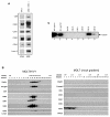
The tetraspanins CD63, CD81 and CD82 are associated with purified HIV-1 virions. (A). Cell lysate from MOLT/HIV-1 cells was run on SDS-PAGE and probed with antibodies against CD45, CD9, CD63, CD81, CD82, and Lamp2 as indicated ("cells"). Purified viral pellet from MOLT/HIV-1 was immunoblotted with the same antibodies ("virus"). (B). Purified virions produced by MOLT/HIV-1 cells (left panel) were loaded on 20–70% sucrose density gradient. After ultracentrifugation at equilibrium, the gradient was fractionated and the density (g/ml) of each fraction was determined, as indicated. Immunoblots of all fractions were performed using antibodies against Gag and Env, the tetraspanins CD63, CD81, CD82, or CD9, and CD45 or Lamp2 as controls. HIV-1 virions, as seen by the CAp24 and TMgp41, appeared in fractions with a density between 1.15–1.17 g/ml. In the same viral fractions, signals were obtained for the tetraspanins CD63, CD81 and CD82. Control gradient from uninfected MOLT cells is presented on the right panel. (C). Purified HIV-1 virions from MOLT/HIV-1 were submitted to immunoprecipitation with CD45, CD9, CD63, CD81 and CD82 antibodies (lane 4 to 7), or with HIV-1 serum and Env gp120 antibody as positive controls (lane 1 and 2) or without antibody as a negative control (No Ab – lane 3). Immunoprecipitated virions were run on SDS-PAGE gels and revealed with an anti-CAp24 antibody.
Figure 5
Figure 5
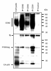
HIV-1 Gag proteins form intracellular complexes with endogenous CD81 and CD82 tetraspanins. Cell lysates from MOLT/HIV-1 cells were immunoprecipitated with antibodies directed against CD81, CD63 and CD82 or a control antibody. Non-immunoprecipitated (supernatants) or immunoprecipitated (IP) proteins were resolved by SDS-PAGE and blotted with an anti-HIV-1 human serum or an anti-CD82 antibody as indicated. The positions of Gag products and CD82 are indicated. The anti-CD82 blot shows the integrity of the tetraspanin web. The anti-HIV-1 blot shows intracellular Gag-tetraspanin interactions.
Figure 6
Figure 6
Partial inhibition of HIV-1 release using anti-tetraspanin antibodies. (A) MOLT/HIV-1 cells were incubated for one hour with anti-tetraspanin antibodies, or with anti-VSVg, anti-CD45 or anti-Lamp2; the antibodies were then removed, and virus release was measured in the supernatant 3 hours post incubation with the antibodies. The results of two independent experiments are presented on the chart. The percentage of virus release is evaluated by RT assay in comparison to the release in the absence of antibodies, normalized to 100%. (B) Cell surface tetraspanin inaccessibility after treatment of MOLT/HIV-1 cells with the anti-tetraspanin antibodies was evaluated by FACS analysis. The histograms present the surface staining of untreated cells and cells treated with anti-CD81 (first panel) and anti-CD45 (last panel), as indicated, at 3 hours post-viral release. When the proteins are expressed at the cell surface (i.e. CD81 or CD45), the antibody treatment leads to a decrease of the Mean Fluorescent Intensity measured. (C) Infectivity of the released virus after the treatment with anti-tetraspanin antibodies. The same amount of virus was inoculated on SupT1 cells, and the resulting RT activity from de novo produced virions was detected (See Materials and Methods). The infectivity obtained from the "No Ab" control virus was referred as 100%. (D) Virus maturation (and/or retention) as well as virus release at 3 hours after anti-tetraspanin treatment in MOLT/HIV-1 cells were evaluated by immunoblotting using an anti-CAp24 monoclonal antibody. Cells were lysed and 50 μg of proteins were deposited on a gel. On the upper panel ("cell lysate") two expositions of the film are presented: at 15 seconds, only the viral capsid could be detected; at 1 hour, all maturation products appeared. Partial inhibition of virus release could be observed on the lower panel ("virus release") which is consistent with that observed by the RT assay on Fig. 4A.
Figure 7
Figure 7
Effects of CD81 downregulation by shRNA on viral production, HIV infectivity and Gag localization. (A) Virus release was determined by measuring RT activity in the supernatant of both control and CD81 silenced MOLT/HIV-1 cells. It is expressed as a percentage of the control. Transduction of MOLT/HIV-1 cells with a CD81 shRNA led to an inhibition of HIV-1 production up to 70% (3-fold). (B) Immunoblots showing intracellular CD81 silencing and its effect on viral particle release. MOLT/HIV-1 cells were transduced by HIV-1 based lentivectors containing a shRNA against CD81 or a control shRNA. Three days later, the cells were washed, and resuspended in new medium for 6 hours to allow HIV-1 virion accumulation. The resulting viral particles were run on a SDS-PAGE gel and immunoblotted with an anti-CAp24 antibody to reveal virus particle release. The cells treated with the control shRNA (lane, "sh control") or with the anti-CD81 shRNA (lane "sh CD81") were lysed and total cell protein content were deposited on SDS-PAGE. Resulting immunoblots were probed with different antibodies as indicated. (C) Infectivity of virions issued from shRNA control or CD81 silenced MOLT/HIV-1. The same amount of virus was innoculated on SupT1 cells, and the resulting RT activity from de novo produced virions was detected (See Materials and Methods). The infectivity obtained from the shRNA control virus was referred as 100%. (D) Gag localization at the cell surface by immunofluorescence microscopy. After treatment with lentiviral vectors expressing shCD81 or control shRNA, MOLT/HIV-1 cells were fixed, permeabilized and stained for Gag (using anti-MAp17 antibody) as described in Materials and Methods. Two major phenotypes of Gag were observed: "clustered" – Gag is located in a cluster at one side of the cell surface; or "dispersed" – Gag is distributed all over the cell periphery as punctuated small dots. Patterns were quantified for CD81(-) cells and for the control cells; the numbers were reported on the chart.

Similar articles

Cited by

References

    1. Charrin S, Manie S, Billard M, Ashman L, Gerlier D, Boucheix C, Rubinstein E. Multiple levels of interactions within the tetraspanin web. Biochem Biophys Res Commun. 2003;304:107–112. doi: 10.1016/S0006-291X(03)00545-X. - DOI - PubMed
    1. Stipp CS, Kolesnikova TV, Hemler ME. Functional domains in tetraspanin proteins. Trends Biochem Sci. 2003;28:106–112. doi: 10.1016/S0968-0004(02)00014-2. - DOI - PubMed
    1. Boucheix C, Rubinstein E. Tetraspanins. Cell Mol Life Sci. 2001;58:1189–1205. doi: 10.1007/PL00000933. - DOI - PMC - PubMed
    1. Charrin S, Manie S, Thiele C, Billard M, Gerlier D, Boucheix C, Rubinstein E. A physical and functional link between cholesterol and tetraspanins. Eur J Immunol. 2003;33:2479–2489. doi: 10.1002/eji.200323884. - DOI - PubMed
    1. Delaguillaumie A, Harriague J, Kohanna S, Bismuth G, Rubinstein E, Seigneuret M, Conjeaud H. Tetraspanin CD82 controls the association of cholesterol-dependent microdomains with the actin cytoskeleton in T lymphocytes: relevance to co-stimulation. J Cell Sci. 2004;117:5269–5282. doi: 10.1242/jcs.01380. - DOI - PubMed

Publication types

MeSH terms