. 2009 Mar;5(3):e1000412.
doi: 10.1371/journal.pgen.1000412. Epub 2009 Mar 13.

Multifactorial regulation of a hox target gene

Affiliations

Multifactorial regulation of a hox target gene

Petra Stöbe et al. PLoS Genet. 2009 Mar.

Erratum in

  • PLoS Genet. 2009 Mar;5(3). doi: 10.1371/annotation/f89de76b-0e06-4086-994c-20d64c238a46. Stein, Sokrates M A [corrected to Stein, M A Sokrates]

Abstract

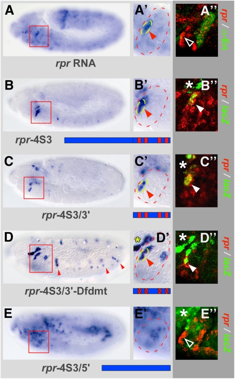
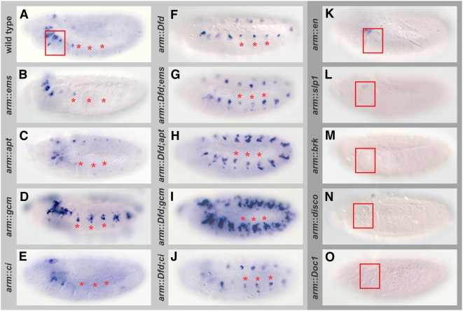
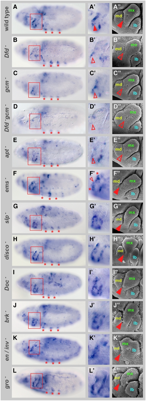
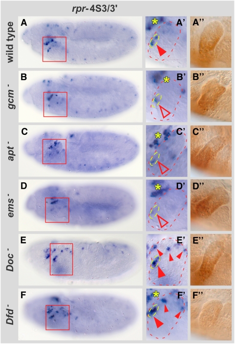
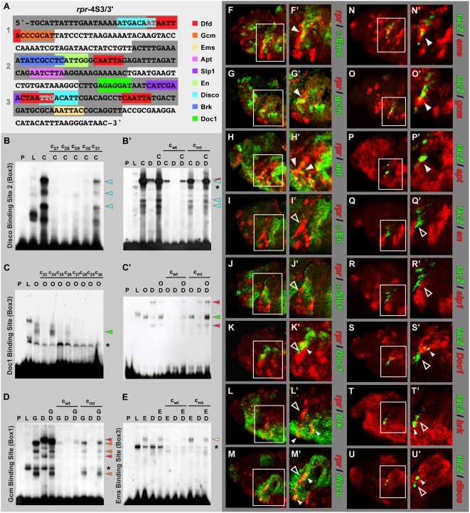
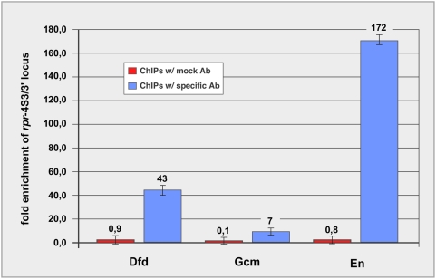
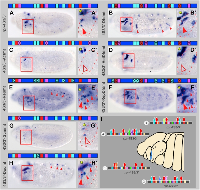
Hox proteins play fundamental roles in controlling morphogenetic diversity along the anterior-posterior body axis of animals by regulating distinct sets of target genes. Within their rather broad expression domains, individual Hox proteins control cell diversification and pattern formation and consequently target gene expression in a highly localized manner, sometimes even only in a single cell. To achieve this high-regulatory specificity, it has been postulated that Hox proteins co-operate with other transcription factors to activate or repress their target genes in a highly context-specific manner in vivo. However, only a few of these factors have been identified. Here, we analyze the regulation of the cell death gene reaper (rpr) by the Hox protein Deformed (Dfd) and suggest that local activation of rpr expression in the anterior part of the maxillary segment is achieved through a combinatorial interaction of Dfd with at least eight functionally diverse transcriptional regulators on a minimal enhancer. It follows that context-dependent combinations of Hox proteins and other transcription factors on small, modular Hox response elements (HREs) could be responsible for the proper spatio-temporal expression of Hox targets. Thus, a large number of transcription factors are likely to be directly involved in Hox target gene regulation in vivo.


Conflict of interest statement

The authors have declared that no competing interests exist.

Figures

Figure 1
Figure 1. Identification of minimal Dfd response element in the rpr enhancer using stage 11 wild-type embryos.
(A and A′) rpr RNA is strongly expressed in the anterior part of the maxillary segment. (A″) Double-labelling of rpr RNA and Engrailed (En) protein. Arrowhead marks rpr transcripts, mostly excluded from the posterior part of the maxillary segment (highlighted by En expression). (B and B′) lacZ RNA expression in the rpr-4S3 reporter line. (C and C′) In the rpr-4S3/3′ reporter line, lacZ expression recapitulates endogenous rpr transcription in the maxillary segment. (D and D′) In the rpr-4S3/3′-Dfdmt reporter line all four Dfd binding sites are mutated, resulting in strong lacZ activation in the anterior part of the maxillary segment. Small, red arrowheads in (D) indicate ectopic lacZ expression in trunk segments. (E and E′) In the rpr-4S3/5′ reporter line, lacZ is expressed in a broad stripe close to the posterior end. (B″ to E″) Double-labelling of rpr and lacZ RNA in the rpr-4S3 (B″), rpr-4S3/3′ (C″), rpr-4S3/3′-Dfdmt (D″) and rpr-4S3/5′ (E″) transgenic lines. The closed arrowheads in (B″ to D″) mark areas of co-localization of rpr and lacZ transcripts, the open arrowhead in (E″) marks area of rpr expression in the anterior part of the maxillary segment without any lacZ transcripts. Red boxes in (A to E) mark the maxillary segment, close-ups of which are shown in (A′ to E′). Asterisks in (B″, C″, D″ and E″) indicate area of lacZ expression in procephalic lobe. Blue bars in (B to E) represent different parts of rpr enhancer, Dfd binding sites are indicated as small red boxes.
Figure 2
Figure 2. Approach to identify factors for Dfd-dependent rpr expression.
lacZ RNA in situ hybridizations in stage 11 embryos ubiquitously mis-expressing different genes in rpr-4S3/3′ reporter line using the arm-GAL4 driver are shown: (A) rpr-4S3/3′ control, (B) arm::ems, (C) arm::apt, (D) arm::gcm, (E) arm::ci, (F) arm::Dfd, (G) arm::Dfd;ems, (H) arm::Dfd;apt, (I) arm::Dfd;gcm, (J) arm::Dfd;ci, (K) arm::en, (L) arm::slp1, (M) arm::brk, (N) arm::disco, (O) arm::Doc1. The screen is based on the observation that ubiquitous mis-expression of Dfd in the rpr-4S3/3′ line leads to ectopic lacZ expression in anterior part of every segment (shown in F). In (A to J) asterisks mark three spots of lacZ expression in trunk, box in (A, K to O) highlights the maxillary segment.
Figure 3
Figure 3. Requirement of transcription factors for rpr expression and development of the maxillary segment.
(A–L) rpr RNA expression in stage 11 wild-type (A), Dfdw21 (B), gcmN7-4 (C), Dfdw21; gcmN7-4 (D), apt03041 (E), ems9G/ems7D99 (F), Df(2L)slp2-Δd66C (G), Df(1)XR14 (H), Df(3L)DocA (I), brkM68 (J), Df(2R)enE (K) and groB48 (L) mutant embryos. To select identical stages, two criteria were used: 1) overall morphology of embryos; 2) three spots of rpr expression in thoracic segments characteristic for stage 11 wild-type embryos (marked by three asterisks). Red boxes in (A to L) highlight maxillary segments. (A′ to L′) Close-up of maxillary segments in respective mutants. In gcmN7-4 and apt03041 mutants, rpr expression is reduced (C′ and E′), in Dfdw21; gcmN7-4 double mutants expression is lost (D′) (open arrowhead). In ems9G/ems7D99 mutants, levels of rpr transcripts are reduced in middle part of anterior rpr expression area (small open arrowhead), in ventral-anterior and dorsal-anterior part rpr transcript levels are increased (highlighted by asterisks). In embryos mutant for repressing transcription factor genes, cells ectopically expressing rpr are observed in various parts of the maxillary segment (G′ to K′). In groB48 mutants, rpr expression in anterior and posterior parts is increased (L and L′). (A″ to L″) Scanning electron micrographs of gnathal segments of late stage 12 embryos of respective mutants. Mandibular (md), maxillary (mx) and labial (lb) segments are indicated in this panel. In mutants for the activating transcription factor genes, the boundary between the maxillary and mandibular segments is reduced or abolished (C″ to E″) (open arrowhead), reminiscent to the effects seen in Dfd mutants (B″), in mutants for the repressing transcription factor genes this boundary is unaffected (G″ to K″) (closed arrowhead).
Figure 4
Figure 4. Co-regulatory transcription factors are required for proper lacZ expression in stage 11 rpr-4S3/3′ reporter line.
(A to F) In rpr-4S3/3′ reporter line, β-galactosidase expression in the following genetic backgrounds is shown: (A) rpr-4S3/3′ control, (B) gcmN7-4, (C) apt03041, (D) ems9G/ems7D99, (E) Df(3L)DocA, (F) Dfdw21 mutant embryos. Red boxes in (A to F) highlight maxillary segments. (A′ to F′) Close-up of maxillary segments in respective mutants. The yellow asterisks in (A′, B′, C′, D′ and F′) mark expression of lacZ in procephalic lobes. (A″ to F″) Dfd protein expression in the respective genotypes. Note that although the morphology of the maxillary segment is changed, the expression domain and intensity of Dfd protein in the respective mutants (B″ to E″) is very similar to wild-type Dfd protein expression (A″).
Figure 5
Figure 5. Co-regulatory transcription factors directly interact with rpr-4S3/3′ enhancer.
(A) Sequence of rpr-4S3/3′ enhancer fragment with binding sites for Dfd (shown in red) and all identified co-regulatory transcription factors (highlighted in different colours) is shown. Conserved regions 1 to 3 within the rpr-4S3/3′ enhancer are highlighted as dark grey boxes. (B and C) EMSAs for mapping of Disco binding site 2 (B) and Doc1 binding site (C) using box 3 as shift probe. EMSA was performed using no protein (P), translation lysate only (L), lysate with Dfd protein (D), lysate with Disco protein (C) and lysate with Doc1 protein (O). c27 to c31 in (B) and c23 to c30 in (C) represent consecutive competitor oligonucleotides with their middle base-pairs mutated. Competition experiments revealed that sequences mutated in the oligonucleotide c31 include binding site for the Disco protein (B), whereas oligonucleotides c24 include binding site for the Doc1 protein (C). The turquoise and green arrowheads indicate specific DNA-protein complexes containing either Disco or Doc1 protein, respectively. (B′, C′, D, E) EMSAs using no protein (P), translation lysate (L), lysate with Dfd protein (D), Doc1 protein (O), Gcm protein (G), Ems protein (E) and lysate with Dfd protein (D). To test specificity of binding of the proteins to the DNA fragments, competitor oligonucleotides for the mapped binding sites were used either in their wild-type (cwt) or mutant (cmt) sequence versions. Red arrowheads indicate specific DNA-protein complexes containing Dfd protein, turquoise, green, orange or light-yellow arrowheads indicate specific DNA-protein complexes containing Disco, Doc1, Gcm or Ems proteins, respectively. Note that in all competitor oligonucleotides only binding site sequences for co-regulatory transcription factors are mutated, but not for Dfd binding sites. (F to U′) Protein or RNA co-localization of co-regulatory transcription factors and rpr (F to M′) or lacZ RNA (N to U′) in head of stage 11 wild-type (F to M′) or rpr-4S3 reporter line (N to U′) embryos. Boxes mark maxillary segment with rpr or lacZ RNAs present in anterior part. In (F′ to M′ and N′ to U′) close-ups of maxillary segments are shown. Co-localization of rpr or lacZ RNAs and co-regulator RNA is observed in individual cells for Doc1, Brk, Disco (K′ to M′ and S′ to U′; small, closed arrowheads). Closed arrowheads mark cells co-expressing rpr and lacZ RNAs and RNA or protein of activating co-regulators, open arrowheads highlight areas of rpr or lacZ transcription and missing expression of repressing co-regulators in anterior part of maxillary segments. Asterisks in (B′, C, D and E) indicate complexes with lysate protein seen also in the controls.
Figure 6
Figure 6. Chromatin immuno-precipitation (ChIP) for Dfd, En and Gcm confirms interaction with rpr-4S3/3′ enhancer in vivo.
Specific enrichment of binding sites within the rpr-4S3/3′ enhancer was assayed by quantitative real-time PCR and compared to negative control locus. All ChIPs performed with specific antibodies (blue) yield at least 7-fold enrichment over the negative control, precipitations with mock antibodies (red) yield no enrichment (ratios below 1). Fold enrichment were normalized against input chromatin sample and to negative control region for primer normalization (for details: see Materials and Methods).
Figure 7
Figure 7. Binding sites for co-regulatory transcription factors are required for rpr enhancer activity in stage 11 embryos.
(A and A′) β-galactosidase is expressed in anterior part of maxillary segment in rpr-4S3/3′ reporter line. Closed, red arrowhead marks anterior part of maxillary segment. (B and B′) In the rpr-4S3/3′-Dfdmt reporter, with all Dfd binding sites mutated, lacZ expression is increased in anterior part of maxillary segment (closed, red arrowhead). lacZ expression is ectopically induced in anterior part of every segment (small, closed arrowheads). (C and C′) In the rpr-4S3/3′-Actmt line, with all sites for activating co-regulators mutated, reporter gene expression in maxillary segment is lost (open arrowhead). (D and D′) In the rpr-4S3/3′-ActDfdmt reporter, with all Dfd binding sites and sites for activating co-regulators mutated, lacZ expression in the anterior part is lost (open arrowhead). (E and E′) In the rpr-4S3/3′-Repmt line, with all sites for repressing co-regulators mutated, additional cells in maxillary segment express reporter gene. In rest of embryo, lacZ expression is ectopically induced (small, closed arrowheads). (F and F′) In the rpr-4S3/3′-RepDfdmt reporter, with all Dfd binding sites and sites for repressing co-regulators mutated, lacZ expression in anterior and posterior parts is strongly induced (closed arrowheads). (G and G′) In stage 11 embryos of rpr-4S3/3′-Gcmmt line, with the Gcm binding site mutated, reporter gene expression in anterior part of maxillary segment is reduced (open arrowhead). (H and H′) In the rpr-4S3/3′-Docmt line, with the Doc1 binding site mutated, reporter gene expression is observed in additional cells in maxillary segment. In the rest of the embryo, lacZ expression is ectopically induced (small, closed arrowheads). (I) Model of rpr regulation through the rpr-4S3/3′ enhancer. Expression of rpr in the anterior part of the maxillary segment (highlighted in blue) is achieved through a combinatorial interaction of the Hox protein Dfd and co-regulatory transcription factors (represented as different-coloured triangles) to specific binding sites in the rpr-4S3/3′ enhancer. Each cell of the maxillary segment expresses different combinations of Dfd and the co-regulatory transcription factors, which is reflected in a cell type-specific occupancy of the rpr-4S3/3′ enhancer, as shown exemplarily for four different cells (marked 1 to 4). According to the model, the decision whether rpr transcription is activated or repressed in individual maxillary cells depends on the nature and combination of regulatory factors interacting with the rpr-4S3/3′ enhancer. Boxes in (A to F) highlight maxillary segments, yellow asterisks in (A′ to H′) mark lacZ expressing cells in procephalic lobes. rpr-4S3/3′ enhancer in (A to H) is represented as blue bar, Dfd binding sites as red, sites for activating co-regulators as pink and sites for repressing co-regulators as turquoise boxes.

Similar articles

Cited by

References

    1. McGinnis W, Krumlauf R. Homeobox genes and axial patterning. Cell. 1992;68:283–302. - PubMed
    1. Mann RS, Morata G. The developmental and molecular biology of genes that subdivide the body of Drosophila. Annu Rev Cell Dev Biol. 2000;16:243–271. - PubMed
    1. Pearson JC, Lemons D, McGinnis W. Modulating Hox gene functions during animal body patterning. Nature Genetics Reviews. 2005;6:893–904. - PubMed
    1. Botas J. Control of morphogenesis and differentiation by HOM/Hox genes. Curr Opin Cell Biol. 1993;5:1015–1022. - PubMed
    1. Graba Y, Aragnol D, Pradel J. Drosophila Hox complex downstream targets and the function of homeotic genes. Bioessays. 1997;19:379–388. - PubMed

Publication types

MeSH terms

LinkOut - more resources