. 2009 Mar 31;106(13):5153-8.
doi: 10.1073/pnas.0901104106. Epub 2009 Mar 10.

FOXO3a-dependent regulation of Pink1 (Park6) mediates survival signaling in response to cytokine deprivation

Affiliations

FOXO3a-dependent regulation of Pink1 (Park6) mediates survival signaling in response to cytokine deprivation

Yang Mei et al. Proc Natl Acad Sci U S A. .

Abstract

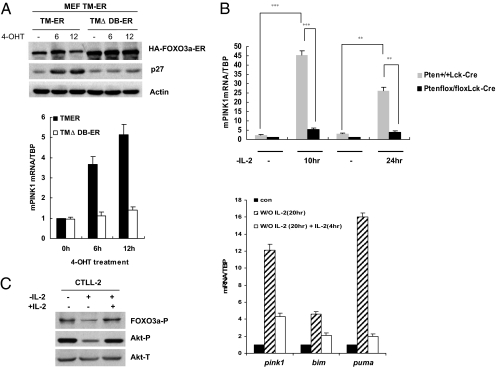
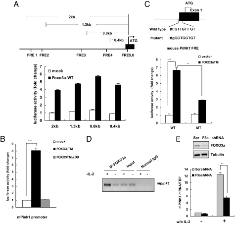
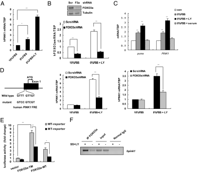
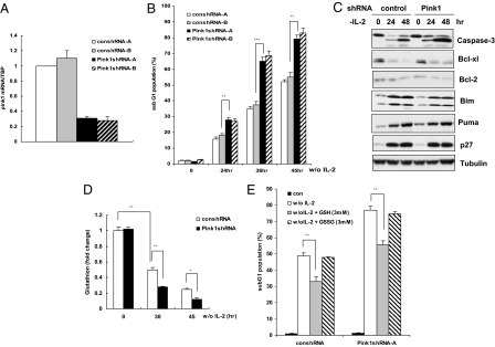
Loss-of-function mutations of phosphatase/tensin homolog deleted on chromosome 10 (PTEN)-induced putative kinase 1 (Pink1) (also known as Park6) identified in familial forms of Parkinson's disease (PD) are associated with compromised mitochondrial function. Emerging data suggest that Pink1 is an essential pro-survival factor that is induced in response to oxidative stress. However, the mechanisms regulating Pink1 expression under stress conditions remain unknown. Forkhead box, subgroup O (FOXO) transcription factors carry out distinct biological functions in response to different extracellular signals. Notably, FOXO factors possess evolutionarily conserved roles in protecting cells from oxidative stress-induced death. Here we report that the FOXO family member FOXO3a controls Pink1 transcription in both mouse and human cells subjected to growth factor deprivation and that this regulation is exerted through evolutionarily conserved FOXO binding elements. Induction of Pink1 by FOXO3a is crucial for survival signals in lymphocytes, as depletion of Pink1 sensitizes these cells to death induced by deprivation of an essential growth factor. Our data reveal that the role of FOXO factors in protecting cells from growth factor deprivation-triggered apoptosis has been underestimated and that FOXOs mediate this protection by transactivating anti-apoptotic effectors like Pink1. Given the essential role of Pink1 in combating cell death, our findings may help to dissect the mechanisms by which FOXO proteins function as anti-oxidative stress factors.


Conflict of interest statement

The authors declare no conflict of interest.

Figures

Fig. 1.
Fig. 1.
Regulation of mPink1 by PI3k/Akt/FOXO signaling. (A) FOXO3a induces Pink1. (Upper) Validation of FOXO3a-expressing cell lines. p53−/− TM-ER or TMΔDB-ER FOXO3a-expressing MEFs were exposed to 4-OHT (1 μM) for 6 h or 12 h, and lysates were subjected to Western blotting using antibodies directed against the indicated proteins. Actin, loading control. (Lower) Pink1 mRNA levels were assessed in the MEFs in A Upper by qRT-PCR. TBP, normalization control. Unless otherwise stated, all results shown are one trial representative of at least 3 independent experiments. (B) Pten loss impairs Pink1 upregulation induced by IL-2 deprivation. RNA extracted from Pten+/+Lck-Cre or Ptenflox/floxLck-Cre activated T cells that were either left untreated or deprived of IL-2 for 10 or 24 h was subjected to qRT-PCR. The results presented are the mean and SEM of 2 independent experiments conducted in triplicate. **, P < 0.01; ***, P < 0.001. (C) Pink1 mRNA expression correlates inversely with PI3K/Akt activation. CTLL-2 cells were subjected to IL-2 withdrawal for 20 h and then treated with IL-2 for an additional 4 h. (Left) Lysates were prepared and analyzed by Western blotting with antibodies against the indicated proteins. P, phosphorylated; T, total. (Right) IL-2 restoration decreases Pink1 mRNA. qRT-PCR was performed to detect expression levels of Pink1 mRNA and Bim and Puma mRNAs (positive controls). Fold induction relative to TBP was determined as for A.
Fig. 2.
Fig. 2.
Murine Pink1 is a direct FOXO3a target. (A) Structure and validation of luciferase reporters driven by murine Pink1 promoter. (Upper) Schematic diagram of the WT murine Pink1 gene, showing the putative FBE sites and the segments of the candidate mPink1 promoter region that were cloned into the pGL3 basic luciferase reporter plasmid. (Lower) Transactivation of the Pink1 promoter by WT FOXO3a. 293T cells were cotransfected with a plasmid bearing a Pink1 promoter region fragment plus a plasmid expressing WT FOXO3a or were mock transfected. Luciferase activity was measured in triplicate after 48 h and normalized to an internal transfection efficiency control (β-galactosidase). (B) Functional FOXO is required for Pink1 induction. 293T cells were cotransfected with the 0.4-kb Pink1 promoter luciferase reporter plus vector alone, or FOXO3aTM, or FOXO3a TMΔDB. Luciferase activity was assessed 48 h after transfection as for A. The results presented correspond to the mean and SEM of 3 independent experiments conducted in triplicate. ***, P < 0.001. (C) FOXO3a transactivates the Pink1 promoter. (Upper) The sequence of the putative FBEs in the mouse Pink1 0.4-kb promoter region and the substitution mutations introduced into this FBE sequence are shown. (Lower) Mutations of the FBEs impair transactivation of the Pink1 promoter by FOXO3a. 293T cells were cotransfected with either control vector or WT FOXO3a plus 0.4-kb Pink1 promoter luciferase reporters containing either intact (WT) or mutated (MT) FBEs. Results were expressed as for A and represent the mean and error of 3 independent experiments conducted in triplicate. **, P < 0.01; ***, P < 0.001. (D) FOXO3a physically binds to the mPink1 promoter. CTLL-2 cells were cultured in IL-2−free medium for 12 h and ChIP assays to determine the binding of FOXO3a to the mPink1 FBE were conducted as described in Materials and Methods. (E) Knockdown of FOXO3a attenuates Pink1 induction. (Upper) CTLL-2 cells expressing either scrambled shRNA (Scr) or FOXO3a shRNA (F3a) were subjected to IL-2 withdrawal and protein lysates were assessed by Western blotting to detect FOXO3a protein. Tubulin, loading control. (Lower) RNA from the CTLL-2 cells in E Upper was analyzed by qRT-PCR to detect mPink1 transcripts. TBP, internal control. The data correspond to the mean and SEM of 2 independent experiments conducted in triplicate. **, P < 0.01.
Fig. 3.
Fig. 3.
FOXO regulates the human Pink1 promoter. (A) Inactivation of Akt signaling increases hPink1 transcription. MCF-7 cells were cultured for 24 h under standard conditions or in medium containing 0% serum or 0% serum plus 25 μM LY294002 (LY). RNA was extracted and levels of hPink1 expression were assessed by qRT-PCR. The data represent the mean and SEM of 4 independent experiments. *, P < 0.05; **, P < 0.01. (B) Depleting FOXO3a abrogates Pink1 induction in response to inactivated PI3K/Akt signaling. (Top) Protein lysates from MCF-7 cells expressing scrambled shRNA (Scr) or FOXO3a shRNA (F3a) were assessed by Western blotting to detect FOXO3a. (Middle and Bottom) RNA from the cells in A was analyzed by qRT-PCR to detect FOXO3a (Middle) or Pink1 (Bottom). Data represent the mean and SEM of 3 independent experiments conducted in triplicate. **, P < 0.01. (C) FOXO3a-dependent induction of Pink1 in SHSY5Y neuroblastoma cells. (Upper) SHSY5Y cells were cultured for 24 h under standard conditions or in medium containing 0% serum, or 0% serum plus 25 μM LY294002 (LY), or serum starved for 24 h and then cultured in 10% FBS for additional 3 h. qRT-PCR was performed to detect expression levels of Pink1 mRNA and Puma mRNAs (positive control). Fold induction relative to TBP was determined as for A. (Lower) SHSY5Y cells were treated as in B Bottom, and RNA was analyzed by qRT-PCR to detect Pink1. The results presented correspond to the mean and SEM of 3 independent quantitative RT-PCR experiments. **, P < 0.01. (D) The sequence of a highly conserved putative FBE in the human Pink1 promoter (WT) and substitution mutations introduced into this FBE (mutant) are shown. (E) FBE mutation impairs hPink1 promoter transactivation by FOXO3a. 293T cells were transfected with hPink1 promoter reporter plasmids containing an intact (WT) or mutated (MT) FBE, in the presence of vector alone, or FOXO3a-TM, or WT FOXO3a (FOXO3a-WT). Data represent the mean and error of 3 independent experiments conducted in triplicate. *, P < 0.05; **, P < 0.01. (F) FOXO3a physically binds to the hPink1 promoter. ChIP assays were performed as for Fig. 2D using MCF-7 cells that were left untreated or serum-starved (SS) for 24 h in the presence of 25 μM LY294002 (LY).
Fig. 4.
Fig. 4.
Pink1 depletion sensitizes T cells to cytokine withdrawal-induced death. (A) Validation of mPink1 shRNAs. CTLL-2 cells were transfected with 2 independent sets of control (con) shRNA or mPink1 shRNA constructs and levels of mPink1 mRNA were determined by qRT-PCR. (B) Reduction of Pink1 increases apoptosis. CTLL-2 cells expressing the indicated shRNAs were cultured in the presence or absence of IL-2 for the indicated times and apoptosis was determined by PI staining and flow cytometry. Data shown are the mean percentage of apoptotic cells ± SD from 3 independent experiments. **, P < 0.01; ***, P < 0.001. (C) Reduction of mPink1 increases caspase-3 cleavage. CTLL-2 cells expressing either control shRNA or Pink1 shRNA were deprived of IL-2 for the indicated time point and cell lysates were subjected to Western blotting to detect the indicated proteins. (D) Reduction of mPink1 correlates with decreased GSH levels. CTLL-2 cells expressing either control shRNA or Pink1 shRNA were cultured in the presence or absence of IL-2 for 30 or 45 h and intracellular GSH levels were measured in triplicate as described in Materials and Methods. Results represent the mean and error of 4 independent experiments conducted in triplicate each time. *, P < 0.05; **, P < 0.01. (E) GSH is a cause, not a consequence, of cell death associated with Pink1 depletion. CTLL-2 cells expressing control or Pink1 shRNA were cultured for 45 h in medium lacking IL-2 but containing 3 mM reduced GSH or oxidized GSSG. Cell death was measured in triplicate by PI staining and flow cytometry. Results shown are the mean percentage of apoptotic cells ± SD from 4 independent experiments. **, P < 0.01.

Similar articles

Cited by

References

    1. Unoki M, Nakamura Y. Growth-suppressive effects of BPOZ and EGR2, two genes involved in the PTEN signaling pathway. Oncogene. 2001;20(33):4457–4465. - PubMed
    1. Petit A, et al. Wild-type PINK1 prevents basal and induced neuronal apoptosis, a protective effect abrogated by Parkinson disease-related mutations. J Biol Chem. 2005;280(40):34025–34032. - PubMed
    1. MacKeigan JP, Murphy LO, Blenis J. Sensitized RNAi screen of human kinases and phosphatases identifies new regulators of apoptosis and chemoresistance. Nat Cell Biol. 2005;7(6):591–600. - PubMed
    1. Deng H, Jankovic J, Guo Y, Xie W, Le W. Small interfering RNA targeting the PINK1 induces apoptosis in dopaminergic cells SH-SY5Y. Biochem Biophys Res Commun. 2005;337(4):1133–1138. - PubMed
    1. Valente EM, et al. Hereditary early-onset Parkinson's disease caused by mutations in PINK1. Science. 2004;304(5674):1158–1160. - PubMed

Publication types

LinkOut - more resources