. 2009 Apr 24;284(17):11216-23.
doi: 10.1074/jbc.M901229200. Epub 2009 Mar 3.

Tristetraprolin mediates interferon-gamma mRNA decay

Affiliations

Tristetraprolin mediates interferon-gamma mRNA decay

Rachel L Ogilvie et al. J Biol Chem. .

Abstract

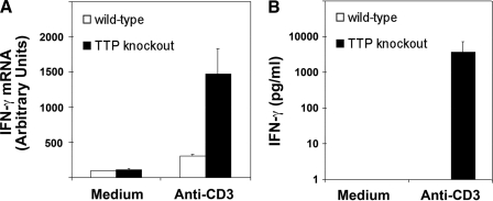
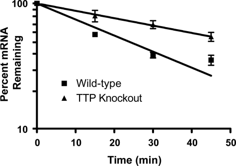
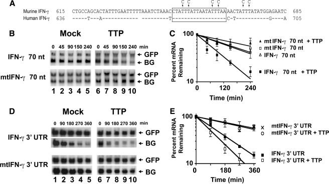
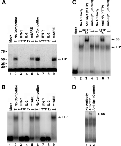
Tristetraprolin (TTP) regulates expression at the level of mRNA decay of several cytokines, including the T cell-specific cytokine, interleukin-2. We performed experiments to determine whether another T cell-specific cytokine, interferon-gamma (IFN-gamma), is also regulated by TTP and found that T cell receptor-activated T cells from TTP knock-out mice overproduced IFN-gamma mRNA and protein compared with activated T cells from wild-type mice. The half-life of IFN-gamma mRNA was 23 min in anti-CD3-stimulated T cells from wild-type mice, whereas it was 51 min in anti-CD3-stimulated T cells from TTP knock-out mice, suggesting that the overexpression of IFN-gamma mRNA in TTP knock-out mice was due to stabilization of IFN-gamma mRNA. Insertion of a 70-nucleotide AU-rich sequence from the murine IFN-gamma 3'-untranslated region, which contained a high affinity binding site for TTP, into the 3'-untranslated region of a beta-globin reporter transcript conferred TTP-dependent destabilization on the beta-globin transcript. Together these results suggest that TTP binds to a functional AU-rich element in the 3'-untranslated region of IFN-gamma mRNA and mediates rapid degradation of the IFN-gamma transcript. Thus, TTP plays an important role in turning off IFN-gamma expression at the appropriate time during an immune response.


Figures

FIGURE 1.
FIGURE 1.
IFN-γ mRNA and protein are overproduced in T cells from TTP knock-out mice. Purified murine T cells from homozygous TTP knock-out mice or wild-type littermates were either unstimulated (medium) or stimulated for 6 h with anti-CD3 antibodies. A, total cellular RNA was isolated, and IFN-γ mRNA levels were measured by real time PCR. For each sample, the expression levels for IFN-γ mRNA were normalized to hypoxanthine phosphoribosyltransferase mRNA levels. Each point represents the mean and S.E. of triplicate samples. B, IFN-γ protein levels were measured in culture supernatants by enzyme-linked immunosorbent assay. The graphed data show the mean and S.E. of triplicate samples for each condition.
FIGURE 2.
FIGURE 2.
IFN-γ mRNA is stabilized in TTP knock-out mice. Purified murine T cells from wild-type or TTP knock-out mice were stimulated for 6 h with anti-CD3 antibodies. Actinomycin D (10 μg/ml) was then added to stop transcription, and total RNA was isolated after 0, 15, 30, and 45 min. IFN-γ mRNA was measured using real time reverse transcription-PCR, and expression levels were normalized to hypoxanthine phosphoribosyltransferase mRNA levels. The normalized level of IFN-γ mRNA at time 0 was set at 100%, and all other normalized mRNA levels were graphed relative to that value. Each point represents the mean and S.E. from triplicate samples. A one-phase model of exponential decay was used to derive the indicated mRNA decay curves.
FIGURE 3.
FIGURE 3.
TTP regulates the decay of mRNA containing the IFN-γ ARE. A, the top line shows a 70-nucleotide AT-rich sequence from the 3′-UTR of the murine IFN-γ cDNA (RefSeq transcript ID NM_008337) that was cloned into the 3′-UTR of the pTetBBB β-globin reporter construct. The arrows above the line indicate the six single nucleotide mutations (T to C) that were introduced to create a mutated sequence. The corresponding sequence from the human IFN-γ cDNA (RefSeq transcript ID NM_000619) is shown on the bottom line to illustrate the cross-species sequence conservation. The boxed sequence indicates an 18-nucleotide AU-rich IFN-γ ribo-oligonucleotide that was identical in mouse and human sequences and was used for the binding reactions shown in Figs. 4 and 5. In B and C, HeLa Tet-Off cells were transfected with tet-responsive β-globin reporter plasmids in which the 70-nucleotide murine IFN-γ ARE sequence (IFN70 nt) or the mutated sequence shown in A (mtIFN70 nt) was inserted into the β-globin 3′-UTR. In D and E, HeLa Tet-Off cells were transfected with tet-responsive β-globin reporter plasmids in which the β-globin 3′-UTR was replaced with the entire 3′-UTR from murine IFN-γ (IFN3-UTR) or a mutated murine IFN-γ 3′-UTR that contained the mutations shown in A (mtIFN3-UTR). The cells were co-transfected with the pCMV.TTP.Tag plasmid (TTP) or a pcDNA3 control plasmid (Mock), and the pTracer GFP expression construct was co-transfected in each group as a control for transfection efficiency. After 72 h, doxycycline was added to stop transcription from the tet-responsive promoter, and total RNA was isolated after the indicated times. Total RNA (10 μg) from each sample was analyzed by Northern blot using a 32P-labeled rabbit β-globin probe (BG) and a 32P-labeled GFP probe (GFP). The experiments shown in B and D were both performed four times. Band intensities were measured using a phosphorimaging system. For each condition, the hybridization intensity values of β-globin mRNA were normalized to GFP mRNA levels. The normalized value for β-globin expression at time 0 was then set to 100%, and all other mRNA levels were graphed over time relative to that value. The graphs in C and E correspond to the experiments shown in B and D, respectively. Each point represents the mean and S.E. from the four experiments.
FIGURE 4.
FIGURE 4.
TTP binds specifically to a conserved AU-rich sequence in the 3′-UTR of IFN-γ mRNA. A, HeLa cells were transiently transfected with the pcDNA-3 plasmid (Mock), the murine pCMV.mTTP.myc-Tag expression plasmid (mTTP Tx), or the human pCMV.hTTP.Tag expression plasmid (hTTP Tx), and cytoplasmic protein extracts were prepared 48 h later. An RNA-protein UV cross-linking assay was performed by mixing cytoplasmic extracts (10 μg of protein) with 20 fmol of a 32P-end-labeled RNA probe containing an AU-rich sequence from the IFN-γ 3′-UTR. A 200-fold molar excess of unlabeled IFN-γ ARE (IFN-γ), IL-2 ARE (IL-2), or mutated ARE (mtARE) competitor RNA was added to the indicated reactions. The reaction mixtures were treated with UV radiation and separated by electrophoresis on a 10% SDS-polyacrylamide gel, and bands were visualized using a phosphorimaging system. The positions of migration of molecular mass markers in kilodaltons are shown to the left of the figure, and the position of migration of the major band present only in reactions from TTP-transfected cells is labeled “TTP.” B, an experiment was performed identically to the experiment described in A except the reactions mixtures were not treated with UV energy but instead were separated by electrophoresis on a 10% polyacrylamide gel under nondenaturing conditions. C, HeLa cells were transiently transfected with the pcDNA-3 plasmid (Mock), the murine pCMV.mTTP.myc-Tag expression plasmid (mTTP Tx), or the human pCMV.hTTP.Tag expression plasmid (hTTP Tx), and cytoplasmic protein extracts were prepared 48 h later. RNA-protein gel shift assays were performed by mixing the cytoplasmic extracts with 20 fmol of a 32P-end-labeled RNA probe containing an AU-rich sequence from the IFN-γ 3′-UTR. A control anti-Sp1 antibody (lanes 4 and 7), an anti-Myc antibody (lane 5), which recognizes Myc-tagged murine TTP, and an anti-HA antibody (lane 6), which recognizes HA-tagged human TTP, were added to the indicated reactions. The binding reactions were separated by electrophoresis on a 10% polyacrylamide gel under non-denaturing conditions, and bands were visualized using a phosphorimaging system. The major band present only in reactions from TTP-transfected cells is labeled TTP, and the TTP-containing supershifted band is labeled “SS.” D, cytoplasmic extracts (10 μg of protein) prepared from purified primary human T cells that had been stimulated for 6 h with an anti-CD3 antibody were incubated with 20 fmol of a 32P-end-labeled RNA probe containing an AU-rich sequence from the IFN-γ 3′-UTR. An anti-TTP antibody (lane 2) or a control anti-Sp1 antibody (lane 3) was added to the indicated reactions. The binding reactions were separated by electrophoresis on a 10% polyacrylamide gel under non-denaturing conditions, and bands were visualized using a phosphorimaging system. The supershifted band present only in the reaction that contained an anti-TTP antibody is labeled SS.
FIGURE 5.
FIGURE 5.
TTP binds to the IFN-γ ARE with high affinity. Cytoplasmic extracts from HeLa cells (10 μg of protein) that were transfected with the murine pCMV.mTTP.myc-Tag expression plasmid (mTTP) or the human pCMV.hTTP.Tag expression plasmid (hTTP) were incubated with 20 fmol of a 32P end-labeled IFN-γ ARE probe in the presence of increasing amounts of unlabeled IFN-γ ARE RNA (40 fmol to 5 pmol) RNA oligonucleotides. Binding reactions were then separated by electrophoresis on 10% polyacrylamide gels under non-denaturing conditions. The bands representing the free probe and the TTP-containing RNA-protein complex were visualized and quantified using a phosphorimaging system. The position of migration of the TTP-containing complex is indicated with an arrow. The graph to the right of each gel represents binding data from three identical independent experiments. Percent maximal binding (y axis) was plotted against the log of the total RNA concentration in molar (x axis) using GraphPad Prism version 4.03 software. Each point represents the mean and S.E. from three experiments.

Similar articles

Cited by

References

    1. Boehm, U., Klamp, T., Groot, M., and Howard, J. C. (1997) Annu. Rev. Immunol. 15, 749-795 - PubMed
    1. Wang, Z. E., Reiner, S. L., Zheng, S., Dalton, D. K., and Locksley, R. M. (1994) J. Exp. Med. 179, 1367-1371 - PMC - PubMed
    1. Schroder, K., Hertzog, P. J., Ravasi, T., and Hume, D. A. (2004) J. Leukoc. Biol. 75, 163-189 - PubMed
    1. Muller, U., Steinhoff, U., Reis, L. F., Hemmi, S., Pavlovic, J., Zinkernagel, R. M., and Aguet, M. (1994) Science 264, 1918-1921 - PubMed
    1. Huang, S., Hendriks, W., Althage, A., Hemmi, S., Bluethmann, H., Kamijo, R., Vilcek, J., Zinkernagel, R. M., and Aguet, M. (1993) Science 259, 1742-1745 - PubMed

Publication types

LinkOut - more resources