Neuronal activity in narcolepsy: identification of cataplexy-related cells in the medial medulla
- PMID: 1925546
- PMCID: PMC8784798
- DOI: 10.1126/science.1925546
Neuronal activity in narcolepsy: identification of cataplexy-related cells in the medial medulla
Abstract
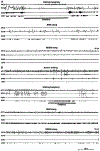
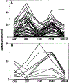
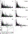
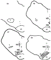
Narcolepsy is a neurological disorder characterized by sleepiness and episodes of cataplexy. Cataplexy is an abrupt loss of muscle tone, most often triggered by sudden, strong emotions. A subset of cells in the medial medulla of the narcoleptic dog discharged at high rates only in cataplexy and rapid eye movement (REM) sleep. These cells were noncholinergic and were localized to ventromedial and caudal portions of the nucleus magnocellularis. The localization and discharge pattern of these cells indicate that cataplexy results from a triggering in waking of the neurons responsible for the suppression of muscle tone in REM sleep. However, most medullary cells were inactive during cataplexy but were active during REM sleep. These data demonstrate that cataplexy is a distinct behavioral state, differing from other sleep and waking states in its pattern of brainstem neuronal activity.
Figures


Similar articles
-
A Discrete Glycinergic Neuronal Population in the Ventromedial Medulla That Induces Muscle Atonia during REM Sleep and Cataplexy in Mice.J Neurosci. 2021 Feb 17;41(7):1582-1596. doi: 10.1523/JNEUROSCI.0688-20.2020. Epub 2020 Dec 28. J Neurosci. 2021. PMID: 33372061 Free PMC article.
-
Activity of medial mesopontine units during cataplexy and sleep-waking states in the narcoleptic dog.J Neurosci. 1992 May;12(5):1640-6. doi: 10.1523/JNEUROSCI.12-05-01640.1992. J Neurosci. 1992. PMID: 1578258 Free PMC article.
-
The neuronal network responsible for paradoxical sleep and its dysfunctions causing narcolepsy and rapid eye movement (REM) behavior disorder.Sleep Med Rev. 2011 Jun;15(3):153-63. doi: 10.1016/j.smrv.2010.08.002. Epub 2010 Nov 5. Sleep Med Rev. 2011. PMID: 21115377 Review.
-
Is narcolepsy a REM sleep disorder? Analysis of sleep abnormalities in narcoleptic Dobermans.Neurosci Res. 2000 Dec;38(4):437-46. doi: 10.1016/s0168-0102(00)00195-4. Neurosci Res. 2000. PMID: 11164570
-
Physiology of REM sleep, cataplexy, and sleep paralysis.Adv Neurol. 1995;67:245-71. Adv Neurol. 1995. PMID: 8848973 Review.
Cited by
-
Amygdala lesions reduce cataplexy in orexin knock-out mice.J Neurosci. 2013 Jun 5;33(23):9734-42. doi: 10.1523/JNEUROSCI.5632-12.2013. J Neurosci. 2013. PMID: 23739970 Free PMC article.
-
Narcolepsy: a key role for hypocretins (orexins).Cell. 1999 Aug 20;98(4):409-12. doi: 10.1016/s0092-8674(00)81969-8. Cell. 1999. PMID: 10481905 Free PMC article. Review. No abstract available.
-
Activity of dorsal raphe cells across the sleep-waking cycle and during cataplexy in narcoleptic dogs.J Physiol. 2004 Jan 1;554(Pt 1):202-15. doi: 10.1113/jphysiol.2003.052134. J Physiol. 2004. PMID: 14678502 Free PMC article.
-
Narcolepsy and orexins: an example of progress in sleep research.Front Neurol. 2011 Apr 18;2:26. doi: 10.3389/fneur.2011.00026. eCollection 2011. Front Neurol. 2011. PMID: 21541306 Free PMC article.
-
Neuronal degeneration in canine narcolepsy.J Neurosci. 1999 Jan 1;19(1):248-57. doi: 10.1523/JNEUROSCI.19-01-00248.1999. J Neurosci. 1999. PMID: 9870955 Free PMC article.
References
Publication types
MeSH terms
Substances
Grants and funding
LinkOut - more resources
Full Text Sources

