

Morphological and ultrastructural features of Iba1-immunolabeled microglial cells in the hippocampal dentate gyrus
- PMID: 19249294
- PMCID: PMC2677570
- DOI: 10.1016/j.brainres.2009.02.031
Morphological and ultrastructural features of Iba1-immunolabeled microglial cells in the hippocampal dentate gyrus
Abstract
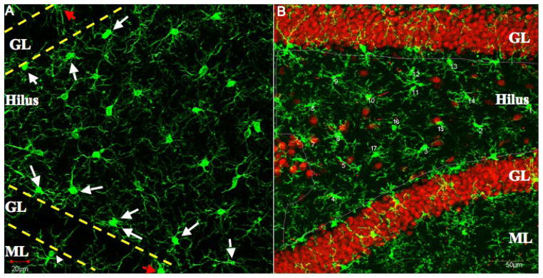
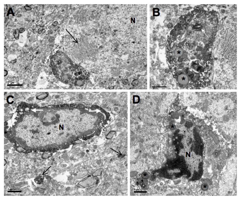
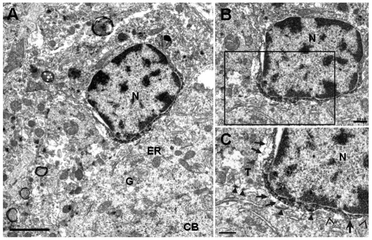
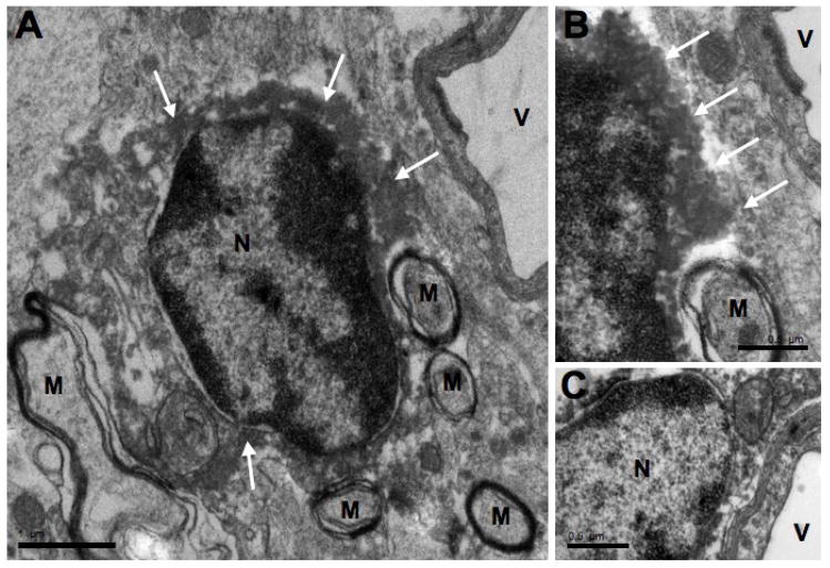
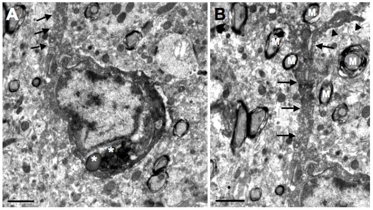
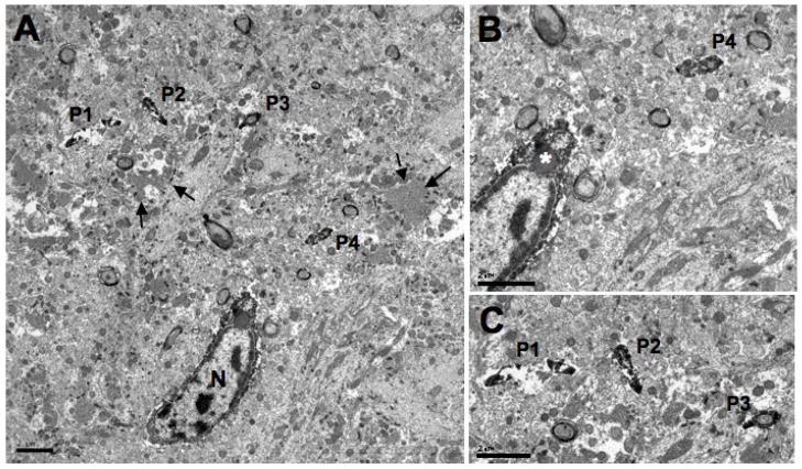
Microglia are found throughout the central nervous system, respond rapidly to pathology and are involved in several components of the neuroinflammatory response. Iba1 is a marker for microglial cells and previous immunocytochemical studies have utilized this and other microglial-specific antibodies to demonstrate the morphological features of microglial cells at the light microscopic level. However, there is a paucity of studies that have used microglial-specific antibodies to describe the ultrastructural features of microglial cells and their processes. The goal of the present study is to use Iba1 immuno-electron microscopy to elucidate the fine structural features of microglial cells and their processes in the hilar region of the dentate gyrus of adult Sprague-Dawley rats. Iba1-labeled cell bodies were observed adjacent to neurons and capillaries, as well as dispersed in the neuropil. The nuclei of these cells had dense heterochromatin next to the nuclear envelope and lighter chromatin in their center. Iba1-immunolabeling was found within the thin shell of perikaryal cytoplasm that contained the usual organelles, including mitochondria, cisternae of endoplasmic reticulum and Golgi complexes. Iba1-labeled cell bodies also commonly displayed an inclusion body. Iba1-labeled cell bodies gave rise to processes that often had a small side branch arise within 5 mum of the microglial cell body. These data showing "resting" Iba-1 labeled microglial cells in the normal adult rat dentate gyrus provide a basis for comparison with the morphology of microglial cells in disease and injury models where they are activated or phagocytotic.
Figures
Similar articles
-
Microglia-associated granule cell death in the normal adult dentate gyrus.Brain Struct Funct. 2009 Dec;214(1):25-35. doi: 10.1007/s00429-009-0231-7. Epub 2009 Nov 21. Brain Struct Funct. 2009. PMID: 19936784 Free PMC article.
-
Morphological features of microglial cells in the hippocampal dentate gyrus of Gunn rat: a possible schizophrenia animal model.J Neuroinflammation. 2012 Mar 16;9:56. doi: 10.1186/1742-2094-9-56. J Neuroinflammation. 2012. PMID: 22424389 Free PMC article.
-
Rare contacts between synapses and microglial processes containing high levels of Iba1 and actin--a postembedding immunogold study in the healthy rat brain.Eur J Neurosci. 2013 Jul;38(1):2030-40. doi: 10.1111/ejn.12213. Epub 2013 Apr 17. Eur J Neurosci. 2013. PMID: 23590220
-
[Activated microglial cells trigger neurogenesis following neuronal loss in the dentate gyrus of adult mice].Nihon Shinkei Seishin Yakurigaku Zasshi. 2012 Nov;32(5-6):281-5. Nihon Shinkei Seishin Yakurigaku Zasshi. 2012. PMID: 23373316 Review. Japanese.
-
A Brief History of Microglial Ultrastructure: Distinctive Features, Phenotypes, and Functions Discovered Over the Past 60 Years by Electron Microscopy.Front Immunol. 2018 Apr 25;9:803. doi: 10.3389/fimmu.2018.00803. eCollection 2018. Front Immunol. 2018. PMID: 29922276 Free PMC article. Review.
Cited by
-
Visual Outcomes in Experimental Rodent Models of Blast-Mediated Traumatic Brain Injury.Front Mol Neurosci. 2021 Apr 15;14:659576. doi: 10.3389/fnmol.2021.659576. eCollection 2021. Front Mol Neurosci. 2021. PMID: 33935648 Free PMC article. Review.
-
Microglia-associated granule cell death in the normal adult dentate gyrus.Brain Struct Funct. 2009 Dec;214(1):25-35. doi: 10.1007/s00429-009-0231-7. Epub 2009 Nov 21. Brain Struct Funct. 2009. PMID: 19936784 Free PMC article.
-
Brain Ultrastructure: Putting the Pieces Together.Front Cell Dev Biol. 2021 Feb 18;9:629503. doi: 10.3389/fcell.2021.629503. eCollection 2021. Front Cell Dev Biol. 2021. PMID: 33681208 Free PMC article. Review.
-
Age and sex drive differential behavioral and neuroimmune phenotypes during postoperative pain.Neurobiol Aging. 2023 Mar;123:129-144. doi: 10.1016/j.neurobiolaging.2022.09.008. Epub 2022 Nov 23. Neurobiol Aging. 2023. PMID: 36577640 Free PMC article.
-
Microglial Activation in the Retina of a Triple-Transgenic Alzheimer's Disease Mouse Model (3xTg-AD).Int J Mol Sci. 2020 Jan 27;21(3):816. doi: 10.3390/ijms21030816. Int J Mol Sci. 2020. PMID: 32012676 Free PMC article.
References
-
- Batchelor PE, Liberatore GT, Wong JYF, Porritt MJ, Frerichs F, Donnan JA, Howells DW. Activated macrophages and microglia induce dopaminergic sprouting in the injured striatum and express brain-derived neurotrophic factor and glial cell line-derived neurotrophic factor. J Neurosci. 1999;19:1708–1716. - PMC - PubMed
-
- Biber K, Neumann H, Inoue K, Boddeke HW. Neuronal ‘On’ and ‘Off’ signals control microglia. Trends Neurosci. 2007;30:596–602. - PubMed
-
- Borges K, Gearing M, McDermott DL, Smith AB, Almonte AG, Wainer BH, Dingledine R. Neuronal and glial pathological changes during epileptogenesis in the mouse pilocarpine model. Exp Neurol. 2003;182:21–34. - PubMed
-
- Chen S, Pickard JD, Harris NG. Time course of cellular pathology after controlled cortical impact injury. Exp Neurol. 2003;182:87–102. - PubMed
Publication types
MeSH terms
Substances
Grants and funding
LinkOut - more resources
Full Text Sources
Molecular Biology Databases
