RANTES release by human adipose tissue in vivo and evidence for depot-specific differences
- PMID: 19240255
- PMCID: PMC2692396
- DOI: 10.1152/ajpendo.90511.2008
RANTES release by human adipose tissue in vivo and evidence for depot-specific differences
Abstract
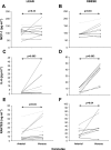
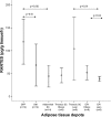
Obesity is associated with elevated inflammatory signals from various adipose tissue depots. This study aimed to evaluate release of regulated on activation, normal T cell expressed and secreted (RANTES) by human adipose tissue in vivo and ex vivo, in reference to monocyte chemoattractant protein-1 (MCP-1) and interleukin-6 (IL-6) release. Arteriovenous differences of RANTES, MCP-1, and IL-6 were studied in vivo across the abdominal subcutaneous adipose tissue in healthy Caucasian subjects with a wide range of adiposity. Systemic levels and ex vivo RANTES release were studied in abdominal subcutaneous, gastric fat pad, and omental adipose tissue from morbidly obese bariatric surgery patients and in thoracic subcutaneous and epicardial adipose tissue from cardiac surgery patients without coronary artery disease. Arteriovenous studies confirmed in vivo RANTES and IL-6 release in adipose tissue of lean and obese subjects and release of MCP-1 in obesity. However, in vivo release of MCP-1 and RANTES, but not IL-6, was lower than circulating levels. Ex vivo release of RANTES was greater from the gastric fat pad compared with omental (P = 0.01) and subcutaneous (P = 0.001) tissue. Epicardial adipose tissue released less RANTES than thoracic subcutaneous adipose tissue in lean (P = 0.04) but not obese subjects. Indexes of obesity correlated with epicardial RANTES but not with systemic RANTES or its release from other depots. In conclusion, RANTES is released by human subcutaneous adipose tissue in vivo and in varying amounts by other depots ex vivo. While it appears unlikely that the adipose organ contributes significantly to circulating levels, local implications of this chemokine deserve further investigation.
Figures


Similar articles
-
Expression and secretion of RANTES (CCL5) in human adipocytes in response to immunological stimuli and hypoxia.Horm Metab Res. 2009 Mar;41(3):183-9. doi: 10.1055/s-0028-1093345. Epub 2008 Oct 27. Horm Metab Res. 2009. PMID: 18956302
-
Physical exercise reduces the expression of RANTES and its CCR5 receptor in the adipose tissue of obese humans.Mediators Inflamm. 2014;2014:627150. doi: 10.1155/2014/627150. Epub 2014 Apr 17. Mediators Inflamm. 2014. PMID: 24895488 Free PMC article.
-
Inverse regulation of inflammation and mitochondrial function in adipose tissue defines extreme insulin sensitivity in morbidly obese patients.Diabetes. 2013 Mar;62(3):855-63. doi: 10.2337/db12-0399. Epub 2012 Dec 6. Diabetes. 2013. PMID: 23223024 Free PMC article.
-
Amyloid precursor protein expression is upregulated in adipocytes in obesity.Obesity (Silver Spring). 2008 Jul;16(7):1493-500. doi: 10.1038/oby.2008.267. Epub 2008 May 15. Obesity (Silver Spring). 2008. PMID: 18483477
-
Interleukin-1β and prostaglandin-synthesizing enzymes as modulators of human omental and subcutaneous adipose tissue function.Prostaglandins Leukot Essent Fatty Acids. 2019 Feb;141:9-16. doi: 10.1016/j.plefa.2018.11.015. Epub 2018 Nov 29. Prostaglandins Leukot Essent Fatty Acids. 2019. PMID: 30661603
Cited by
-
The Alzheimer's Disease Exposome.Alzheimers Dement. 2019 Sep;15(9):1123-1132. doi: 10.1016/j.jalz.2019.06.3914. Epub 2019 Sep 10. Alzheimers Dement. 2019. PMID: 31519494 Free PMC article.
-
Adipose tissue heterogeneity: implication of depot differences in adipose tissue for obesity complications.Mol Aspects Med. 2013 Feb;34(1):1-11. doi: 10.1016/j.mam.2012.10.001. Epub 2012 Oct 13. Mol Aspects Med. 2013. PMID: 23068073 Free PMC article. Review.
-
The Effect of Intensive Dietary Intervention on the Level of RANTES and CXCL4 Chemokines in Patients with Non-Obstructive Coronary Artery Disease: A Randomised Study.Biology (Basel). 2021 Feb 16;10(2):156. doi: 10.3390/biology10020156. Biology (Basel). 2021. PMID: 33669450 Free PMC article.
-
Mechanisms and metabolic implications of regional differences among fat depots.Cell Metab. 2013 May 7;17(5):644-656. doi: 10.1016/j.cmet.2013.03.008. Epub 2013 Apr 11. Cell Metab. 2013. PMID: 23583168 Free PMC article. Review.
-
Contribution of Adipose Tissue to the Chronic Immune Activation and Inflammation Associated With HIV Infection and Its Treatment.Front Immunol. 2021 Jun 18;12:670566. doi: 10.3389/fimmu.2021.670566. eCollection 2021. Front Immunol. 2021. PMID: 34220817 Free PMC article. Review.
References
-
- Berg AH, Scherer PE. Adipose tissue, inflammation, and cardiovascular disease. Circ Res 96: 939–949, 2005. - PubMed
-
- Braunersreuther V, Zernecke A, Arnaud C, Liehn EA, Steffens S, Shagdarsuren E, Bidzhekov K, Burger F, Pelli G, Luckow B, Mach F, Weber C. Ccr5 but not Ccr1 deficiency reduces development of diet-induced atherosclerosis in mice. Arterioscler Thromb Vasc Biol 27: 373–379, 2007. - PubMed
-
- Friedewald WT, Levy RI, Fredrickson DS. Estimation of the concentration of low-density lipoprotein cholesterol in plasma, without use of the preparative ultracentrifuge. Clin Chem 18: 499–502, 1972. - PubMed
-
- Frayn KN, Coppack SW, Humphreys SM, Whyte PL. Metabolic characteristics of human adipose tissue in vivo. Clin Sci (Lond) 76: 509–516, 1989. - PubMed
-
- Fontana L, Eagon JC, Trujillo ME, Scherer PE, Klein S. Visceral fat adipokine secretion is associated with systemic inflammation in obese humans. Diabetes 56: 1010–1013, 2007. - PubMed
Publication types
MeSH terms
Substances
Grants and funding
LinkOut - more resources
Full Text Sources
Research Materials
Miscellaneous

