. 2009 Apr 24;284(17):11485-97.
doi: 10.1074/jbc.M809233200. Epub 2009 Feb 23.

Kindlin-1 and -2 directly bind the C-terminal region of beta integrin cytoplasmic tails and exert integrin-specific activation effects

Affiliations

Kindlin-1 and -2 directly bind the C-terminal region of beta integrin cytoplasmic tails and exert integrin-specific activation effects

David S Harburger et al. J Biol Chem. .

Abstract

Integrin activation, the rapid conversion of integrin adhesion receptors from low to high affinity, occurs in response to intracellular signals that act on the short cytoplasmic tails of integrin beta subunits. Talin binding to integrin beta tails provides one key activation signal, but additional factors are likely to cooperate with talin to regulate integrin activation. The integrin beta tail-binding proteins kindlin-2 and kindlin-3 were recently identified as integrin co-activators. Here we report an analysis of kindlin-1 and kindlin-2 interactions with beta1 and beta3 integrin tails and describe the effect of kindlin expression on integrin activation. We demonstrate a direct interaction of kindlin-1 and -2 with recombinant integrin beta tails in pulldown binding assays. Our mutational analysis shows that the second conserved NXXY motif (Tyr(795)), a preceding threonine-containing region (Thr(788) and Thr(789)) of the integrin beta1A tail, and a conserved tryptophan in the F3 subdomain of the kindlin FERM domain (kindlin-1 Trp(612) and kindlin-2 Trp(615)) are required for direct kindlin-integrin interactions. Similar interactions were observed for integrin beta3 tails. Using fluorescence-activated cell sorting we further show that transient expression of kindlin-1 or -2 in Chinese hamster ovary cells inhibits the activation of endogenous alpha5beta1 or stably expressed alphaIIbbeta3 integrins. This inhibition is not dependent on direct kindlin-integrin interactions because mutant kindlins exhibiting impaired integrin binding activity effectively inhibit integrin activation. Consistent with previous reports, we find that when co-expressed with the talin head, kindlin-1 or -2 can activate alphaIIbbeta3. This effect is dependent on an intact integrin-binding site in kindlin. Notably however, even when co-expressed with activating levels of talin head, neither kindlin-1 or -2 can cooperate with talin to activate beta1 integrins; instead they strongly inhibit talin-mediated activation. We suggest that kindlins are adaptor proteins that regulate integrin activation, that kindlin expression levels determine their effects, and that kindlins may exert integrin-specific effects.


Figures

FIGURE 1.
FIGURE 1.
Kindlin-1 and -2 bind directly to β1A integrin tails. A, schematic diagram of the human kindlin-1 and -2 proteins and fragments generated. The shaded regions correspond to the predicted FERM domain. The two upper lines signify a portion of the FERM domain with high similarity to the FERM domain of talin. The white PH region corresponds to a region with high homology to Pleckstrin homology domains. B, pulldown assays using recombinant β1A tails mixed with CHO cell lysates containing FLAG-tagged kindlin-1 or -2. Binding of kindlins, endogenous talin (positive control), and the major vault protein (negative control), was assessed by Western blotting. αIIb tails serve as controls for binding specificity to the β tails. Loading of each tail protein was judged by protein staining. The 10% lanes represent 10% of the starting material in the binding assay. C and D, direct binding of GST-tagged kindlin-1 and -2 to β1A but not αIIb tails. IB, immunoblot.
FIGURE 2.
FIGURE 2.
The FERM domain in kindlin-1 and -2 is required for binding toβ1A integrin tails. A and B, pulldown assays examining the ability of FLAG-tagged fragments with and without the FERM domain of kindlin-1 and -2 to bind β1A tails. C, kindlin-1 and -2 binding was quantified by densitometry, normalized to starting material loading, and then calculated relative to binding of wild-type full-length constructs (means ± S.E.; n ≥ 4). IB, immunoblot.
FIGURE 3.
FIGURE 3.
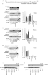
A partially conserved site in the F3 subdomain of the kindlin-1 and -2 FERM domains is required for binding to β1A tails. A, pulldown assays mapping the F3 subdomain of kindlin-1 for mutants that inhibit association with β1A tails. Lysates from CHO cells expressing FLAG-tagged kindlin-1 wild-type and mutant constructs were mixed with recombinant β1A tails and assessed for binding. B, the binding of kindlin-1 and mutants was quantified by densitometry, normalized to starting material loading, and then calculated relative to binding of wild-type kindlin-1 (means ± S.E.; n = 10). C, pulldown assays mapping the F3 subdomain of kindlin-2 for mutants that inhibit association with β1A tails. D, the binding of kindlin-2 and mutants was quantified by densitometry, normalized to starting material loading, and then calculated relative to binding of wild-type kindlin-2 (means ± S.E.; n = 3). E and F, purified GST-tagged kindlin-1 and -2 constructs with mutations of a critical tryptophan residue, W612A and W615A for kindlin-1 and -2, respectively, were tested for binding to β1A tails. IB, immunoblot; WT, wild type.
FIGURE 4.
FIGURE 4.
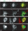
Integrin binding activity correlates with kindlin-1 targeting to focal adhesions. Images of NIH 3T3 cells transiently transfected with GFP-tagged kindlin-1 wild-type and mutant expression constructs. Kindlin-1 co-localizes with vinculin at focal adhesions. Recruiting kindlin-1 to vinculin-rich focal adhesions is blocked by W612A but not Q612A mutations.
FIGURE 5.
FIGURE 5.
Kindlin-1 and -2 bind to the membrane-distal NPKY motif in β1A tails. A, the protein sequence for the cytoplasmic tail of human integrin β1A. Asterisks denote residues required for kindlin-1 and -2 binding. B-D, pulldown assays mapping the binding site on β1A tails for kindlin-1 and -2 binding. Lysates from CHO cells either untransfected or transiently expressing FLAG-tagged kindlin-1 or -2 were mixed with wild-type and mutant β1A tails and assessed for binding. The binding of kindlin-1 and -2 was quantified by densitometry, normalized to starting material loading, and then calculated relative to binding of wild-type β1A (means ± S.E.; n ≥ 3). E and F, bacterially purified GST-tagged kindlin-1 and -2 were examined for their ability to bind to β1A tail mutants. IB, immunoblot; WT, wild type.
FIGURE 6.
FIGURE 6.
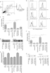
Kindlin-1 and -2 inhibit talin mediated-activation of α5β1 integrin. A, wild-type CHO cells were co-transfected with GFP-tagged mouse-talin-head (1-433) and DsRed-tagged kindlin-1 cDNAs. Cells expressing both GFP and DsRed were selected (gate G) and assessed for activation of endogenous α5β1 integrin (FN9-11 binding) or α5β1 integrin expression (PB1 binding). B, representative histogram panels of FN9-11 binding to active α5β1 integrin in gated co-expressing CHO cells. Upper left panel, FN9-11 binding to GFP and DsRed gated expressing cells (shaded, native; dashed, in the presence RGD inhibitor). In the remaining panels, the shaded regions represent native FN9-11 binding to gated cells expressing GFP and DsRed, whereas the bold histograms represent FN9-11 binding in the presence of indicated tagged proteins. Careful gating ensured comparable levels of GFP and DsRed fluorescence in the analyzed population. C, activation indices of α5β1 integrin from gated CHO cells co-expressing GFP-talin head and DsRed-kindlin-1 were calculated and normalized for integrin expression (see “Experimental Procedures”). The results represent the means ± S.E. (n ≥ 3). **, p < 0.01; *, p < 0.05. D, total lysates from double transfected CHO cells were separated by SDS-PAGE and analyzed by immunoblot for tagged proteins. E, integrin expression ratios from gated CHO cells co-expressing GFP-talin head and DsRed-kindlin-1 were calculated relative to untransfected cells (see “Experimental Procedures”). The results represent means ± S.E. (n ≥ 3). ***, p < 0.0001; **, p < 0.01; *, p < 0.05. F, activation indices of α5β1 integrin from gated CHO cells co-expressing GFP or GFP-talin head and DsRed or DsRed-kindlin-2 constructs as indicated were calculated and normalized for integrin expression (see “Experimental Procedures”). The results represent the means ± S.E. (n ≥ 3). **, p < 0.01; *, p < 0.05. G, total lysates from double transfected CHO cells were separated by SDS-PAGE and analyzed by immunoblot for tagged proteins.
FIGURE 7.
FIGURE 7.
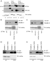
Kindlin-1 and -2 bind directly to β3 integrin tails. A, pulldown assays using recombinant β3 tails mixed with CHO cell lysates containing FLAG-tagged kindlin-1 or -2. Binding of kindlins, endogenous talin (positive control), and the major vault protein (negative control) was assessed by Western blotting. αIIb tails serve as a control for binding specificity to the β tails. Loading of each tail protein was judged by protein staining. The 10% lanes represent 10% of the starting material in the binding assay. B and C, direct binding of GST-tagged kindlin-1 and -2 to wild-type and mutant β3 tails. D and E, purified GST-tagged kindlin-1 and -2 constructs with mutations of a critical tryptophan residue, W612A and W615A for kindlin-1 and -2, respectively, were tested for binding to β3 tails. IB, immunoblot.
FIGURE 8.
FIGURE 8.
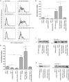
Kindlin-1 and -2 cooperate with talin to activate αIIbβ3. CHO cells stably expressing αIIbβ3 integrin were co-transfected with GFP or GFP-tagged mouse-talin-head (1-433) and DsRed or DsRed-tagged kindlin-1 cDNAs as indicated. Co-expressing cells with similar fluorescence of GFP and DsRed tags were assessed for αIIbβ3 integrin activation (PAC1 binding) or αIIbβ3 integrin expression (D57 Binding). A, representative histogram panels of PAC1 binding to active αIIbβ3 integrin in gated co-expressing CHO cells. Upper left panel, PAC1 binding to GFP and DsRed gated expressing cells (shaded, native; dashed, in the presence RGD inhibitor). In the remaining panels the shaded regions represent native PAC1 binding to gated cells expressing GFP and DsRed, and the bold histograms represent PAC1 binding in the presence of indicated tagged proteins. B, activation indices of αIIbβ3 integrin from gated CHO cells co-expressing GFP-talin head and DsRed-kindlin-1 were calculated and normalized for integrin expression (see “Experimental Procedures”). The results represent the means ± S.E. (n ≥ 3). **, p < 0.01; *, p < 0.05. C, total lysates from double transfected CHO cells were separated by SDS-PAGE and analyzed by immunoblot for tagged proteins. D, activation indices of αIIbβ3 integrin from gated CHO cells co-expressing GFP-talin head and DsRed-kindlin-2 were calculated and normalized for integrin expression (see “Experimental Procedures”). The results represent the means ± S.E.(n≥3). **, p < 0.01; *, p < 0.05. E, total lysates from double transfected CHO cells were separated by SDS-PAGE and analyzed by immunoblot for tagged proteins. IB, immunoblot.

Similar articles

Cited by

References

    1. Calderwood, D. A. (2004) J. Cell Sci. 117 657-666 - PubMed
    1. Hynes, R. O. (2002) Cell 110 673-687 - PubMed
    1. Luo, B., Carman, C., and Springer, T. (2007) Annu. Rev. Immunol. 25 619-647 - PMC - PubMed
    1. Harburger, D., and Calderwood, D. (2009) J. Cell Sci. 122 159-163 - PMC - PubMed
    1. Zaidel-Bar, R., Itzkovitz, S., Ma'ayan, A., Iyengar, R., and Geiger, B. (2007) Nat. Cell Biol. 9 858-867 - PMC - PubMed

Publication types

LinkOut - more resources