. 2009 Apr;149(4):1824-37.
doi: 10.1104/pp.108.132092. Epub 2009 Feb 20.

Subcellular localization and functional analysis of the Arabidopsis GTPase RabE

Affiliations

Subcellular localization and functional analysis of the Arabidopsis GTPase RabE

Elena Bray Speth et al. Plant Physiol. 2009 Apr.

Abstract

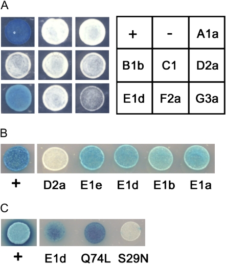
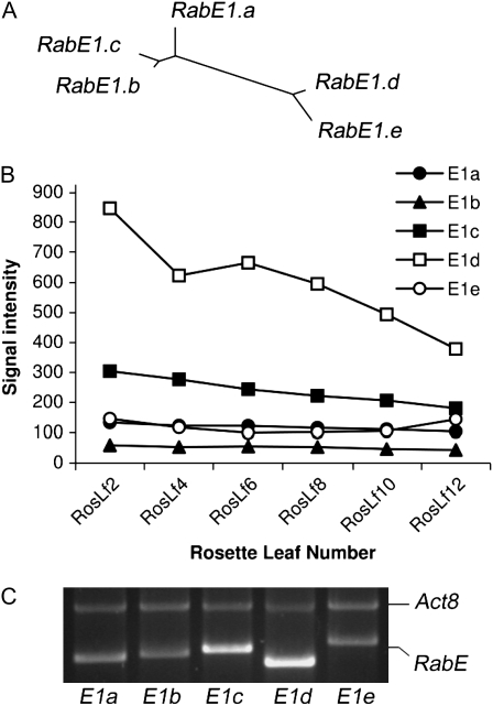
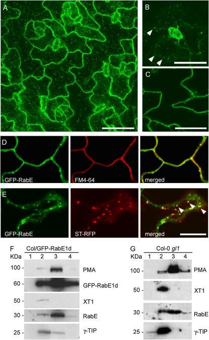
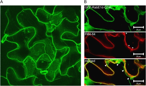
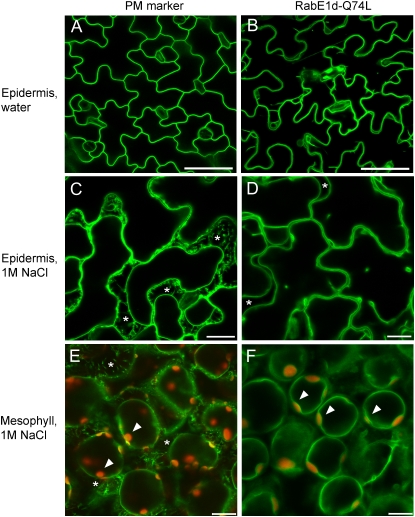
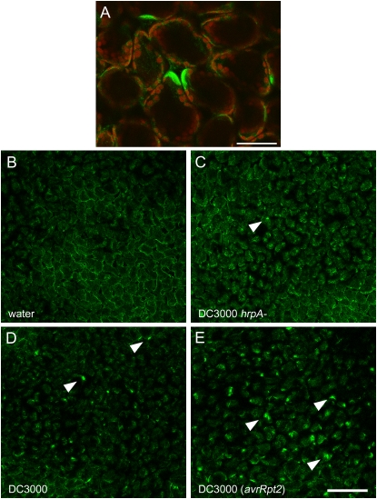
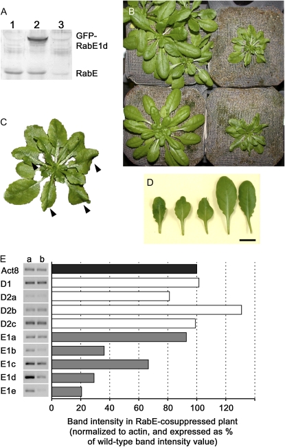
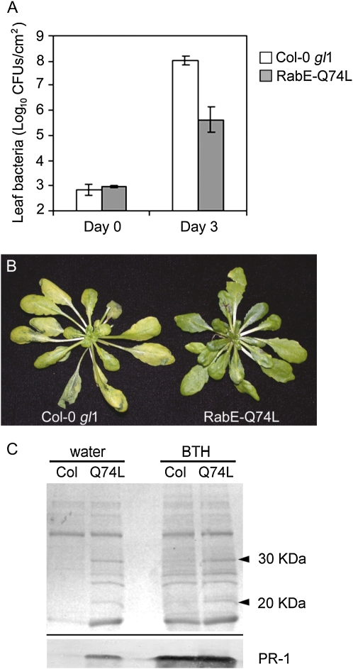
Membrane trafficking plays a fundamental role in eukaryotic cell biology. Of the numerous known or predicted protein components of the plant cell trafficking system, only a relatively small subset have been characterized with respect to their biological roles in plant growth, development, and response to stresses. In this study, we investigated the subcellular localization and function of an Arabidopsis (Arabidopsis thaliana) small GTPase belonging to the RabE family. RabE proteins are phylogenetically related to well-characterized regulators of polarized vesicle transport from the Golgi apparatus to the plasma membrane in animal and yeast cells. The RabE family of GTPases has also been proposed to be a putative host target of AvrPto, an effector protein produced by the plant pathogen Pseudomonas syringae, based on yeast two-hybrid analysis. We generated transgenic Arabidopsis plants that constitutively expressed one of the five RabE proteins (RabE1d) fused to green fluorescent protein (GFP). GFP-RabE1d and endogenous RabE proteins were found to be associated with the Golgi apparatus and the plasma membrane in Arabidopsis leaf cells. RabE down-regulation, due to cosuppression in transgenic plants, resulted in drastically altered leaf morphology and reduced plant size, providing experimental evidence for an important role of RabE GTPases in regulating plant growth. RabE down-regulation did not affect plant susceptibility to pathogenic P. syringae bacteria; conversely, expression of the constitutively active RabE1d-Q74L enhanced plant defenses, conferring resistance to P. syringae infection.


Figures

Figure 1.
Figure 1.
Arabidopsis RabE proteins interact with P. syringae AvrPto in the Y2H system. Interaction is visualized by development of blue color on medium containing X-Gal. Negative control (−) = pNLexA (bait) and pB42AD (prey) vectors; positive control (+) = pLexA-A53 and pB42AD-T. A, AvrPto (in pNLexA) interacts with Arabidopsis RabE1d but not with other members of the Rab superfamily (in pB42AD). B, AvrPto (in pNLexA) interacts with all four tested Arabidopsis RabE proteins (in pB42AD). Yeast expressing AvrPto in pNLexA and RabD2a in pB42AD is shown as a negative control. C, AvrPto (in pB42AD) interacts with RabE1d or RabE1d-Q74L but not with RabE1d-S29N. For this experiment only, wild-type RabE1d and the mutated RabE1d proteins were cloned in the bait vector pGILDA and modified by replacing the two C-terminal conserved Cys residues (sites of geranylgeranylation [Supplemental Fig. S1]) with Gly and Ser to prevent prenylation and membrane association.
Figure 2.
Figure 2.
A, Phylogenetic relationship among RabE family members; E1d and E1e, as well as E1b and E1c, are predicted to be the result of genome duplication events. B, E-northern of RabE gene expression in Arabidopsis Col-0 rosette leaves at different developmental stages. Data were obtained from AtGenExpress at the Genevestigator site (https://www.genevestigator.ethz.ch/); signal intensities were averaged across three biological replicates. C, RT-PCR showing RabE gene expression in rosette leaves. RT-PCR products represent the five RabE genes, reverse transcribed and amplified from Arabidopsis Col-0 gl1 rosette leaf total RNA. A single RT reaction was performed, and equal amounts of the resulting cDNA were used as template in PCR. Each PCR sample contained primers for the Actin8 gene in addition to primers for one of the RabE genes (primers are listed in Supplemental Table S2). Equal volumes of each PCR product were loaded on a 1% agarose gel.
Figure 3.
Figure 3.
GFP-RabE1d localization in Arabidopsis leaf cells. Confocal microscopy images of the leaf epidermis of transgenic Arabidopsis plants expressing GFP-RabE1d are shown. A, Projection along the z axis of several optical planes intersecting the leaf epidermal cell layer. GFP-RabE1d is visible in intracellular punctate structures and at the cell periphery. B, Single focal plane intersecting a stomate and the cortical layer of epidermal cells. Arrowheads point at intracellular punctate structures. C, Single focal plane intersecting epidermal cells. The same pattern of distribution of GFP-RabE1d was observed in at least five independent transgenic lines. Bars = 50 μm. D and E, Confocal microscopy images illustrating GFP-RabE1d colocalization with the FM4-64 dye at the plasma membrane (D) and with sialyl transferase (ST-RFP) at the Golgi apparatus (E). D, Single focal plane crossing adjacent epidermal cells (40× oil-immersion objective, 4× zoom). Left, GFP-RabE1d fluorescence, green; center, FM4-64 fluorescence, red; right, merged image, in which the yellow color results from the overlap of red and green. E, Single focal plane crossing the cytoplasm of a cell (40× oil-immersion objective, 4× zoom). Left, GFP-RabE1d fluorescence, green; center, ST-RFP fluorescence, red; right, merged image, in which the yellow color results from the overlap of red and green. Arrowheads point at some of the colabeled Golgi stacks in the merged image. Bars = 10 μm. F and G, Western blots of cellular membrane fractions separated by ultracentrifugation on a Suc step gradient. Two different samples are shown: transgenic Arabidopsis overexpressing GFP-RabE1d (F) and nontransgenic Arabidopsis Col-0 gl1 (G). Lanes 1 through 4 represent the four membrane fractions collected at the interfaces between layers of different Suc concentrations: 18% to 25% (lane 1), 25% to 34% (lane 2), 34% to 40% (lane 3), and 40% to 50% (lane 4). Equal volumes of each fraction were loaded on SDS-PAGE gels. PM-ATPase (PMA) is a plasma membrane marker, XT1 is a trans-Golgi resident protein, and γ-TIP is a marker for the tonoplast. GFP-RabE1d and endogenous RabE proteins were detected with a polyclonal anti-RabE antibody (courtesy of Dr. K. Nomura).
Figure 4.
Figure 4.
Intracellular localization of the mutant GFP- RabE1d-Q74L protein. A, Confocal microscopy image of a representative Arabidopsis leaf expressing GFP-RabE1d-Q74L. Projection along the z axis of several focal planes crossing the epidermal cell layer. Several independent transgenic lines were analyzed with similar results. B, GFP-RabE1d-Q74L is mostly localized in the tonoplast. Leaves were stained with FM4-64, to visualize the plasma membrane, and immediately observed. The image represents a single focal plane (40× oil-immersion objective). Top, GFP fluorescence; middle, FM4-64 fluorescence (asterisks indicate autofluorescent chloroplasts in the mesophyll layer, below the epidermis); bottom, merged image (arrowheads indicate where the tonoplast is most clearly distinct from the plasma membrane). Invaginations and the formation of membranous structures are typical of the highly dynamic vacuolar membrane. Even in the areas where the plasma membrane and tonoplast are closest, green and red fluorescence are visibly distinct. Bars = 20 μm.
Figure 5.
Figure 5.
Localization pattern of GFP-RabE1d-Q74L compared with the localization of a PM marker protein. A, C, and E, Arabidopsis expressing a GFP fusion to the PM integral protein PIP2A (line Q8). B, D, and F, Arabidopsis expressing GFP-RabE1d-Q74L. In C to F, samples were plasmolyzed in 1 m NaCl. Asterisks indicate areas between plasmolyzed cells; Hechtian strands are noticeable in C and E (PM) but not in D and F (RabE-Q74L). Arrowheads indicate chloroplast autofluorescence (red); chloroplasts are on the inside of the PM (E), while GFP-RabE1d-Q74L fluorescence is on the cytoplasmic side of the chloroplasts. All images are single focal planes. Bars = 50 μm (A and B), 20 μm (C), and 10 μm (D–F).
Figure 6.
Figure 6.
Focal accumulation of GFP-RabE1d in response to bacteria. Leaves of transgenic GFP-RabE1d Arabidopsis were syringe inoculated with various strains of Pst DC3000 at a density of 1 × 10⁸ cfu mL−1. A, Confocal microscopy observation at 6 h after inoculation revealed focal accumulation of the fluorescent GFP-RabE1d in mesophyll cells. Such accumulation is limited to a few cells in response to virulent and nonpathogenic bacteria, whereas it is widespread in response to avirulent bacteria, which trigger gene-for-gene resistance. Bar = 50 μm. B, No bacteria. C, Pst DC3000 hrpA mutant bacteria, nonpathogenic. D, Wild-type Pst DC3000, virulent. E, Pst DC3000 (avrRpt2), avirulent. Three independent transgenic lines (A1, A5, and A14) yielded similar results. For B to E, all panels represent Z stacks of the same number of optical planes intersecting the leaf mesophyll (arrowheads indicate some of the points of accumulation). A second, independent set of images is shown in Supplemental Figure S4. Bar = 200 μm.
Figure 7.
Figure 7.
RabE cosuppression adversely affects Arabidopsis plant size and leaf morphology. A, Western blot (with anti-RabE antibody) illustrating how RabE-cosuppressed plants (lane 3) have a considerably lower amount of endogenous RabE protein compared with both the wild type (lane 1) and GFP-RabE1d overexpressors (lane 2). B, RabE-cosuppressed plants (top and bottom right) have smaller size and altered morphology compared with wild-type Arabidopsis (top left) and RabE1d-overexpressing plants (bottom left). C, Enlarged image of the RabE-cosuppressed plant in B (top right); arrowheads point to the wavy leaves. D, Size comparison of rosette leaves from 5-week-old RabE-cosuppressed (three left) and wild-type (two right) Arabidopsis plants. Bar = 10 mm. E, RT-PCR analysis of expression of the five RabE and four RabD genes in wild-type (lane a) and in RabE-cosuppressed (lane b) Arabidopsis plants. Equal volumes of the PCR samples were loaded on a 1% agarose gel. The gel was photographed with a Bio-Rad imager, and Quantity One software was used to quantify the bands. Intensity values, normalized to those of Actin8, are represented in the chart as percentages of the wild-type value.
Figure 8.
Figure 8.
Constitutive expression of RabE-Q74L confers resistance to Pst DC3000. A, Bacterial multiplication in GFP-RabE1d-Q74L-expressing plants (gray bars) compared with that in wild-type Arabidopsis (white bars). Pst DC3000 was syringe infiltrated at a density of 1 × 10⁵ cfu mL−1. B, Disease symptoms 3 d after vacuum inoculation with Pst DC3000 at a density of 1 × 10⁶ cfu mL−1. Left, Arabidopsis Col-0 gl1 (wild type); right, Arabidopsis expressing GFP-RabE-Q74L. C, Accumulation of extracellular proteins in plants expressing GFP-RabE1d-Q74L. Proteins in the IWF from wild-type (Col) and RabE1d-Q74L-expressing (Q74L) plants were separated by SDS-PAGE. Top, Coomassie Brilliant Blue-stained gel representing total proteins; the arrowheads indicate bands that seem to be exclusive to the Q74L plants. Bottom, Western blot with the anti-PR-1 antibody (a gift of Dr. X. Dong).

Similar articles

Cited by

References

    1. Abramovitch RB, Anderson JC, Martin GB (2006) Bacterial elicitation and evasion of plant innate immunity. Nat Rev Mol Cell Biol 7 601–611 - PMC - PubMed
    1. Alfano JR, Collmer A (2004) Type III secretion system effector proteins: double agents in bacterial disease and plant defense. Annu Rev Phytopathol 42 385–414 - PubMed
    1. Assaad FF, Qiu JL, Youngs H, Ehrhardt D, Zimmerli L, Kalde M, Wanner G, Peck SC, Edwards H, Ramonell K, et al (2004) The PEN1 syntaxin defines a novel cellular compartment upon fungal attack and is required for the timely assembly of papillae. Mol Biol Cell 15 5118–5129 - PMC - PubMed
    1. Batoko H, Zheng HQ, Hawes C, Moore I (2000) A Rab1 GTPase is required for transport between the endoplasmic reticulum and Golgi apparatus and for normal Golgi movement in plants. Plant Cell 12 2201–2218 - PMC - PubMed
    1. Block A, Li G, Fu ZQ, Alfano JR (2008) Phytopathogen type III effector weaponry and their plant targets. Curr Opin Plant Biol 11 396–403 - PMC - PubMed

Publication types

MeSH terms

LinkOut - more resources