. 2009 May 1;329(1):44-54.
doi: 10.1016/j.ydbio.2009.02.011. Epub 2009 Feb 20.

Neuron-derived FGF9 is essential for scaffold formation of Bergmann radial fibers and migration of granule neurons in the cerebellum

Affiliations

Neuron-derived FGF9 is essential for scaffold formation of Bergmann radial fibers and migration of granule neurons in the cerebellum

Yongshun Lin et al. Dev Biol. .

Abstract

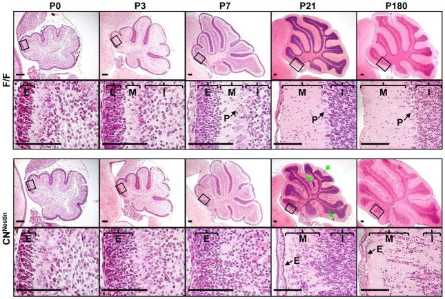
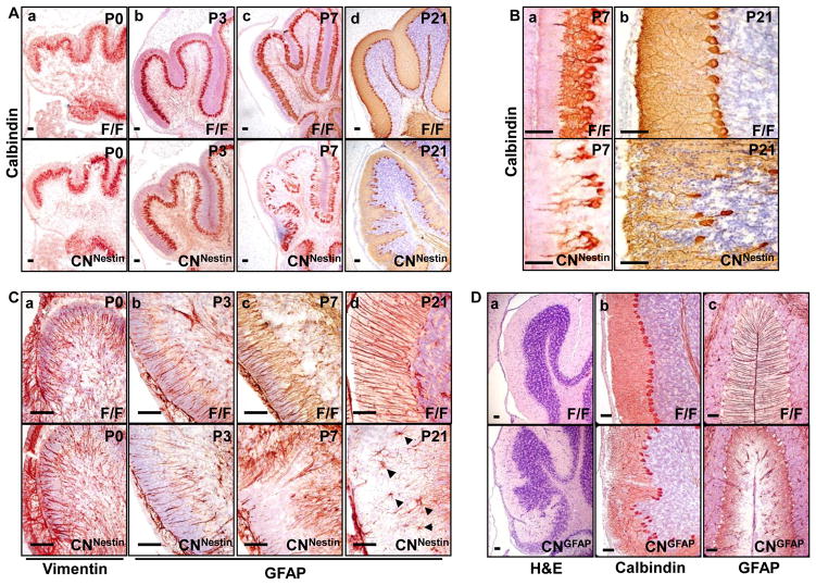
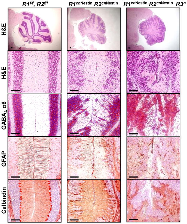
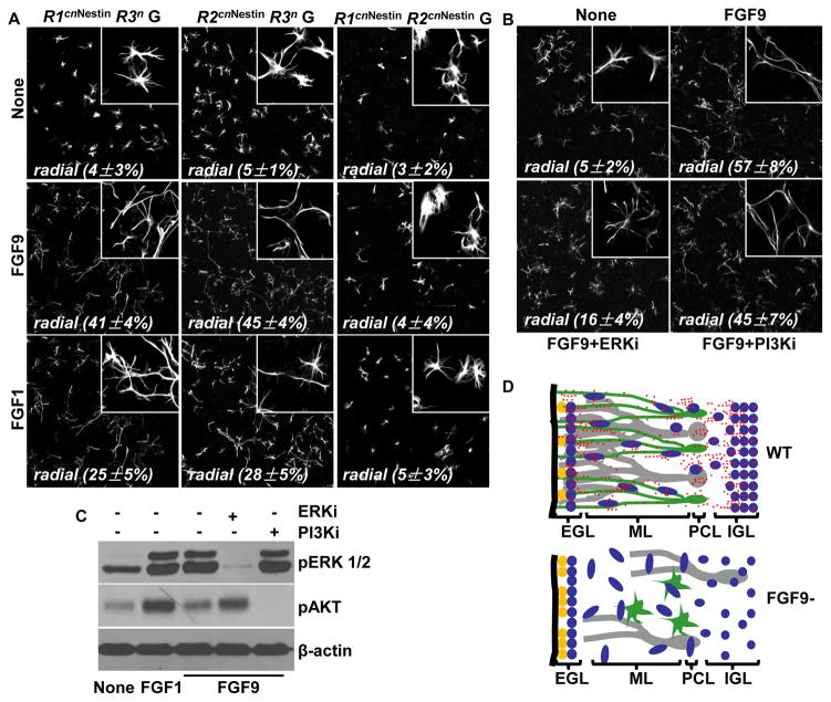
Although fibroblast growth factor 9 (FGF9) is widely expressed in the central nervous system (CNS), the function of FGF9 in neural development remains undefined. To address this question, we deleted the Fgf9 gene specifically in the neural tube and demonstrated that FGF9 plays a key role in the postnatal migration of cerebellar granule neurons. Fgf9-null mice showed severe ataxia associated with disrupted Bergmann fiber scaffold formation, impaired granule neuron migration, and upset Purkinje cell maturation. Ex vivo cultured wildtype or Fgf9-null glia displayed a stellate morphology. Coculture with wildtype neurons, but not Fgf9-deficient neurons, or treating with FGF1 or FGF9 induced the cells to adopt a radial glial morphology. In situ hybridization showed that Fgf9 was expressed in neurons and immunostaining revealed that FGF9 was broadly distributed in both neurons and Bergmann glial radial fibers. Genetic analyses revealed that the FGF9 activities in cerebellar development are primarily transduced by FGF receptors 1 and 2. Furthermore, inhibition of the MAP kinase pathway, but not the PI3K/AKT pathway, abrogated the FGF activity to induce glial morphological changes, suggesting that the activity is mediated by the MAP kinase pathway. This work demonstrates that granule neurons secrete FGF9 to control formation of the Bergmann fiber scaffold, which in turn, guides their own inward migration and maturation of Purkinje cells.


Figures

Fig. 1
Fig. 1. Conditional knockout of Fgf9 in the CNS causes ataxic phenotypes
A. Schematic diagram describes the design of floxed and conditional null Fgf9 allele. Coding exons are shown in black boxes and non-coding exon sequences in open boxes. Primers f1, f2 and f3 are used for genotyping; f4 and f5 are used for RT-PCR analyses. B. RT-PCR analyses demonstrate a decreased Fgf9 expression in the Fgf9cnNestin (CNNestin) cerebellum compared to the wild-type (WT) and Fgf9flox/flox (F/F) controls at 3 weeks of age. Gapdh was used as internal loading controls. C. Fgf9cnNestin mice have paroxysmal hyperextended hind limbs with chronic spasms. The arrows indicate that hind limbs of 3-week-old CNNestin mice were extended outward. D. When suspended by the tail, the limbs and digits of CNNestin mice were often retracted and clasped, whereas the F/F mice usually extended their limbs and digits. E. CNNestin mice show ataxic gaits that lack the forefoot-hindfoot correspondence seen in control mice. The front paws were labeled with red ink and rear paws with blue ink. Arrows indicate the direction of movement. F. CNNestin mice exhibit growth retardation from postnatal day 15. Data represent means ± sd of triplicate samples.
Fig. 2
Fig. 2. Fgf9 ablation disrupts the migration and alignment of cerebellar granule neurons
H&E staining of midsagittal sections of the F/F (top two rows) and CNNestin (bottom two rows) cerebellum from postnatal day 0 (P0) to P180. Higher magnification views of the framed areas in the low-magnification images in rows 1 and 3 are shown in rows 2 and 4. Clusters of granule cells were found in the molecular layer (M) of CNNestin mice (indicated by green arrows). The alignment of Purkinje cells was also disturbed in mature cerebella. E, external granule layer; internal granule layer; P, Purkinje cells. Scale bar, 20 um.
Fig. 3
Fig. 3. Fgf9 knockout prolongs the proliferation window of granule neurons without affecting their final maturation
A&B, Midsagittal sections of the F/F and CNNestin cerebellum at the indicated age were immunolabeled with (A) anti-PCNA antibody for proliferative cells or (B) anti-p27kip1 antibody for postmitotic cells and counterstained with hematoxylin for the nuclei. At P21, the CNNestin cerebellum still had a thin layer of PCNA-positive cells in the EGL (Ac) and displaced postmitotic granule cells in the ML (Bb). C. Immunostaining of anti-GABAA α6 revealed that the ectopic granule cells in the ML also expressed a mature granule cell marker, GABAA α6 (indicated by arrows) at P21. D. TO-PRO3 staining shows the nuclei of neuron and glia cells. The nucleus of both F/F and CNNestin granule cells, which makes up the majority of cells, is elongated in the molecular layer (indicated by arrows).
Fig. 4
Fig. 4. Ablation of Fgf9 also affects the development of Purkinje neurons and Bergmann glia
A. Impaired Purkinje cell alignment and arborization in cerebella deficient in Fgf9. Purkinje cells of control and mutant cerebellum from P0 to P21 were immunostained with anti-Calbindin antibody. B. Enlarged views of the P7 and P21 cerebellum. The alignment of mutant Purkinje cells (CNNestin) was disorganized. They had elongated primary dendritic bundles and reduced dendritic arborization compared to the control (F/F). C. Midsagittal sections of cerebella were immunostained with anti-vimentin or anti-GFAP antibody for Bergmann glia. The radial fibers of Bergmann glia in the CNNestin cerebellum were poorly developed and failed to reach the pial surface. At P21, some glia in the CNNestin cerebellum exhibited a stellate morphology (arrowheads). D. Defective cerebellum in Fgf9cnGFAP (CNGFAP) mice, in which the Fgf9 alleles were intact in Purkinje cells but deleted in the Bergmann glia and granule neurons. Midsagittal sections of the CNGFAP cerebellum at P21 were stained with H&E or the indicated antibodies. Note that the granule cell migration, Purkinje cell alignment and arborization, and Bergmann glial radial fiber formation were all impaired, indicating that the Purkinje cell-made FGF9 alone is not enough to guarantee normal cerebellar development.
Fig. 5
Fig. 5. Granule neuron-derived FGF9 is essential for the development of radial morphology of cerebellar glia
A. Glia cells were isolated from the F/F and CNNestin cerebellum, cultured with or without exogenous FGF9 (20 ng/ml) for 48 hours, and stained with anti-GFAP antibody. (a) Without FGF9, both control and mutant glial cells exhibited a stellate astrocytic morphology. (b) FGF9 treatment induced a morphological change in both F/F and CNNestin glia from stellate to radial shapes. Inserts, high magnification views from the same slides. B. Control and mutant granule neurons (N+ and N) or glia (G+ and G) were prepared separately from the F/F and CNNestin cerebellum and cocultured for 48 hours in different combinations. Glia and neurons were stained with anti-GFAP and Tuj1 antibodies, respectively. Both F/F and CNNestin glia exhibited a radial morphology when being cocultured with F/F granule cells (N+), and a stellate morphology when being cocultured with CNNestin granule cells (N). The percentages of glia with radial morphologies were measured from 3 individual experiments (mean ± sd). C. Expression of neuronal and glial markers was assessed by real-time RT-PCR. Total RNAs were extracted from the control or mutant cerebellar neurons or glia. β-actin expression was used as an internal control. Each bar represents the average (± sd) of 3 independent experiments. NRG, neuregulin; Ngn1, neurogenin 1; Ngn2, Neurogenin 2; ASTN, astrotactin. D. Expression of FGF9 in the cerebellum at the indicated ages. In situ hybridization showed that Fgf9 mRNA was predominantly located in neurons including Purkinje and granule cells. Immunostaining demonstrated that FGF9 protein was broadly distributed in the cerebellum, including neurons and Bergmann glial radial fiber scaffold. Note that both in situ and immunostaining signals were significantly reduced in Fgf9 mutants.
Fig. 6
Fig. 6. FGFR1 and FGFR2 redundantly regulate formation of the Bergmann radial fiber scaffold
The floxed Fgfr1 and Fgfr2 alleles were deleted by the Nestin-Cre driver. Midsagittal sections of the cerebellum prepared from the homozygous Fgfr1flox/flox and Fgfr2floxflox (R1f/f, R2f/f), Fgfr1/Fgfr2 double conditional null (R1cnNestin/R2cnNestin), and Fgfr1/Fgfr2/Fgfr3 triple null (R1cnNestin/R2cnNestin/R3n) mice at P21. Each panel was labeled with H&E, anti-GABAA α6, anti-GFAP, and anti-calbindin antibodies. A combined deletion of Fgfr1 and Fgfr2, but not that of Fgfr1/Fgfr3 or Fgfr2/Fgfr3, caused defects in the cerebellar development similar to but more severe than those caused by Fgf9 knockout.
Fig. 7
Fig. 7. The activity of FGF9 to induce radial morphology in cerebellar glia requires either FGFR1 or FGFR2, and the MAP kinase pathway
A. Glia cells were prepared from R1cnNestin/R3n, R2cnNestin/R3n, or R1cnNestin/R2cnNestin cerebellum, cultured in the absence or presence of FGF9 (20 ng/ml) or FGF1 (20 ng/ml) for 48 hours, and stained with anti-GFAP antibody. Both FGF9 and FGF1 could induce the exhibition of the radial morphology in R1cnNestin/R3n and R2cnNestin/R3n, but not R1cnNestin/R2cnNestin, cerebellar glia, which normally would exhibit a stellate astrocytic morphology when being cultured alone. B. Inhibition of ERK1/2, but not AKT, phosphorylation inhibited FGF9 activity to induce glia to adopt the radial morphology. C. FGF induced phosphorylation of ERK1/2 and AKT in primary culture of cerebellum glia. Western blot with the indicated antibodies demonstrate that FGF1 induces strong activation of both ERK1/2 and AKT, whereas FGF9 induces strong ERK1/2 activation and weak AKT activation. pAKT, phosphorylated AKT; PI3Ki, PI3K inhibitor; pERK1/2, phosphorylated ERK1/2; ERKi, ERK kinase inhibitor. D. Diagrammatic depiction of FGF9 role in cerebellar development. In the normal cerebellum, FGF9 is secreted by granule neurons and acts on FGFR1 or FGFR2 on Bergmann glia (top panel). The FGF9 activity is essential for the maturation and formation of radial fiber scaffolds of Bergmann glia. Fgf9 knockout results in defects in the differentiation of Bergmann glia, the migration of granule neurons, and the alignment and dendritic arborization of Purkinje neurons (bottom panel).

Similar articles

Cited by

References

    1. Adams NC, Tomoda T, Cooper M, Dietz G, Hatten ME. Mice that lack astrotactin have slowed neuronal migration. Development. 2002;129:965–72. - PubMed
    1. Basson MA, Echevarria D, Petersen Ahn C, Sudarov A, Joyner AL, Mason IJ, Martinez S, Martin GR. Specific regions within the embryonic midbrain and cerebellum require different levels of FGF signaling during development. Development. 2008;135:889–98. - PMC - PubMed
    1. Blaess S, Corrales JD, Joyner AL. Sonic hedgehog regulates Gli activator and repressor functions with spatial and temporal precision in the mid/hindbrain region. Development. 2006;133:1799–809. - PubMed
    1. Blak AA, Naserke T, Saarimaki-Vire J, Peltopuro P, Giraldo-Velasquez M, Vogt Weisenhorn DM, Prakash N, Sendtner M, Partanen J, Wurst W. Fgfr2 and Fgfr3 are not required for patterning and maintenance of the midbrain and anterior hindbrain. Dev Biol. 2007;303:231–43. - PubMed
    1. Chanas-Sacre G, Rogister B, Moonen G, Leprince P. Radial glia phenotype: origin, regulation, and transdifferentiation. J Neurosci Res. 2000;61:357–63. - PubMed

Publication types

MeSH terms

Substances

LinkOut - more resources