. 2009 Feb 18;29(7):1962-76.
doi: 10.1523/JNEUROSCI.5351-08.2009.

Bacterial artificial chromosome transgenic mice expressing a truncated mutant parkin exhibit age-dependent hypokinetic motor deficits, dopaminergic neuron degeneration, and accumulation of proteinase K-resistant alpha-synuclein

Affiliations

Bacterial artificial chromosome transgenic mice expressing a truncated mutant parkin exhibit age-dependent hypokinetic motor deficits, dopaminergic neuron degeneration, and accumulation of proteinase K-resistant alpha-synuclein

Xiao-Hong Lu et al. J Neurosci. .

Abstract

Recessive mutations in parkin are the most common cause of familial early-onset Parkinson's disease (PD). Recent studies suggest that certain parkin mutants may exert dominant toxic effects to cultured cells and such dominant toxicity can lead to progressive dopaminergic (DA) neuron degeneration in Drosophila. To explore whether mutant parkin could exert similar pathogenic effects to mammalian DA neurons in vivo, we developed a BAC (bacterial artificial chromosome) transgenic mouse model expressing a C-terminal truncated human mutant parkin (Parkin-Q311X) in DA neurons driven by a dopamine transporter promoter. Parkin-Q311X mice exhibit multiple late-onset and progressive hypokinetic motor deficits. Stereological analyses reveal that the mutant mice develop age-dependent DA neuron degeneration in substantia nigra accompanied by a significant loss of DA neuron terminals in the striatum. Neurochemical analyses reveal a significant reduction of the striatal dopamine level in mutant mice, which is significantly correlated with their hypokinetic motor deficits. Finally, mutant Parkin-Q311X mice, but not wild-type controls, exhibit age-dependent accumulation of proteinase K-resistant endogenous alpha-synuclein in substantia nigra and colocalized with 3-nitrotyrosine, a marker for oxidative protein damage. Hence, our study provides the first mammalian genetic evidence that dominant toxicity of a parkin mutant is sufficient to elicit age-dependent hypokinetic motor deficits and DA neuron loss in vivo, and uncovers a causal relationship between dominant parkin toxicity and progressive alpha-synuclein accumulation in DA neurons. Our study underscores the need to further explore the putative link between parkin dominant toxicity and PD.


Figures

Figure 1.
Figure 1.
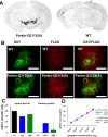
Generation of Parkin-Q311X transgenic mice. A, Schematic representation of the mouse DAT gene structure. 5′-FLAG-tagged mutant parkin (Q311X) followed by a polyadenylation signal (PA) was inserted into exon 2 of the DAT gene preceding the endogenous translation initiation codon. B, Determination of transgene copy number using semiquantitative PCR. Equal amounts of genomic DNA from two Parkin-Q311X line A mice and two Parkin-Q311X line D mice were amplified using a human parkin-specific primer [A, Parkin-Q311X(A); D, Parkin-Q311X(D)]. The loading per lane was controlled using PCR amplicons of a ribosomal 18S DNA. Band densities were normalized to human genomic DNA (assumed to have 2 copies of endogenous parkin) amplification band. C, parkin-Q311X is a human mutation of parkin that lacks the second RING finger motif and IBR domain. This truncated parkin protein migrates in SDS-PAGE gel as a 30 kDa band as shown previously (Hyun et al., 2002; Henn et al., 2005). BAC transgenic mice (lines A, B, C, and D) and wild-type mice were killed at 3 months of age. Mouse striatal and midbrain sections were dissected and homogenized. Parkin was detected by Western blotting using a FLAG antibody. The asterisk (*) indicates nonspecific staining of FLAG antibody in wild-type mouse brain tissue. D, Immunoprecipitation of mutant protein using FlagM2 affinity gel and detected with FLAG-M2 antibody in brains of Parkin-Q311X(A) and wild-type littermates.
Figure 2.
Figure 2.
Selective expression of transgene in DA neurons. A, Representative photomicrograph of in situ hybridizations using 35S-labeled parkin-Q311X antisense RNA probe on midbrain sections from transgenic mice (line A) and wild-type mice at 3 months of age. B, DAT and FLAG double immunofluorescence. Midbrain sections from Parkin-Q311X(A) and wild-type mice were immunostained using DAT (green) and FLAG (red) antibodies. Scale bar, 12.5 μm. C, D, qPCR analyses of parkin expression in laser-capture microdissected dopaminergic neurons of the SNc. Error bars indicate SEM. C, Absolute quantification of human and mouse parkin mRNA levels in WT, transgenic line A (tgA), and line D (tgD) animals. mRNA quantities are given in picograms. Note that there is no human parkin PCR product in WT animals. D, The amplification efficiencies for the mouse and human parkin cDNAs, calculated as increase in PCR product for each PCR cycle, are virtually identical. Both values are close to the theoretical value of 2.0.
Figure 3.
Figure 3.
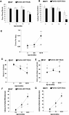
Progressive behavioral deficits in Parkin-Q311X transgenic mice. A, B, Locomotor activity in independent groups of naive Parkin-Q311X(A) (n = 12, 6, 7, 8 for different time points) and wild-type littermate (n = 6, 5, 7, 13 for different time points) mice was measured in the open field at 6, 12, 16, and 21 months of age. Parkin-Q311X mice demonstrate significant progressive hypoactivity as measured by floor plane moves and vertical plane entries in open field (significant genotype and time difference revealed by two-way ANOVA, p < 0.001; significant genotype difference at the same time point is indicated; *p < 0.05, independent sample Student's t test). C, Motor performance and coordination were repeatedly measured in Parkin-Q311X(A) (n = 12) and wild-type (n = 9) mice using the challenging beam. Errors per step were measured at 3, 6–9, and 16–19 months of age. Parkin-Q311X(A) mice made more errors per steps while traversing the beam compared with wild-type mice; **p < 0.01 compared with wild-type mice at the same age. D, E, Spontaneous activity of Parkin-Q311X(A) mice and wild-type littermates in the cylinder was measured at 3, 6–9, and 16–19 months. Rearing (D) and hindlimb (E) steps were measured. Transgenic mice were less active in each measure compared with wild-type mice at the older ages. *p < 0.05 compared with wild-type mice. F, G, Motor response to sensory stimuli was measured in the adhesive removal test at 3–4, 12, and 16–19 months of age [Parkin-Q311X(A) (n = 11) and wild-type (n = 8)]. Parkin-Q311X(A) mice had significantly slower contact (F) and removal time (G) compared with wild-type mice at 16–19 months of age (*p < 0.05, Mann–Whitney U test). Error bars indicate SEM.
Figure 4.
Figure 4.
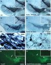
Progressive loss of SNc DA neurons in Parkin-Q311X transgenic mice. A, B, Photomicrographs of TH immunostaining of representative midbrain sections showing DA neurons in the SNc of Parkin-Q311X(A) and WT littermates at 3 months of age. Scale bar, 250 μm. The insets on the right top corner are higher-power views of the SNc area in A and B. Scale bar, 125 μm. C, D, Photomicrographs of TH immunostaining of midbrain sections for Parkin-Q311X(A) mice and wild-type littermates at 16 months of age (scale bar, 250 μm) and the higher-power view of the SNc area in C and D (scale bar, 50 μm). E, F, Higher-power view of the TH-immunoreactive neurons in Parkin-Q311X(A) mice and wild-type littermates at 16 months of age. Scale bar, 12.5 μm. G, H, Photomicrographs of direct visualization of GFP-expressing neurons in the representative midbrain sections from TH-GFP/Parkin-Q311X(D) mice and TH-GFP control mice at 16 months of age. Scale bar, 250 μm. The white arrows in the photomicrographs indicate SNc area. The white stars indicate the VTA.
Figure 5.
Figure 5.
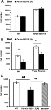
SNc DA neuron stereology analyses. A, TH-immunoreactive neurons were estimated using design-based stereology (Gundersen et al., 1988; West et al., 1991). Normal numbers of TH-expressing neurons in the SNc of Parkin-Q311X(A) mice at 3 months of age [n = 4 per genotype; TH, tyrosine hydroxylase-positive neuron numbers in the SNc; total number of neurons in SNc was estimated by counting of both TH(+) and Nissl(+)/TH(−) neurons in the SNc; p > 0.05 compared with wild-type littermates, independent sample Student's t test]. B, Significant loss of TH-immunoreactive neurons in the SNc of Parkin-Q311X(A) mice at 16 months of age (n = 4 per genotype; *p < 0.05 compared with wild-type littermates, independent sample Student's t test). C, Normal numbers of TH-immunoreactive neurons in the SNc of TH-GFP mice at 16 months of age [Parkin-Q311X(A), n = 6; WT, n = 6; TH-GFP, n = 3; **p < 0.01 compared with Parkin-Q311X(A) mice; ##p < 0.01 compared with Parkin-Q311X(A) mice, independent sample Student's t test]. Error bars indicate SEM.
Figure 6.
Figure 6.
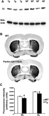
Striatum DA terminal loss in Parkin-Q311X mice. A, Representative Western blot demonstrating striatal TH levels in transgenic mice from line A and wild-type littermate controls at 12 months of age. Quantification of TH bands confirmed that striatal TH levels were significantly lower in Parkin-Q311X(A) mice than in wild-type littermate controls. n = 8 per genotype. *p < 0.05 compared with wild-type littermates. B, Representative photographs showing the TH immunohistochemistry staining in striatum of wild-type (top) and Parkin-Q311X(A) mice at 16–18 months of age. C, Quantification of TH immunofluorescence using a microarray scanner in 16-month-old mice. In rostral striatum, dorsolateral and ventrolateral striatal TH levels were significantly lower in Parkin-Q311X(A) mice than in wild-type littermate controls. n = 4 per genotype. *p < 0.05 compared with wild-type littermates. Error bars indicate SEM.
Figure 7.
Figure 7.
Reduced striatal DA and metabolite content in Parkin-Q311X mice and the correlation with behavioral deficits. A–C, Striatal DA, DOPAC, and HVA content measured using HPLC-ECD, in Parkin-Q311X(A) mice at 19–21 months of age. Results are expressed as picomoles per milligram of tissue [WT, n = 10; Parkin-Q311X(A), n = 7; *p < 0.05 compared with wild-type littermates]. Error bars indicate SEM. D, E, Total activity (D) and vertical plane entries (E) in open-field test were examined in wild-type littermates (blue squares) and Parkin-Q311X(A) mice (red squares) at 19–21 months of age and correlated with striatal DA levels.
Figure 8.
Figure 8.
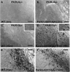
Accumulation of proteinase K-resistant α-synuclein in nigra of Parkin-Q311X(A) mice. A, B, Photomicrographs of the midbrain sections of Parkin-Q311X(A) mice and wild-type littermates at 3 months of age immunostained for proteinase K-resistant α-synuclein. Scale bar, 50 μm. C–F, Adjacent midbrain sections of Parkin-Q311X(A) mice and wild-type littermates at 16–18 months of age were immunostained for TH (C, D) (scale bar, 50 μm), proteinase K-resistant α-synuclein (E, F) (scale bar, 50 μm), and higher-magnification microphotographs of α-synuclein-immunopositive cells in nigra (insets in C and D) (scale bar, 12.5 μm).
Figure 9.
Figure 9.
Colocalization of nitrotyrosine and proteinase K-resistant α-synuclein in nigra of the Parkin-Q311X(A) mice. Confocal microphotographs of double immunofluorescence labeling proteinase K-resistant α-synuclein (green) and 3-NT (red) in nigra of Parkin-Q311X(A) (D–F) mice and wild-type littermates (A–C) at 16–18 months of age. Scale bar, 18 μm.

Comment in

Similar articles

Cited by

References

    1. Beffert U, Rosenberg RN. Increased risk for heterozygotes in recessive Parkinson disease. Arch Neurol. 2006;63:807–808. - PubMed
    1. Binkofski F, Reetz K, Gaser C, Hilker R, Hagenah J, Hedrich K, van Eimeren T, Thiel A, Büchel C, Pramstaller PP, Siebner HR, Klein C. Morphometric fingerprint of asymptomatic Parkin and PINK1 mutation carriers in the basal ganglia. Neurology. 2007;69:842–850. - PubMed
    1. Braak H, Del Tredici K, Rüb U, de Vos RA, Jansen Steur EN, Braak E. Staging of brain pathology related to sporadic Parkinson's disease. Neurobiol Aging. 2003;24:197–211. - PubMed
    1. Caudle WM, Richardson JR, Wang MZ, Taylor TN, Guillot TS, McCormack AL, Colebrooke RE, Di Monte DA, Emson PC, Miller GW. Reduced vesicular storage of dopamine causes progressive nigrostriatal neurodegeneration. J Neurosci. 2007;27:8138–8148. - PMC - PubMed
    1. Chesselet MF. In vivo alpha-synuclein overexpression in rodents: a useful model of Parkinson's disease? Exp Neurol. 2008;209:22–27. - PMC - PubMed

Publication types

MeSH terms