

Notch signaling contributes to the pathogenesis of human osteosarcomas
- PMID: 19228774
- PMCID: PMC2733809
- DOI: 10.1093/hmg/ddp057
Notch signaling contributes to the pathogenesis of human osteosarcomas
Abstract
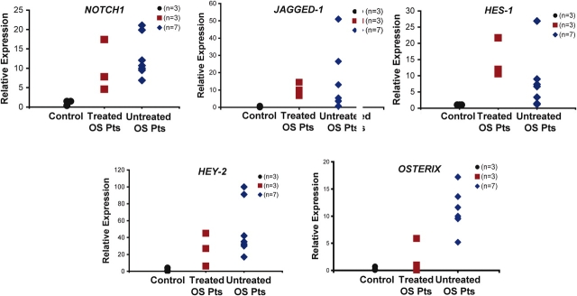
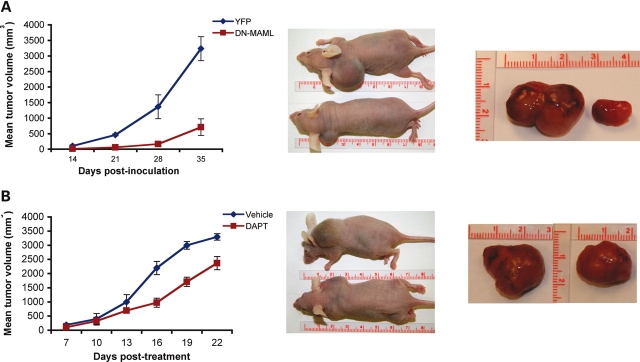
Notch signaling plays an important role in developmental processes and adult tissue homeostasis. Altered Notch signaling has been associated with various diseases including cancer. While the importance of altered Notch signaling in cancers of hematopoietic and epithelial origins has been established, its role in tumors of mesenchymal origin is less clear. Here, we report that human osteosarcoma cell lines and primary human osteosarcoma tumor samples show significant up-regulation of Notch, its target genes and Osterix. Notch inhibition by gamma-secretase inhibitors or by using lentiviral mediated expression of dominant negative Mastermind-like protein (DN-MAML) decreases osteosarcoma cell proliferation in vitro. In vivo, established human tumor xenografts in nude mice show decreased tumor growth after chemical or genetic inhibition of Notch signaling. Finally, transcriptional profiling of osteosarcomas from p53 mutant mice confirmed up-regulation of Notch1 target genes Hes1, Hey1 and its ligand Dll4. Our data suggest that activation of Notch signaling contributes to the pathogenesis of human osteosarcomas and its inhibition may be a therapeutic approach for the treatment of this mesenchymal tumor.
Figures


Similar articles
-
MAML1 regulates cell viability via the NF-κB pathway in cervical cancer cell lines.Exp Cell Res. 2011 Aug 1;317(13):1830-40. doi: 10.1016/j.yexcr.2011.05.005. Epub 2011 May 24. Exp Cell Res. 2011. PMID: 21640102
-
Inhibition of Notch pathway prevents osteosarcoma growth by cell cycle regulation.Br J Cancer. 2009 Jun 16;100(12):1957-65. doi: 10.1038/sj.bjc.6605060. Epub 2009 May 19. Br J Cancer. 2009. PMID: 19455146 Free PMC article.
-
Inhibition of the Notch-Hey1 axis blocks embryonal rhabdomyosarcoma tumorigenesis.Clin Cancer Res. 2011 Dec 1;17(23):7324-36. doi: 10.1158/1078-0432.CCR-11-1004. Epub 2011 Sep 23. Clin Cancer Res. 2011. PMID: 21948088 Free PMC article.
-
Understanding the role of Notch in osteosarcoma.Adv Exp Med Biol. 2014;804:67-92. doi: 10.1007/978-3-319-04843-7_4. Adv Exp Med Biol. 2014. PMID: 24924169 Review.
-
How the NOTCH pathway contributes to the ability of osteosarcoma cells to metastasize.Cancer Treat Res. 2009;152:479-96. doi: 10.1007/978-1-4419-0284-9_28. Cancer Treat Res. 2009. PMID: 20213410 Review.
Cited by
-
TNFα enhances cancer stem cell-like phenotype via Notch-Hes1 activation in oral squamous cell carcinoma cells.Biochem Biophys Res Commun. 2012 Jul 20;424(1):58-64. doi: 10.1016/j.bbrc.2012.06.065. Epub 2012 Jun 20. Biochem Biophys Res Commun. 2012. PMID: 22728043 Free PMC article.
-
Genomic heterogeneity of osteosarcoma - shift from single candidates to functional modules.PLoS One. 2015 Apr 7;10(4):e0123082. doi: 10.1371/journal.pone.0123082. eCollection 2015. PLoS One. 2015. PMID: 25848766 Free PMC article.
-
Integrated approaches to miRNAs target definition: time-series analysis in an osteosarcoma differentiative model.BMC Med Genomics. 2015 Jun 30;8:34. doi: 10.1186/s12920-015-0106-0. BMC Med Genomics. 2015. PMID: 26123714 Free PMC article.
-
Establishment and characterization of a new highly metastatic human osteosarcoma cell line derived from Saos2.Int J Clin Exp Pathol. 2014 May 15;7(6):2871-82. eCollection 2014. Int J Clin Exp Pathol. 2014. PMID: 25031706 Free PMC article.
-
γ-Secretase inhibitors and modulators.Biochim Biophys Acta. 2013 Dec;1828(12):2898-907. doi: 10.1016/j.bbamem.2013.06.005. Epub 2013 Jun 17. Biochim Biophys Acta. 2013. PMID: 23791707 Free PMC article. Review.
References
-
- Bray S.J. Notch signalling: a simple pathway becomes complex. Nat. Rev. Mol. Cell. Biol. 2006;7:678–689. - PubMed
-
- Miele L., Golde T., Osborne B. Notch signaling in cancer. Curr. Mol. Med. 2006;6:905–918. - PubMed
-
- Roy M., Pear W.S., Aster J.C. The multifaceted role of Notch in cancer. Curr. Opin. Genet. Dev. 2007;17:52–59. - PubMed
-
- Gridley T. Notch signaling and inherited disease syndromes. Hum. Mol. Genet. 2003;12(Spec no. 1):R9–R13. - PubMed
Publication types
MeSH terms
Substances
Grants and funding
LinkOut - more resources
Full Text Sources
Other Literature Sources
Research Materials
Miscellaneous
