. 2009 Mar;276(6):1610-28.
doi: 10.1111/j.1742-4658.2009.06893.x. Epub 2009 Feb 7.

Talin 2 is a large and complex gene encoding multiple transcripts and protein isoforms

Affiliations

Talin 2 is a large and complex gene encoding multiple transcripts and protein isoforms

Emmanuel Debrand et al. FEBS J. 2009 Mar.

Abstract

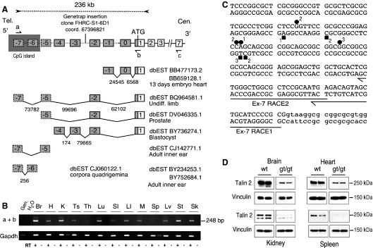
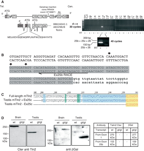
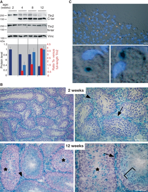
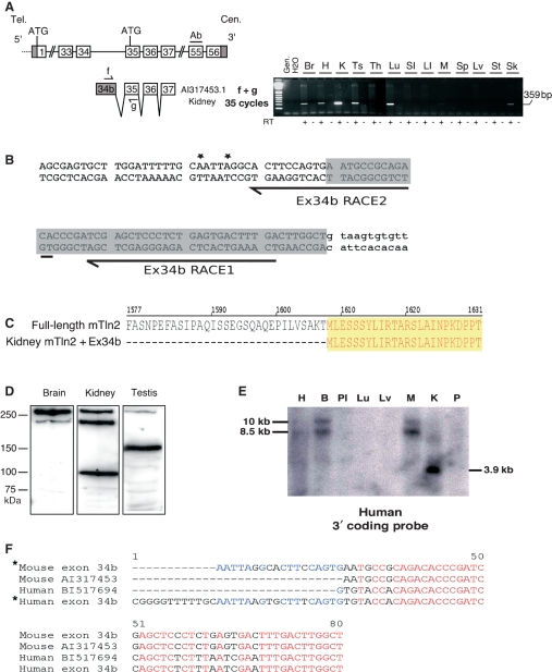
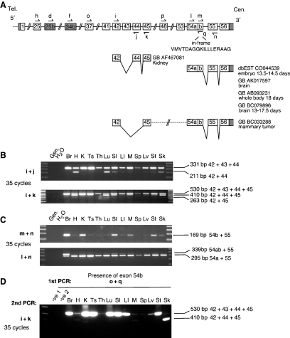
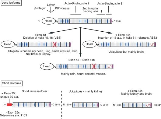
Talins are large adaptor proteins that link the integrin family of adhesion molecules to F-actin. In vertebrates, there are two talin genes. Talin 1 is essential for integrin-mediated cell adhesion; the role of talin 2 is unclear. Here we report a detailed analysis of mammalian talin 2. This reveals the existence of a previously unrecognized promoter associated with a CpG island, and separated from the first coding exon by numerous alternatively spliced noncoding exons spanning > 200 kb. Analysis of a mouse gene trap line shows that this promoter accounts for most of the talin 2 expression in adult tissues. We also demonstrate that testis and kidney express truncated talin 2 isoforms that lack the N-terminal half of the protein, and provide evidence for the developmentally regulated expression of the short testis-specific talin 2 isoform in elongating spermatids. Finally, we identify four tissue-specific alternative splicing events within the coding region of talin 2.


Figures

Fig. 1
Fig. 1
The 5′-end of mouse Tln2 is associated with a CpG island and contains a large number of 5′-UTR exons scattered over 200 kb. (A) Schematic diagram of the 5′-region of mouse Tln2 and corresponding ESTs. Tln2 is transcribed from the minus strand; consequently, the mouse chromosome 9 telomere is on the left (Tel.) and the centromere is on the right (Cen.). Boxes represent previously known exons; dashed boxes denote new putative exons and are linked to the rest of Tln2 by a dashed line. The first codon (ATG) is in exon 1, which contains both noncoding and coding sequences. Noncoding exons (grey) are numbered from 0 to −7. The CpG island associated with exon −7 is represented by a grey rectangle. The identity and position of the gene trap insertion downstream of exon −3 are indicated. Below the genomic structure, ESTs are shown that contain different combinations of Tln2 exons. The distance between the exons is indicated in bp, and the oblique lines represent splicing events. (B) RT-PCR between exon −7 and exon 1 (primers a + b) revealed widespread expression of Tln2 from the CpG island region in adult mouse tissues. Gen., genomic DNA; H2O, negative water control; RT+, reversed-transcribed RNA; RT−, non-reversed transcribed control. Gapdh primers were used to assess the amount of cDNA used in PCR reactions. Br., brain; H., heart; K, kidney; Ts, testis; Th, thymus; Lu, lung; SI, small intestine; LI, large intestine; M, skeletal muscle; Sp, spleen; Lv, liver; St, stomach; Sk, skin. (C) 5′-RACE anchored in exon −7 confirmed the 5′-end of Tln2. The sequence of exon −7 and flanking 5′ and 3′ (lower-case) sequences are shown. The position of the RACE primers in exon −7 is indicated by half-arrows. The RACE products obtained were cloned and sequenced. The position of the transcriptional start sites is indicated by squares and circles in brain and kidney respectively. The number of clones obtained at each position is also shown. (D) The gene trap insertion downstream of exon −3 dramatically reduces levels of talin 2 in mouse adult tissues. Proteins from wild-type (wt) and homozygous gene trap (gt/gt) mice were analysed by western blotting. Equal amount of total proteins were loaded, and talin 2 was detected with a monoclonal antibody against talin 2 (epitope within residues 482–991). Vinculin was used as an additional loading control and as a normalization reporter to quantify the reduction in Tln2 expression by luminometry (% of residual talin 2). The reduction in talin 2 levels was ∼ 80% in brain, 63% in heart, 93% in kidney, and 97% in spleen.
Fig. 2
Fig. 2
A shorter Tln2 transcript and protein product are specifically expressed in testis. (A) Schematic diagram of the mouse genomic region between coding exons 25 and 56, showing the gene trap insertion downstream of exon 28. The two testis ESTs containing additional exons 25b and 25c are shown under the genomic region. Grey boxes represent noncoding exons; white boxes are coding sequences. The additional amino acid sequence encoded by exon 25c is indicated. The position of the epitope recognized by the C-terminal polyclonal and monoclonal antibodies (Ab) is shown (encoded by exon 55). Half-arrows represent primers used in RT-PCR. RT-PCR amplification after 30 cycles and 40 cycles (testis) using primers d + e are depicted on the right-hand side. Gen., genomic DNA; H2O, negative water control; RT+, reversed-trascribed RNA; RT−, non-reversed-transcribed control. Br, brain; H, heart; K, kidney; Ts, testis; Th, thymus; Lu, lung; SI, small intestine; LI, large intestine; M, skeletal muscle; Sp, spleen; Lv, liver; St, stomach; Sk, skin. (B) 5′-RACE confirms the 5′-end of short testis-specific Tln2 transcripts. The sequence of exon 25b is shown – the position of ESTs is highlighted in grey; the position of the RACE primer is indicated by a half-arrow; and the position of the 5′-ends of three sequenced RACE amplicons is marked by a star. (C) Alignment of the predicted N-termini of the truncated testis-specific talin 2 isoforms with the full-length talin 2. +Ex25C, translation of transcripts containing exon 25c using the in-frame ATG present in exon 25c; −Ex25c, translation of short transcripts in which exon 25c is skipped (exons 25b + 26 to 56), using the next ATG in exon 26. The colour wrapping indicates identical amino acids in two (blue) or three (yellow) sequences; green represents positions occupied by different but structurally similar amino acids. (D) Western blotting of protein extracts from wild-type (wt) and homozygous gene trap (gt/gt) mice. Left panel: probed with a C-terminal polyclonal anti-talin 2 antibody [see (A) and Experimental procedures]. Right panel: probed with a polyclonal antibody against β-galactosidase. Br, brain; Ts, testis. The table indicates the predicted molecular masses of proteins detected by the two antibodies.
Fig. 3
Fig. 3
Ts-talin 2 is developmentally regulated and is expressed in early elongating spermatids. (A) Western blotting of testis protein extracts from two C57Bl/6 mice prior to (2 weeks old) and after puberty (4, 8 and 12 weeks). A talin 2 monoclonal C-terminal antibody was used to detect both full-length talin 2 and the short Ts-talin 2 isoforms, whereas the N-terminal monoclonal antibody detected only full-length talin 2. The respective levels of Ts-talin 2 (light blue), full-length talin 2 (dark blue) and the Ts-/full-length talin 2 ratio (red) were assessed by luminometry (average of two animals per age group; relative to vinculin loading control). (B) 5-bromo-4-chloro-3-indolyl-β-d-galactopyranoside (X-gal) staining of cryosections of 2-week-old and 12-week-old testis from animals with a gene trap insertion reporting the expression of both full-length and Ts-talin 2 (see Fig. 2). Arrows indicate expression in myoid cells adjacent to the basement membrane of the seminiferous tubule; arrowheads point to the staining of interstitial Leydig cells. Staining in both locations was identical in prepubertal and pubertal mice. An asterisk shows the punctate expression detected only after puberty, restricted to specific stages of the tubule cycle and replaced by a diffuse staining of the adluminal compartment at the point of spermiation (bracket). Left panel: 20× objective. Right panel: 40× objective. Bar = 50 μm. (C) Differential interference contrast image of a stage XII–I seminiferous tubule squash (top panel) from a 3-month-old gene trap animal, stained with DAPI and X-gal, and detail of two elongating spermatids (bottom panel). Arrowheads indicate β-galactosidase staining. The data show that the punctate talin 2 expression in the mature testis, most likely Ts-talin 2, is restricted to the cytoplasm of elongating spermatids. Bar = 20 μm.
Fig. 4
Fig. 4
Evidence for a truncated mouse Tln2 transcript and protein product highly expressed in kidney and conserved in human. (A) Schematic diagram of the mouse Tln2 genomic region between coding exons 33 and 56. An EST containing the additional exon 34b is shown below. Grey boxes represent noncoding exons; white boxes are coding sequences. Half-arrows indicate the primers used in RT-PCR. RT-PCR amplification after 35 cycles using primers f + g is shown. Gen., genomic DNA; H2O, negative water control; RT+, reversed-transcribed RNA; RT−, non-reversed-transcribed control. Br, brain; H, heart; K, kidney; Ts, testis; Th, thymus; Lu, lung; SI, small intestine; LI, large intestine; M, skeletal muscle; Sp, spleen; Lv, liver; St, stomach; Sk, skin. (B) 5′-RACE confirms the 5′-end of the short transcript. The sequence of exon 34b is shown, and the position of the EST is shaded grey; intronic sequences are in lower case; the position of the RACE primers is indicated by an arrow, and the positions of the 5′-ends of two sequenced RACE amplicons are marked by a star. (C) Alignment of the predicted N-terminus of the corresponding truncated protein with the full-length talin 2. (D) Western blotting showing the presence of a 90 kDa talin 2 protein in kidney (*). (E) Conservation of the short ‘kidney’ transcript in humans. A human tissue northern blot was probed with a DNA fragment detecting the 3′ coding region of TLN2. A 3.9 kb transcript strongly expressed in kidney was detected. (F) A kidney cDNA library was screened with primers near the putative 5′-end of the transcript and in the cloning vector. This revealed a new exon that was also present in a human EST. The alignment of the corresponding cDNA sequences with the equivalent mouse sequences is shown. *, 5′-ends determined in this study.
Fig. 5
Fig. 5
Evidence for alternatively spliced mTln2 transcripts. (A) Schematic diagram of two regions showing alternative splicing. ESTs supporting alternative splicing are shown below the genomic region. Primers used to detect alternative splicing (i, j, k, l, m and n) and short transcript isoforms (d, short testis transcript; f, short kidney transcript) are indicated by half-arrows. The amino acid sequence of the in-frame peptide added as a result of inclusion of exon 54b is shown. (B, C) RT-PCR amplification after 35 cycles using various primer combinations. (B) Primer i + j detects splicing out of exon 43; i + k reveals splicing of both exon 43 and exon 44. (C) m + n indicates the presence of exon 54b; l + n indicates the relative abundance of exon 54a + exon55 and exon 54ab + exon 55 products. Gen., genomic DNA; H2O, negative water control; RT+, reversed-transcribed RNA; RT−, non-reversed-transcribed control. Br, brain; H, heart; K, kidney; Ts, testis; Th, thymus; Lu, lung; SI, small intestine; LI, large intestine; M, skeletal muscle; Sp, spleen; Lv, liver; St, stomach; Sk, skin.
Fig. 6
Fig. 6
Diagram of the main talin 2 isoforms. Top: model of full-length talin 2 based on the assumption that the main structural features and ligand-binding sites of talin 1 are conserved in talin 2. Vinculin-binding sites (VBS) are shown in blue. The consequences of the three main alternative splicing events detected by RT-PCR for the structure of the long talin 2 isoforms (Figs 5 and S7) are indicated. A red cross indicates that exon 43 skipping (−exon 43) deletes VBS3. A red curved line shows the 15 amino acids inserted into the C-terminal actin-binding site (ABS3) by the inclusion of exon 54b (+exon 54b). Bottom: structures of the main short talin 2 isoforms predicted to arise from smaller Tln2 transcripts.

Similar articles

Cited by

References

    1. Critchley DR, Gingras AR. Talin at a glance. J Cell Sci. 2008;121:1345–1347. - PubMed
    1. Calderwood DA. Integrin activation. J Cell Sci. 2004;117:657–666. - PubMed
    1. Calderwood DA, Zent R, Grant R, Rees DJG, Hynes RO, Ginsberg MH. The talin head domain binds to integrin b subunit cytoplasmic tails and regulates integrin activation. J Biol Chem. 1999;274:28071–28704. - PubMed
    1. Lee HS, Bellin RM, Walker DL, Patel B, Powers P, Liu H, Garcia-Alvarez B, de Pereda JM, Liddington RC, Volkmann N, et al. Characterization of an actin-binding site within the talin FERM domain. J Mol Biol. 2004;343:771–784. - PubMed
    1. Loer B, Bauer R, Bornheim R, Grell J, Kremmer E, Kolanus W, Hoch M. The NHL-domain protein Wech is crucial for the integrin–cytoskeleton link. Nat Cell Biol. 2008;10:422–428. - PubMed

Publication types

MeSH terms