Early and sustained innate immune response defines pathology and death in nonhuman primates infected by highly pathogenic influenza virus
- PMID: 19218453
- PMCID: PMC2642661
- DOI: 10.1073/pnas.0813234106
Early and sustained innate immune response defines pathology and death in nonhuman primates infected by highly pathogenic influenza virus
Abstract
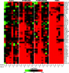
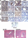
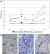
The mechanisms responsible for the virulence of the highly pathogenic avian influenza (HPAI) and of the 1918 pandemic influenza virus in humans remain poorly understood. To identify crucial components of the early host response during these infections by using both conventional and functional genomics tools, we studied 34 cynomolgus macaques (Macaca fascicularis) to compare a 2004 human H5N1 Vietnam isolate with 2 reassortant viruses possessing the 1918 hemagglutinin (HA) and neuraminidase (NA) surface proteins, known conveyors of virulence. One of the reassortants also contained the 1918 nonstructural (NS1) protein, an inhibitor of the host interferon response. Among these viruses, HPAI H5N1 was the most virulent. Within 24 h, the H5N1 virus produced severe bronchiolar and alveolar lesions. Notably, the H5N1 virus targeted type II pneumocytes throughout the 7-day infection, and induced the most dramatic and sustained expression of type I interferons and inflammatory and innate immune genes, as measured by genomic and protein assays. The H5N1 infection also resulted in prolonged margination of circulating T lymphocytes and notable apoptosis of activated dendritic cells in the lungs and draining lymph nodes early during infection. While both 1918 reassortant viruses also were highly pathogenic, the H5N1 virus was exceptional for the extent of tissue damage, cytokinemia, and interference with immune regulatory mechanisms, which may help explain the extreme virulence of HPAI viruses in humans.
Conflict of interest statement
Conflict of interest statement: A.G.-S. owns patent positions for reverse genetics of influenza viruses.
Figures


Similar articles
-
Accumulation of CD11b⁺Gr-1⁺ cells in the lung, blood and bone marrow of mice infected with highly pathogenic H5N1 and H1N1 influenza viruses.Arch Virol. 2013 Jun;158(6):1305-22. doi: 10.1007/s00705-012-1593-3. Epub 2013 Feb 9. Arch Virol. 2013. PMID: 23397329 Free PMC article.
-
Disease severity is associated with differential gene expression at the early and late phases of infection in nonhuman primates infected with different H5N1 highly pathogenic avian influenza viruses.J Virol. 2014 Aug;88(16):8981-97. doi: 10.1128/JVI.00907-14. Epub 2014 Jun 4. J Virol. 2014. PMID: 24899188 Free PMC article.
-
Extrapulmonary tissue responses in cynomolgus macaques (Macaca fascicularis) infected with highly pathogenic avian influenza A (H5N1) virus.Arch Virol. 2010 Jun;155(6):905-14. doi: 10.1007/s00705-010-0662-8. Epub 2010 Apr 7. Arch Virol. 2010. PMID: 20372944 Free PMC article.
-
Innate immune responses to influenza A H5N1: friend or foe?Trends Immunol. 2009 Dec;30(12):574-84. doi: 10.1016/j.it.2009.09.004. Epub 2009 Oct 26. Trends Immunol. 2009. PMID: 19864182 Free PMC article. Review.
-
[Cytokine storm in avian influenza].Mikrobiyol Bul. 2008 Apr;42(2):365-80. Mikrobiyol Bul. 2008. PMID: 18697437 Review. Turkish.
Cited by
-
Phenotypic differences in virulence and immune response in closely related clinical isolates of influenza A 2009 H1N1 pandemic viruses in mice.PLoS One. 2013;8(2):e56602. doi: 10.1371/journal.pone.0056602. Epub 2013 Feb 18. PLoS One. 2013. PMID: 23441208 Free PMC article.
-
DDX3X coordinates host defense against influenza virus by activating the NLRP3 inflammasome and type I interferon response.J Biol Chem. 2021 Jan-Jun;296:100579. doi: 10.1016/j.jbc.2021.100579. Epub 2021 Mar 23. J Biol Chem. 2021. PMID: 33766561 Free PMC article.
-
Systems-level comparison of host-responses elicited by avian H5N1 and seasonal H1N1 influenza viruses in primary human macrophages.PLoS One. 2009 Dec 14;4(12):e8072. doi: 10.1371/journal.pone.0008072. PLoS One. 2009. PMID: 20011590 Free PMC article.
-
Mixed Lineage Kinase 3 deficiency delays viral clearance in the lung and is associated with diminished influenza-induced cytopathic effect in infected cells.Virology. 2010 May 10;400(2):224-32. doi: 10.1016/j.virol.2010.02.001. Epub 2010 Feb 25. Virology. 2010. PMID: 20185156 Free PMC article.
-
Estriol Reduces Pulmonary Immune Cell Recruitment and Inflammation to Protect Female Mice From Severe Influenza.Endocrinology. 2018 Sep 1;159(9):3306-3320. doi: 10.1210/en.2018-00486. Endocrinology. 2018. PMID: 30032246 Free PMC article.
References
-
- Ng WF, To KF. Pathology of human H5N1 infection: New findings. Lancet. 2007;370:1106–1108. - PubMed
-
- Writing Committee of the Second World Health Organization Consultation on Clinical Aspects of Human Infection with Avian Influenza A (H5N1) Virus. Update on avian influenza A (H5N1) virus infection in humans. N Engl J Med. 2008;358:261–273. - PubMed
-
- Uyeki TM. Global epidemiology of human infections with highly pathogenic avian influenza A (H5N1) viruses. Respirology. 2008;13(s1):S2–S9. - PubMed
-
- Morens DM, Fauci AS. The 1918 influenza pandemic: Insights for the 21st century. J Infect Dis. 2007;195:1018–1028. - PubMed
Publication types
MeSH terms
Grants and funding
- R01 AI046954/AI/NIAID NIH HHS/United States
- P51 RR000166/RR/NCRR NIH HHS/United States
- R03 AI075019/AI/NIAID NIH HHS/United States
- U01AI070469/AI/NIAID NIH HHS/United States
- P01AI58113,/AI/NIAID NIH HHS/United States
- K08 AI059106/AI/NIAID NIH HHS/United States
- R01 AI022646-20A1/AI/NIAID NIH HHS/United States
- R01 AI022646/AI/NIAID NIH HHS/United States
- P51 RR00166-45/RR/NCRR NIH HHS/United States
- P01 AI058113/AI/NIAID NIH HHS/United States
- R03 AI075019-01/AI/NIAID NIH HHS/United States
- U01 AI070469/AI/NIAID NIH HHS/United States
- K08 AI059106-02/AI/NIAID NIH HHS/United States
- R24 RR016354/RR/NCRR NIH HHS/United States
- R24 RR16354-04/RR/NCRR NIH HHS/United States
- R01AI46954,/AI/NIAID NIH HHS/United States
LinkOut - more resources
Full Text Sources
Medical
Molecular Biology Databases

