. 2009 Apr 17;284(16):10361-6.
doi: 10.1074/jbc.M900956200. Epub 2009 Feb 13.

Inhibition of SUV39H1 methyltransferase activity by DBC1

Affiliations

Inhibition of SUV39H1 methyltransferase activity by DBC1

Zhenyu Li et al. J Biol Chem. .

Abstract

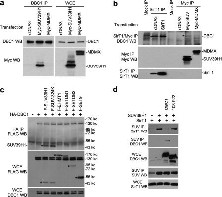
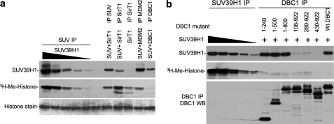
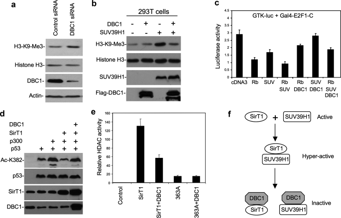
SUV39H1 is a histone H3K9-specific methyltransferase important for heterochromatin formation, regulation of gene expression, and induction of senescence in premalignant cells. SUV39H1 forms a complex with SirT1, and its activity is stimulated by SirT1 binding. Here we present evidence that the product of the DBC1 (deleted in breast cancer 1) gene disrupts the SUV39H1-SirT1 complex. Furthermore, DBC1 binds to the SUV39H1 catalytic domain and inhibits its ability to methylate histone H3 in vitro and in vivo. Knockdown of endogenous DBC1 increased the level of cellular H3K9 methylation. As expected, DBC1 also binds to SirT1 and inhibits the deacetylase activity of SirT1. These results identify DBC1 as a novel cellular inhibitor of SUV39H1 activity. DBC1 may be an important regulator of heterochromatin formation and genomic stability by disrupting the SUV39H1-SirT1 complex and inactivating both enzymes.


Figures

FIGURE 1.
FIGURE 1.
Interaction between DBC1 and SUV39H1. a, H1299 cells were transiently transfected with the indicated plasmids for 48 h. Endogenous DBC1 was immunoprecipitated with anti-DBC1 antibody. Coprecipitated SUV39H1 was detected by anti-Myc Western blotting (WB). DBC1, Myc-MDMX, and Myc-SUV39H1 expression levels were confirmed in WCE by anti-DBC1 and anti-Myc antibodies. b, SirT1 was immunoprecipitated with antibody 10E4, whereas SUV and MDMX were immunoprecipitated with anti-Myc antibody. Coprecipitated DBC1 was detected by anti-DBC1 Western blotting. SUV39H1 and MDMX were detected by anti-Myc Western blotting. c, HA-DBC1 was cotransfected into H1299 cells with FLAG-tagged histone methyltransferases and immunoprecipitated with anti-HA antibody. Coprecipitated enzymes were detected by anti-FLAG Western blotting. Expression of HA-DBC1 in the WCE was detected with anti-DBC1 antibody. d, H1299 cells were transiently transfected with the indicated plasmids. SUV39H1 was immunoprecipitated with mouse anti-SUV39H1 antibody. Coprecipitated SirT1 and DBC1 were detected with antibody 10E4 and anti-DBC1 antibody, respectively. SUV39H1 was detected by reprobing with rabbit anti-SUV antibody.
FIGURE 2.
FIGURE 2.
Mapping of DBC1 domain involved in SUV39H1-DBC1 and SirT1-DBC1 interactions. H1299 cells were transiently transfected with the indicated plasmids for 48 h. a, DBC1 was immunoprecipitated with anti-FLAG antibody. SUV39H1 coprecipitation was detected by anti-SUV39H1 Western blotting (WB). WT, wild-type. b, in vitro-translated SUV39H1 was incubated with GST-DBC1 truncation mutants bound to glutathione beads. The captured SUV39H1 was detected by SDS-PAGE and autoradiography. c, DBC1 and truncation mutants were immunoprecipitated with M2 beads. SirT1 coprecipitation was detected by rabbit anti-SirT1 antibody, whereas DBC1 was detected by rabbit anti-FLAG antibody. d, the diagram summarize results in a–c. NLS, nuclear localization signal; LZ, leucine zipper.
FIGURE 3.
FIGURE 3.
Mapping of SUV39H1 domain involved in SUV39H1-DBC1 interaction. a, DBC1 was immunoprecipitated with anti-HA antibody. SUV39H1 coprecipitation was detected by rabbit anti-FLAG antibody. WT, wild-type; WB, Western blot. b, diagrams summarizing the results are shown. Chromo, chromodomain.
FIGURE 4.
FIGURE 4.
DBC1 inhibits SUV39H1 methyltransferase activity with its N-terminal domain. H1299 cells were transiently transfected with the indicated plasmids for 48 h. a, different amounts of SUV39H1 (relative levels 1×, 2×, 4×, 8×, and 16×) were immunoprecipitated with anti-Myc antibody. SirT1 was immunoprecipitated with antibody 10E4; MDM2 and SUV39H1 were immunoprecipitated with anti-FLAG antibody. The beads were used for in vitro methylation of histone H3 and detected by 3H autoradiography. SUV39H1 was detected by Western blotting. Histone H3 on the membrane was revealed by Coomassie staining. b, SUV39H1 was immunoprecipitated in the same manner as indicated in a. DBC1 and deletion mutants were immunoprecipitated with anti-FLAG antibody. The beads were used for in vitro methylation of histone H3. Expression levels of DBC1 mutants and coprecipitation of SUV39H1 were confirmed by Western blotting (WB). Wt, wild-type.
FIGURE 5.
FIGURE 5.
DBC1 inhibits SUV39H1 in vivo. a, H1299 cells were transfected with DBC1 siRNA or control siRNA. Histones were extracted, and the H3K9me3 level was detected with anti-H3K9me3 antibody and reprobing with anti-H3 antibody. DBC1 in the WCE was detected with anti-DBC1 antibody. b, 293T cells were transiently transfected with the indicated plasmids for 48 h. Histone was extracted, and H3K9me3 was detected with anti-H3K9me3 antibody. DBC1 in the WCE was detected with anti-DBC1 antibody. SUV39H1 in the WCE was detected with anti-Myc antibody. c, H1299 cells were transfected with the indicated plasmids for 48 h. Relative luciferase activity was determined and normalized by cotransfected lacZ control. d, H1299 cells were transiently transfected with the indicated plasmids for 48 h. Acetylation of p53 Lys382 was detected with anti-Ac-K382 antibody and reprobed with p53 antibody DO1. e, H1299 was transfected with the indicated plasmids for 48 h. Cell lysate was analyzed for histone deacetylase (HDAC) activity using a 3H-labeled acetylated H3 peptide as substrate. f, a model of DBC1 regulation of the SUV39H1-SirT1 complex is shown. Error bars represent S.D. from three experiments.

References

    1. Aagaard, L., Laible, G., Selenko, P., Schmid, M., Dorn, R., Schotta, G., Kuhfittig, S., Wolf, A., Lebersorger, A., Singh, P. B., Reuter, G., and Jenuwein, T. (1999) EMBO J. 18 1923–1938 - PMC - PubMed
    1. Peters, A. H., O'Carroll, D., Scherthan, H., Mechtler, K., Sauer, S., Schofer, C., Weipoltshammer, K., Pagani, M., Lachner, M., Kohlmaier, A., Opravil, S., Doyle, M., Sibilia, M., and Jenuwein, T. (2001) Cell 107 323–337 - PubMed
    1. Nielsen, S. J., Schneider, R., Bauer, U. M., Bannister, A. J., Morrison, A., O'Carroll, D., Firestein, R., Cleary, M., Jenuwein, T., Herrera, R. E., and Kouzarides, T. (2001) Nature 412 561–565 - PubMed
    1. Braig, M., Lee, S., Loddenkemper, C., Rudolph, C., Peters, A. H., Schlegelberger, B., Stein, H., Dorken, B., Jenuwein, T., and Schmitt, C. A. (2005) Nature 436 660–665 - PubMed
    1. Imai, S., Armstrong, C. M., Kaeberlein, M., and Guarente, L. (2000) Nature 403 795–800 - PubMed

Publication types

MeSH terms