

Clinical and pathological continuum of multisystem TDP-43 proteinopathies
- PMID: 19204154
- PMCID: PMC2774117
- DOI: 10.1001/archneurol.2008.558
Clinical and pathological continuum of multisystem TDP-43 proteinopathies
Abstract
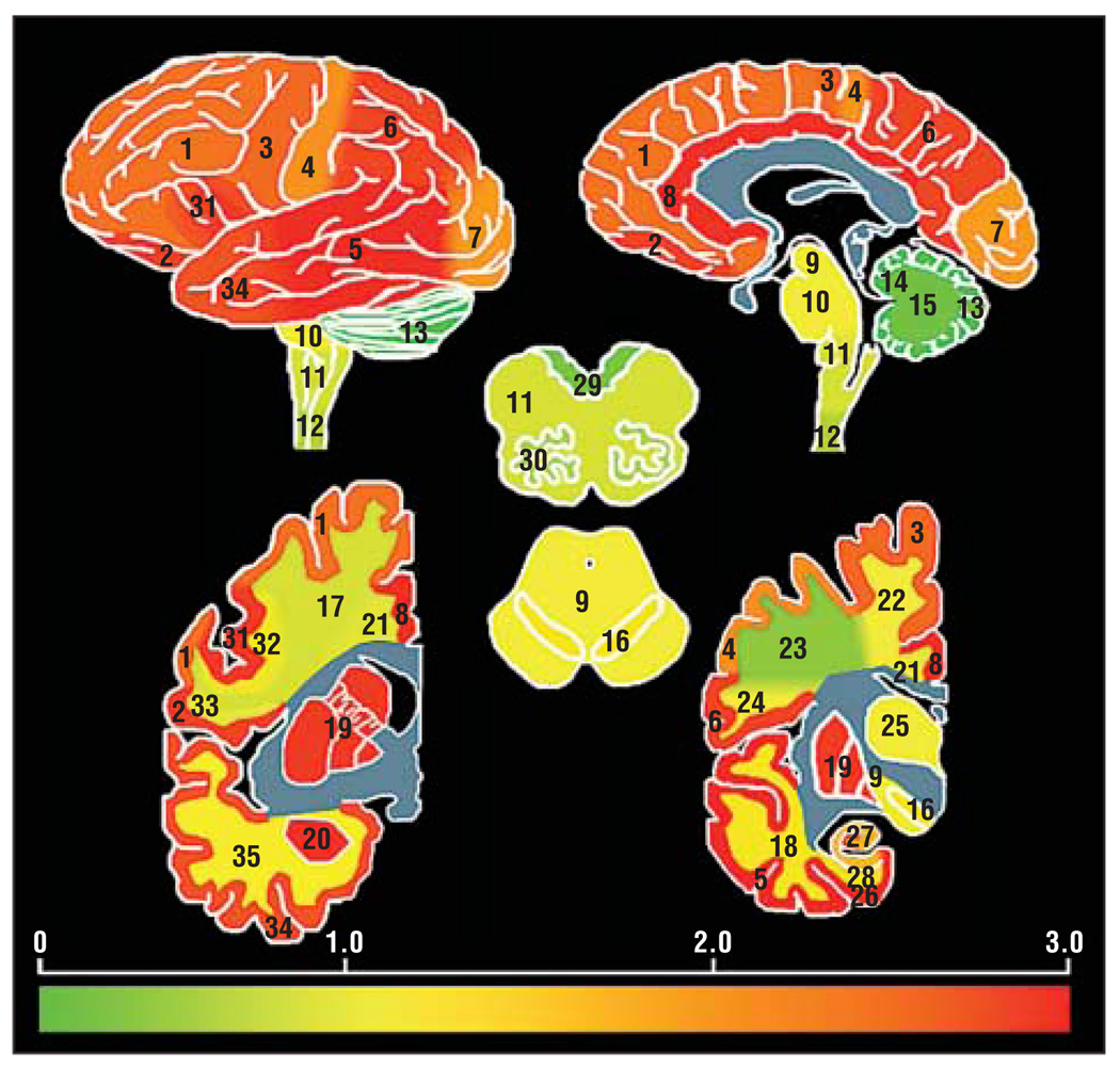
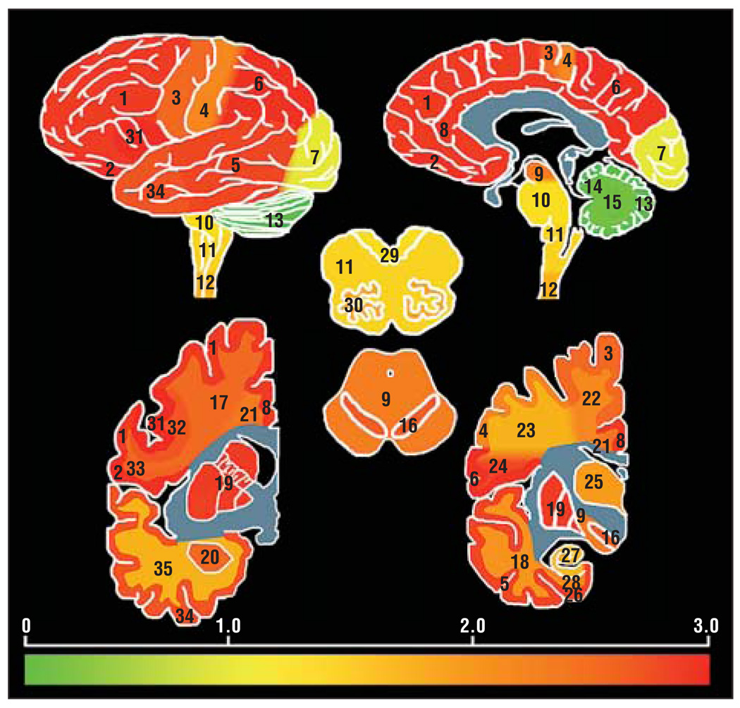
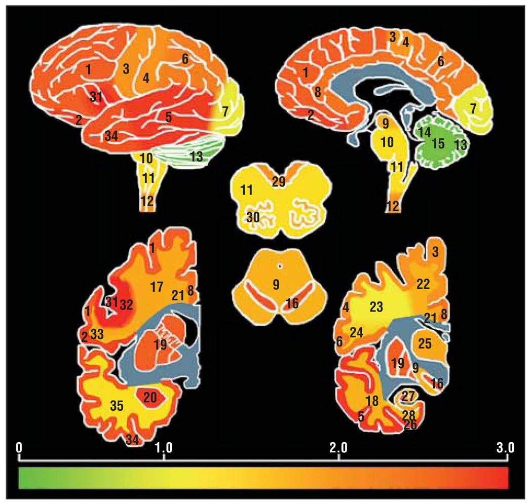
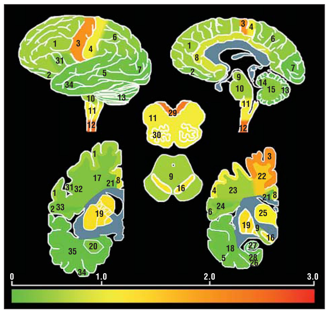
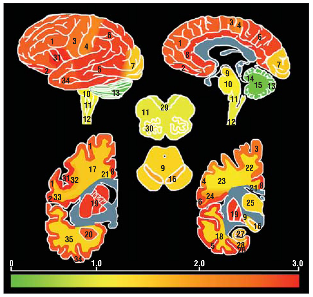
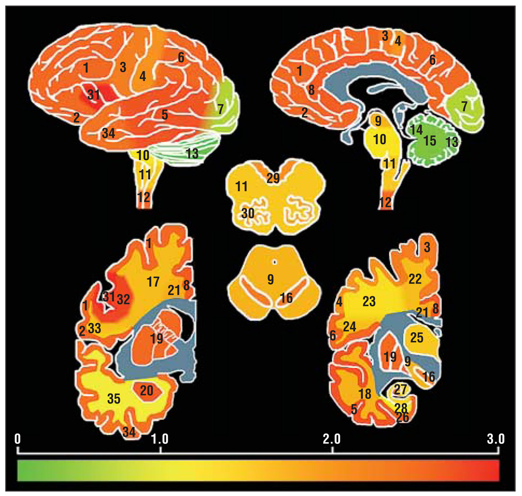
Objective: To determine the extent of transactivation response DNA-binding protein with a molecular weight of 43 kDa (TDP-43) pathology in the central nervous system of patients with clinically and autopsy-confirmed diagnoses of frontotemporal lobar degeneration with and without motor neuron disease and amyotrophic lateral sclerosis with and without cognitive impairment.
Design: Performance of immunohistochemical whole-central nervous system scans for evidence of pathological TDP-43 and retrospective clinical medical record review.
Setting: An academic medical center.
Participants: We included 64 patients with clinically and pathologically confirmed frontotemporal lobar degeneration with ubiquitinated inclusions with or without motor neuron disease and amyotrophic lateral sclerosis with or without cognitive impairment.
Main outcome measure: Neuronal and glial TDP-43 pathology.
Results: We found evidence of neuronal and glial TDP-43 pathology in all disease groups throughout the neuraxis, albeit with variations in the frequency, morphology, and distribution of TDP-43 lesions. Moreover, the major clinical manifestations (eg, cognitive impairments, motor neuron signs, extrapyramidal symptoms, neuropsychiatric features) were reflected by the predominant distribution and burden of TDP-43 pathology.
Conclusion: These findings strongly suggest that amyotrophic lateral sclerosis, frontotemporal lobar degeneration with amyotrophic lateral sclerosis or motor neuron disease, and frontotemporal lobar degeneration with ubiquitinated inclusions are different manifestations of a multiple-system TDP-43 proteinopathy linked to similar mechanisms of neurodegeneration.
Figures

Similar articles
-
Evidence of multisystem disorder in whole-brain map of pathological TDP-43 in amyotrophic lateral sclerosis.Arch Neurol. 2008 May;65(5):636-41. doi: 10.1001/archneur.65.5.636. Arch Neurol. 2008. PMID: 18474740
-
Severe subcortical TDP-43 pathology in sporadic frontotemporal lobar degeneration with motor neuron disease.Acta Neuropathol. 2008 Jan;115(1):123-31. doi: 10.1007/s00401-007-0315-5. Epub 2007 Nov 15. Acta Neuropathol. 2008. PMID: 18004574
-
Sporadic amyotrophic lateral sclerosis: two pathological patterns shown by analysis of distribution of TDP-43-immunoreactive neuronal and glial cytoplasmic inclusions.Acta Neuropathol. 2008 Aug;116(2):169-82. doi: 10.1007/s00401-008-0385-z. Epub 2008 May 15. Acta Neuropathol. 2008. PMID: 18481073
-
Amyotrophic lateral sclerosis and frontotemporal lobar degeneration: a spectrum of TDP-43 proteinopathies.Neuropathology. 2010 Apr;30(2):103-12. doi: 10.1111/j.1440-1789.2009.01091.x. Epub 2010 Jan 25. Neuropathology. 2010. PMID: 20102519 Free PMC article. Review.
-
TDP-43: the relationship between protein aggregation and neurodegeneration in amyotrophic lateral sclerosis and frontotemporal lobar degeneration.FEBS J. 2011 Oct;278(19):3539-49. doi: 10.1111/j.1742-4658.2011.08256.x. Epub 2011 Aug 24. FEBS J. 2011. PMID: 21777387 Free PMC article. Review.
Cited by
-
CSF biomarkers cutoffs: the importance of coincident neuropathological diseases.Acta Neuropathol. 2012 Jul;124(1):23-35. doi: 10.1007/s00401-012-0983-7. Epub 2012 Apr 22. Acta Neuropathol. 2012. PMID: 22526019 Free PMC article.
-
Selective neuronal degeneration in MATR3 S85C knock-in mouse model of early-stage ALS.Nat Commun. 2020 Oct 20;11(1):5304. doi: 10.1038/s41467-020-18949-w. Nat Commun. 2020. PMID: 33082323 Free PMC article.
-
Microstructural changes across different clinical milestones of disease in amyotrophic lateral sclerosis.PLoS One. 2015 Mar 20;10(3):e0119045. doi: 10.1371/journal.pone.0119045. eCollection 2015. PLoS One. 2015. PMID: 25793718 Free PMC article.
-
Head-to-Head Comparison of Tau-PET Radioligands for Imaging TDP-43 in Post-Mortem ALS Brain.Mol Imaging Biol. 2023 Jun;25(3):513-527. doi: 10.1007/s11307-022-01779-1. Epub 2022 Oct 18. Mol Imaging Biol. 2023. PMID: 36258099
-
Impaired cognitive flexibility in amyotrophic lateral sclerosis.Cogn Behav Neurol. 2015 Mar;28(1):17-26. doi: 10.1097/WNN.0000000000000049. Cogn Behav Neurol. 2015. PMID: 25812127 Free PMC article.
References
-
- Neumann M, Sampathu DM, Kwong LK, et al. Ubiquitinated TDP-43 in frontotemporal lobar degeneration and amyotrophic lateral sclerosis. Science. 2006;314(5796):130–133. - PubMed
-
- Brooks BR, Miller RG, Swash M, Munsat TL. World Federation of Neurology Research Group on Motor Neuron Diseases. El Escorial revisited: revised criteria for the diagnosis of amyotrophic lateral sclerosis. Amyotroph Lateral Scler Other Motor Neuron Disord. 2000;1(5):293–299. - PubMed
Publication types
MeSH terms
Substances
Grants and funding
LinkOut - more resources
Full Text Sources
Medical
