. 2009 Apr 17;284(16):10923-34.
doi: 10.1074/jbc.M808760200. Epub 2009 Feb 6.

Reconstitution and dissection of the 600-kDa Srv2/CAP complex: roles for oligomerization and cofilin-actin binding in driving actin turnover

Affiliations

Reconstitution and dissection of the 600-kDa Srv2/CAP complex: roles for oligomerization and cofilin-actin binding in driving actin turnover

Omar Quintero-Monzon et al. J Biol Chem. .

Abstract

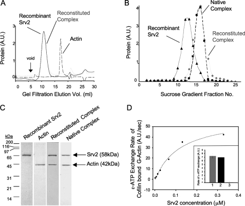
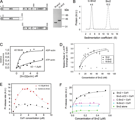
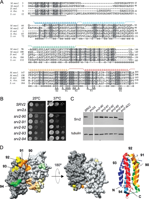
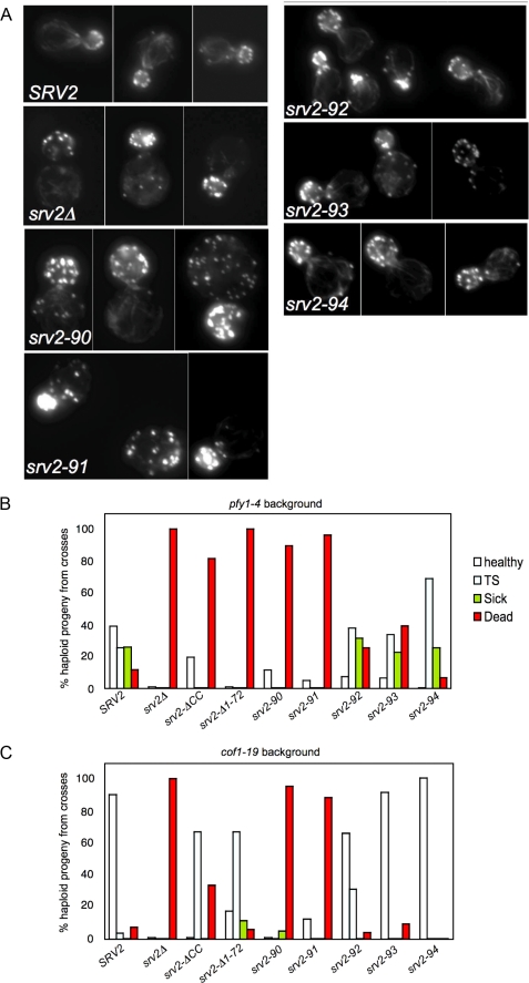
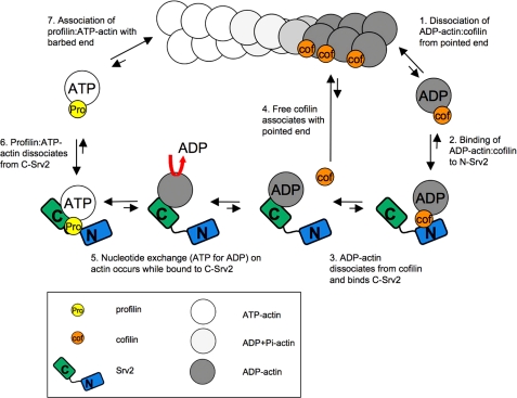
Srv2/cyclase-associated protein is expressed in virtually all plant, animal, and fungal organisms and has a conserved role in promoting actin depolymerizing factor/cofilin-mediated actin turnover. This is achieved by the abilities of Srv2 to recycle cofilin from ADP-actin monomers and to promote nucleotide exchange (ATP for ADP) on actin monomers. Despite this important and universal role in facilitating actin turnover, the mechanism underlying Srv2 function has remained elusive. Previous studies have demonstrated a critical functional role for the G-actin-binding C-terminal half of Srv2. Here we describe an equally important role in vivo for the N-terminal half of Srv2 in driving actin turnover. We pinpoint this activity to a conserved patch of surface residues on the N-terminal dimeric helical folded domain of Srv2, and we show that this functional site interacts with cofilin-actin complexes. Furthermore, we show that this site is essential for Srv2 acceleration of cofilin-mediated actin turnover in vitro. A cognate Srv2-binding site is identified on a conserved surface of cofilin, suggesting that this function likely extends to other organisms. In addition, our analyses reveal that higher order oligomerization of Srv2 depends on its N-terminal predicted coiled coil domain and that oligomerization optimizes Srv2 function in vitro and in vivo. Based on these data, we present a revised model for the mechanism by which Srv2 promotes actin turnover, in which coordinated activities of its N- and C-terminal halves catalyze sequential steps in recycling cofilin and actin monomers.


Figures

FIGURE 1.
FIGURE 1.
Reconstitution of the Srv2-actin complex from purified proteins. A, gel filtration analysis of purified full-length recombinant Srv2, actin, and reconstituted Srv2-actin complex. B, sucrose gradient sedimentation analysis of recombinant full-length Srv2, reconstituted Srv2-actin complex, and native Srv2-actin complex. C, Coomassie-stained gel of peak fractions from gel filtration compared with native Srv2-actin complex isolated from yeast. D, recombinant Srv2 has concentration-dependent effects in relieving cofilin inhibition of ε-ATP nucleotide exchange on actin monomers. Inset, ε-ATP exchange rates for reactions containing 1 μm G-actin, 2 μm Cof1, and either 53 nm recombinant Srv2 (bar 1), 53 nm native Srv2 (bar 2), or no Srv2 (bar 3). A.U., arbitrary units.
FIGURE 2.
FIGURE 2.
Domain requirements for Srv2 oligomerization, G-actin binding, catalysis of actin nucleotide exchange, and acceleration of actin turnover. A, schematic of Srv2 domain organization and Coomassie-stained gel of purified Srv2 fragments. CC, coiled coil domain; HFD, helical folded domain; P, polyproline-rich motifs; W, WASp-homology 2 (WH2) domain; Di, dimerization motif. B, analytical ultracentrifugation sedimentation coefficient distributions for purified full-length Srv2 (dotted line) and C-Srv2 (solid line). Sedimentation coefficients are shown above peaks. C, concentration-dependent binding of full-length Srv2 to 0.2 μm ATP-bound (squares) or ADP-bound (circles) NBD-labeled actin monomers. Values on the x axis are molar ratios of Srv2 to actin. Binding constants were calculated as described (24). D, concentration-dependent effects of full-length Srv2, N-Srv2, and C-Srv2 on rate of ε-ATP exchange on actin monomers. E, concentration-dependent effects of cofilin on rate of steady state turnover of F-actin (7.7 μm) measured by Pi release assay in the absence (black circles) and presence (red circles) of 0.132 μm full-length Srv2. F, concentration-dependent effects of different Srv2 proteins on rate of turnover of F-actin in the presence of a fixed concentration (3.8 μm) of cofilin. A.U., arbitrary units.
FIGURE 3.
FIGURE 3.
Mutational analysis of the Srv2 HFD domain. A, alignment of N-terminal sequences for diverse Srv2/CAP homologues using ClustalW. M. mus1, mouse CAP1; M. mus2, mouse CAP2; D. mel., Drosophila melanogaster CAP; A. tha, Arabidopsis thaliana CAP; and S. cer., S. cerevisiae Srv2. Residues 73-241 in S. cerevisiae Srv2 form the HFD domain (18). Each helix is indicated above the primary sequence and color-coded. Solvent-exposed residues are designated below the primary sequence (e indicates solvent-exposed; - indicates for solvent-inaccessible). Below the alignment, the predicted coiled coil domain (residues 14-34) is underlined, and residues changed to alanine are marked A for each srv2 allele (numbered 90-94). B, SRV2 and srv2 mutant strains were grown to log phase, serially diluted, and plated on YPD plates at 25 and 37 °C to compare cell growth. C, immunoblot of whole cell extracts from SRV2 and srv2 mutant strains probed with anti-Srv2 antibodies and tubulin antibodies as a loading control. D, srv2 mutations modeled on a rendered view of the crystal structure of the dimeric, anti-parallel HFD from Dictyostelium Srv2/CAP (Protein Data Bank code 1S0P). Residues mutated in each allele are color-coded by the relative severity of their cell growth phenotype (orange, severe; yellow, mild; green, pseudo-wild type). Shading is lighter on one Srv2 molecule in the dimer. A ribbon structure is also shown for one molecule in the dimer, with helices color-coded to match those in A. The ribbon view has the same orientation as the rendered structure on the far left.
FIGURE 4.
FIGURE 4.
Cellular actin organization and genetic interactions of srv2 mutants. A, wild type SRV2 and srv2 mutant strains were grown to log phase, fixed, and stained with Alexa488-phalloidin to visualize filamentous actin. B and C, haploid yeast strains carrying integrated srv2 alleles were crossed to haploid pfy1-4 (B) and cof1-19 (C) strains. Diploids were sporulated and tetrads dissected (minimum 20 tetrads, 80 spores), and all haploid progeny were compared for cell growth on YPD plates at 25, 30, 34, and 37 °C. For each cross, we determined the percentage of haploid progeny that grew normally at all temperatures compared with a wild type strain (healthy), exhibited impaired growth at 37 °C (TS), exhibited impaired growth at all temperatures (sick), or were dead at 25 °C (dead).
FIGURE 5.
FIGURE 5.
Srv2 binding to cofilin-actin complexes. A, Coomassie-stained gel of purified wild type and mutant full-length Srv2 proteins. B, concentration-dependent effects of wild type and mutant Srv2 proteins on rate of turnover of F-actin in the presence of Cof1 measured in Pi release assays. C, comparison of the effects of wild type and mutant Srv2 polypeptides and/or Cof1 on rate of actin turnover measured by Pi release for rabbit muscle actin (upper panel) versus yeast actin (lower panel). D, supernatant depletion pulldown assay measuring binding of full-length wild type Srv2 to GST-Cof1 on beads in the presence (+) and absence (-) of ADP-G-actin. Supernatants were analyzed on Coomassie-stained gels to compare levels of unbound Srv2. Lane 1, Srv2 loading control. Lanes 2 and 3, control reactions with GST alone on beads. Lanes 4 and 5, reactions with GST-Cof1 on beads. E, supernatant depletion pulldown assay using GST-N-Srv2 beads and soluble Cof1. Lane 1, actin and Cof1 loading controls. Lane 2, GST-N-Srv2 and Cof1 without actin. Lane 3, GST-N-Srv2 with actin and Cof1. F, supernatant depletion pulldown assays comparing binding of wild type and mutant Srv2 proteins to GST-Cof1 beads in the presence (+) and absence (-) of ADP-G-actin. G, supernatant depletion pulldown assays comparing binding of Srv2 to wild type and mutant GST-Cof1 beads in the presence (+) and absence (-) of ADP-G-actin. Data in E and F were averaged from four independent experiments; standard deviations shown as error bars. H, residues mutated in cof1-5 (orange) and cof1-9 (red) highlighted on the modeled structure of S. cerevisiae cofilin (Protein Data Bank code 1COF) bound to actin (44). The two major actin binding surfaces on Cof1 defined by mutagenesis studies (35) are shaded green and yellow. Note the Srv2-binding surface is separate from the actin-binding interface. A.U., arbitrary units.
FIGURE 6.
FIGURE 6.
Biochemical and in vivo analysis of a coiled coil domain Srv2 mutant. A, schematic of an anti-parallel Srv2 dimer (same domain abbreviations as in Fig. 2A). The HFD is an anti-parallel dimer, which positions the two CC domains on opposite ends of the dimer such that they are unlikely to form an intra-dimer association, and instead they may be used to form inter-dimer associations leading to oligomerization into higher order complexes. Shown above the schematic are residues 1-40 of S. cerevisiae Srv2, highlighting three predicted heptad repeats in its coiled coil domain (boxed, asterisks at positions 1 and 4 of each heptad). B, Coomassie-stained gels of purified full-length Srv2 and Srv2-ΔCC. C, SRV2, srv2Δ, and srv2CC strains were grown to log phase, serially diluted, plated on YPD plates, and compared for growth at 25 and 37 °C. D, same strains were examined for actin organization by staining fixed cells with Alexa488-phalloidin. E, sedimentation velocity profiles for full-length Srv2 and Srv2-ΔCC. Lysates from SRV2 and srv2CC strains were fractionated on sucrose gradients. Blotted fractions were probed with anti-Srv2 antibodies.
FIGURE 7.
FIGURE 7.
Model for Srv2 mechanism in actin turnover. In sequential steps, the activities of the N- and C-terminal halves of Srv2 (shaded blue and green, respectively) are coordinated to accelerate conversion of ADP-actin monomers to ATP-actin monomers and recycle cofilin for new rounds of filament severing (see “Discussion” for details).

Similar articles

Cited by

References

    1. Pollard, T. D. (2007) Annu. Rev. Biophys. Biomol. Struct. 36 451-477 - PubMed
    1. Goode, B. L., and Eck, M. J. (2007) Annu. Rev. Biochem. 76 593-627 - PubMed
    1. Pollard, T. D. (1986) J. Cell Biol. 103 2747-2754 - PMC - PubMed
    1. Mabuchi, I. (1983) J. Cell Biol. 97 1612-1621 - PMC - PubMed
    1. Maciver, S. K., Zot, H. G., and Pollard, T. D. (1991) J. Cell Biol. 115 1611-1620 - PMC - PubMed

Publication types

MeSH terms