Quantification of circulating cell-free DNA in the plasma of cancer patients during radiation therapy
- PMID: 19200259
- PMCID: PMC11158820
- DOI: 10.1111/j.1349-7006.2008.01021.x
Quantification of circulating cell-free DNA in the plasma of cancer patients during radiation therapy
Abstract
Cell-free plasma DNA is elevated in cancer patients and decreases in response to effective treatments. Consequently, these nucleic acids have potential as new tumor markers. In our current study, we investigated whether the plasma DNA concentrations in patients with cancer are altered during the course of radiation therapy. To first determine the origin of cell-free plasma DNA, plasma samples from mice bearing transplanted human tumors were analyzed for human-specific and mouse-specific cell-free DNA. Human-specific DNA was detectable only in plasma from tumor-bearing mice. However, mouse-specific plasma DNA was significantly higher in tumor-bearing mice than in normal mice, suggesting that cell-free plasma DNA originated from both tumor and normal cells. We measured the total cell-free plasma DNA levels by quantitative polymerase chain reaction in 15 cancer patients undergoing radiation therapy and compared these values with healthy control subjects. The cancer patients showed higher pretreatment plasma DNA concentrations than the healthy controls. Eleven of these patients showed a transient increase of up to eightfold in their cell-free plasma DNA concentrations during the first or second week of radiation therapy, followed by decreasing concentrations toward the end of treatment. In two other cancer patients, the cell-free plasma DNA concentrations only decreased over the course of the treatment. The total cell-free plasma DNA levels in cancer patients thus show dynamic changes associated with the progression of radiation therapy. Additional prospective studies will be required to elucidate the potential clinical utility and biological implications of dynamic changes in cell-free plasma DNA during radiation therapy.
Figures

 , SROV‐3;
  , DLD‐1;
  , SQ5‐SLK;
  , KM12C;
  , A431;
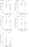
  , RPMI 1788) and in control mice. (a) Sixteen of 24 plasma samples from mice bearing human tumors, but none of the 11 samples from control mice, contained human‐specific genomic DNA (P < 0.001). The concentration of human‐specific DNA in plasma varied according to the implanted tumor cell type. (b) Samples from mice bearing human tumors (n = 24) contained significantly greater concentrations of cell‐free mouse DNA than samples from control mice (n = 11) (P < 0.01). When the concentration of mouse‐specific DNA of each tumor type were compared with the control group, the difference depended on the tumor type implanted.

Similar articles
-
Circulating nucleic acids in plasma and serum as a noninvasive investigation for cancer: time for large-scale clinical studies?Int J Cancer. 2003 Jan 10;103(2):149-52. doi: 10.1002/ijc.10791. Int J Cancer. 2003. PMID: 12455027 Review. No abstract available.
-
Analysis of circulating DNA and protein biomarkers to predict the clinical activity of regorafenib and assess prognosis in patients with metastatic colorectal cancer: a retrospective, exploratory analysis of the CORRECT trial.Lancet Oncol. 2015 Aug;16(8):937-48. doi: 10.1016/S1470-2045(15)00138-2. Epub 2015 Jul 13. Lancet Oncol. 2015. PMID: 26184520 Free PMC article.
-
Detection of free-circulating tumor-associated DNA in plasma of colorectal cancer patients and its association with prognosis.Int J Cancer. 2002 Aug 10;100(5):542-8. doi: 10.1002/ijc.10526. Int J Cancer. 2002. PMID: 12124803
-
Circulating nucleic acids (CNAs) and cancer--a survey.Biochim Biophys Acta. 2007 Jan;1775(1):181-232. doi: 10.1016/j.bbcan.2006.10.001. Epub 2006 Oct 7. Biochim Biophys Acta. 2007. PMID: 17137717 Review.
-
Plasma cell-free DNA in ovarian cancer: an independent prognostic biomarker.Cancer. 2010 Apr 15;116(8):1918-25. doi: 10.1002/cncr.24997. Cancer. 2010. PMID: 20166213 Free PMC article.
Cited by
-
The Role of Circulating Tumor DNA in Ovarian Cancer.Cancers (Basel). 2024 Sep 10;16(18):3117. doi: 10.3390/cancers16183117. Cancers (Basel). 2024. PMID: 39335089 Free PMC article. Review.
-
Clinical value of sequential circulating tumor DNA analysis using next-generation sequencing and epigenetic modifications for guiding thermal ablation for colorectal cancer metastases: a prospective study.Radiol Med. 2024 Oct;129(10):1530-1542. doi: 10.1007/s11547-024-01865-0. Epub 2024 Aug 25. Radiol Med. 2024. PMID: 39183242
-
Clinical potential of circulating free DNA and circulating tumour cells in patients with metastatic non-small-cell lung cancer treated with pembrolizumab.Mol Oncol. 2021 Nov;15(11):2923-2940. doi: 10.1002/1878-0261.13094. Epub 2021 Sep 23. Mol Oncol. 2021. PMID: 34465006 Free PMC article.
-
Radiation Induced Upregulation of DNA Sensing Pathways is Cell-Type Dependent and Can Mediate the Off-Target Effects.Cancers (Basel). 2020 Nov 13;12(11):3365. doi: 10.3390/cancers12113365. Cancers (Basel). 2020. PMID: 33202881 Free PMC article.
-
Circulating tumor nucleic acids: biology, release mechanisms, and clinical relevance.Mol Cancer. 2023 Jan 21;22(1):15. doi: 10.1186/s12943-022-01710-w. Mol Cancer. 2023. PMID: 36681803 Free PMC article. Review.
References
-
- Anker P, Mulcahy H, Chen XQ, Stroun M. Detection of circulating tumour DNA in the blood (plasma/serum) of cancer patients. Cancer Metastasis Rev 1999; 18: 65–73. - PubMed
-
- Sozzi G, Conte D, Leon M et al . Quantification of free circulating DNA as a diagnostic marker in lung cancer. J Clin Oncol 2003; 21: 3902–8. - PubMed
-
- Leon SA, Shapiro B, Sklaroff DM, Yaros MJ. Free DNA in the serum of cancer patients and the effect of therapy. Cancer Res 1977; 37: 646–50. - PubMed
-
- Fleischhacker M, Schmidt B. Circulating nucleic acids (CNAs) and cancer – a survey. Biochim Biophys Acta 2007; 1775: 181–232. - PubMed
-
- Fournie GJ, Courtin JP, Laval F et al . Plasma DNA as a marker of cancerous cell death. Investigations in patients suffering from lung cancer and in nude mice bearing human tumours. Cancer Lett 1995; 91: 221–7. - PubMed
Publication types
MeSH terms
Substances
LinkOut - more resources
Full Text Sources
Other Literature Sources

