Review
. 2009 Jan;10(1):63-73.
doi: 10.1038/nrm2597.

Mechanotransduction gone awry

Affiliations
Review

Mechanotransduction gone awry

Diana E Jaalouk et al. Nat Rev Mol Cell Biol. 2009 Jan.

Abstract

Cells sense their physical surroundings through mechanotransduction - that is, by translating mechanical forces and deformations into biochemical signals such as changes in intracellular calcium concentration or by activating diverse signalling pathways. In turn, these signals can adjust cellular and extracellular structure. This mechanosensitive feedback modulates cellular functions as diverse as migration, proliferation, differentiation and apoptosis, and is crucial for organ development and homeostasis. Consequently, defects in mechanotransduction - often caused by mutations or misregulation of proteins that disturb cellular or extracellular mechanics - are implicated in the development of various diseases, ranging from muscular dystrophies and cardiomyopathies to cancer progression and metastasis.


Figures
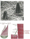
Figure 1
Figure 1. Mechanotransduction in hair cells
(a). Scanning electron micrograph of two hair bundles in the sensory macula of the bull frog saccule, showing the arrangement of stereocilia with increasing heights. These bundles are ~8μm tall and contain 50–60 stereocilia. (b) Schematic drawing of a hair bundle in resting (gray) and deflected (color) configuration. Deflection, i.e., shearing of the stereocilia relative to each other, causes the ~150–200nm long tip links to pull directly on K+ channels in the stereocilia, causing the channels to open. Myosin motors that link the channels to the actin core of the sterocilia can adjust the position to restore resting tension in the tip link, allowing adaptation to persistent stimulation. Mutations in the K+ channel, the linker proteins, or the unconventional myosins (UCM), which keep the tip links under tension, can result in deafness. Figure is modified from Ref (82). Part a of the Figure is reproduced and part b of the Figure is modified with permission from Ref. (83)
Figure 2
Figure 2. Force transmission between the extracellular matrix (ECM) and the nucleus
Extracellular forces are transmitted through the ECM, consisting of tissue-specific proteins such as collagen, laminin, and fibronectin. Adhesion complexes at the cell surface physically link the ECM to the cytoskeleton. For example, focal adhesions, comprised of integrins, talin, vinculin, and other proteins, connect the ECM to actin filaments. In skeletal muscle, the dystrophin-associated protein complex links the ECM to actin filaments. The configuration and binding affinity of these complexes can be modulated through intra- and extracellular signalling. Intracellular forces are then transmitted through the cytoskeletal network (i.e., actin filaments, microtubules, and intermediate filaments). The cytoskeleton is coupled to the nucleus through nesprins and possibly other proteins on the outer nuclear membrane. The giant isoforms of nesprins-1 and -2 bind to actin filaments, whereas nesprin-3 can associate with intermediate filaments through plectin. Nesprins interact across the luminal space with inner nuclear membrane proteins (e.g., SUN1 and SUN2), that are retained there by interaction with other nuclear envelope proteins such as lamins and emerin. Nuclear lamins and SUN proteins also bind to nuclear pore complexes, which could contribute to nuclear cytoskeletal coupling. Finally, lamins form stable nuclear structures and can bind DNA, thus completing the force transmission between the ECM and the nuclear interior. Mutations in any of these components, as well as changes in cellular structure and organization, or changes in the cellular environment, could disturb mechanotransduction signalling and result in altered cellular function; however, this has only been conclusively demonstrated for a subset of these molecules, motivating further research.
Figure 3
Figure 3. Unifying characteristics of mechanotransduction disorders
Altered cellular mechanotransduction signalling can be caused by changes in the extracellular environment (e.g., variation in the mechanical forces or deformations experienced by the tissue, or changes in extracellular matrix composition that affect its stiffness and biochemical properties), the cellular structure and organization, or elements of the mechanotransduction process itself. Changes in cellular structure and organization often result from inherited or de novo mutations in proteins that are part of the force generating machinery, the cytoskeletal network, or the nuclear envelope and interior. This category also includes transmembrane proteins involved in cell-cell or cell-ECM adhesion. Abnormal function of these proteins can alter the intracellular force distribution and thus mechanotransduction signalling. In contrast, defects in the cellular mechanosensors can disturb mechanotransduction signalling even in the case of normal force distribution. Note that many proteins can fall into more than one category, as structural proteins could also have mechanosensing capabilities, and mechanotransduction signalling can in turn cause changes in cellular structure and organization and the extracellular environment. Importantly, mechanical activation often initiates multiple signalling pathways at once that can significantly overlap and cross-talk, making it more difficult to study specific pathways. Furthermore, several of the signalling pathways are often shared with “classical”, receptor-mediated pathways. For example, the mitogen-activated protein kinase (MAPK) pathway can be turned on by mechanical strain as well as by receptor-linked tyrosine kinases (e.g., epidermal growth factor receptor). Ultimately, excessive and prolonged disturbances in the normal mechanotransduction signalling can result in many disease conditions.
Figure 4
Figure 4. Cardiac mechanotransduction signalling
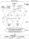
Cardiac myocytes respond to altered haemodynamics by activating multiple intracellular signalling pathways that are implicated in the maintenance and regulation of cardiac myocyte fucntion. Mechanical loading can be sensed by cardiomyocytes through a diverse group of membrane anchored mechanosensors including stretch activated ion channels, cell membrane - spanning G-protein coupled receptors, growth factor receptors, and integrins. This mechanical sensation is then converted to biochemical signals by triggering the multi-step activation of downstream partners in an array of signalling cascades in the cytoplasm. The highlights of such cascades include the three modules of the MAPK family underscored by the activation of Ras, the JAK-STAT pathway, Rac activation, calcium and NO signalling. The convergence of these pathways results in the activation of select transcription factors including NF-AT and NF-kB which then translocate to the nucleus and modulate the expression of a panel of mechanosenstive genes including egr-1 and iex-1. Ultimately, the net sum of gene expression reprogramming in cardiomyocytes dictates the functional response of the cell to mechanical stress.
Figure 5
Figure 5. Mechanotransduction in cancer cells
Schematic representation of how increased extracellular matrix (ECM) stiffness and altered cytoskeletal tension can contribute to tumour formation. Increased ECM stiffness may arise from fibrosis, or in response to increased cytoskeletal tension, caused for examples by oncogene (Ras)-driven ERK activation. The increased ECM stiffness is sensed by focal adhesions and activates integrins and focal adhesion kinase, thereby promoting focal adhesion assembly and stimulating the Rho-ROCK pathway. ROCK activation increases cytoskeletal tension by increasing myosin light chain (MLP) phosphorylation, which could result in further increases in ECM stiffness due to cellular mechanotransduction signalling, completing a self-enforcing (i.e., positive) feedback loop. Cross-talk between the Rho-ROCK pathway and the epidermal growth-factor receptor (EGFR)-Ras-ERK pathway, as well as modulation of Growth factor-dependent ERK activation by integrins, results in increased proliferation. ERK activation can also increase cytoskeletal tension through ROCK, further complementing the cross-talk between cytoskeletal tension and proliferative pathways. In breast cancer cells, the combined action of increased contractility and proliferation, triggered by increased extracellular matrix stiffness, may drive the undifferentiated and proliferative phenotype of mammary epithelial cancer cells and result in tumour formation. Decreasing Rho-mediated cytoskeletal contractility or ERK activity is sufficient to revert EGFR-transformed cells that form disorganized and invasive colonies into phenotypically normal cells that form polarized and growth-arrested acini in 3D culture . Figure modified with permission from Ref. (58).

Similar articles

Cited by

References

    1. Engler AJ, Sen S, Sweeney HL, Discher DE. Matrix elasticity directs stem cell lineage specification. Cell. 2006;126:677–89. - PubMed
    1. Paszek MJ, et al. Tensional homeostasis and the malignant phenotype. Cancer Cell. 2005;8:241–54. This paper presents an elegant study linking matrix stiffness and cytoskeletal tension to cancer progression. As a component of a “mechano-regulatory circuit” that includes integrins, Rho, and ERK, disruption of tensional homeostasis can promote malignant phenotype in a model of breast cancer. - PubMed
    1. Ingber DE. Cellular mechanotransduction: putting all the pieces together again. FASEB J. 2006;20:811–27. - PubMed
    1. Orr AW, Helmke BP, Blackman BR, Schwartz MA. Mechanisms of mechanotransduction. Dev Cell. 2006;10:11–20. - PubMed
    1. Vogel V, Sheetz M. Local force and geometry sensing regulate cell functions. Nat Rev Mol Cell Biol. 2006;7:265–75. This is a comprehensive review of the literature on how physical cues in the microenvironment of a eukaryotic cell are sensed and converted to biochemical signals that define cell shape and regulate cell growth, differentiation, and survival. - PubMed

Publication types