Breast cancer cell lines contain functional cancer stem cells with metastatic capacity and a distinct molecular signature
- PMID: 19190339
- PMCID: PMC2819227
- DOI: 10.1158/0008-5472.CAN-08-2741
Breast cancer cell lines contain functional cancer stem cells with metastatic capacity and a distinct molecular signature
Abstract
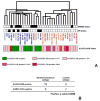
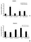
Tumors may be initiated and maintained by a cellular subcomponent that displays stem cell properties. We have used the expression of aldehyde dehydrogenase as assessed by the ALDEFLUOR assay to isolate and characterize cancer stem cell (CSC) populations in 33 cell lines derived from normal and malignant mammary tissue. Twenty-three of the 33 cell lines contained an ALDEFLUOR-positive population that displayed stem cell properties in vitro and in NOD/SCID xenografts. Gene expression profiling identified a 413-gene CSC profile that included genes known to play a role in stem cell function, as well as genes such as CXCR1/IL-8RA not previously known to play such a role. Recombinant interleukin-8 (IL-8) increased mammosphere formation and the ALDEFLUOR-positive population in breast cancer cell lines. Finally, we show that ALDEFLUOR-positive cells are responsible for mediating metastasis. These studies confirm the hierarchical organization of immortalized cell lines, establish techniques that can facilitate the characterization of regulatory pathways of CSCs, and identify potential stem cell markers and therapeutic targets.
Figures


Similar articles
-
Aldehyde dehydrogenase activity of breast cancer stem cells is primarily due to isoform ALDH1A3 and its expression is predictive of metastasis.Stem Cells. 2011 Jan;29(1):32-45. doi: 10.1002/stem.563. Stem Cells. 2011. PMID: 21280157
-
Identification of Breast Cancer Stem Cells Using a Newly Developed Long-acting Fluorescence Probe, C5S-A, Targeting ALDH1A1.Anticancer Res. 2022 Mar;42(3):1199-1205. doi: 10.21873/anticanres.15586. Anticancer Res. 2022. PMID: 35220209
-
Aldehyde dehydrogenase 1-positive cancer stem cells mediate metastasis and poor clinical outcome in inflammatory breast cancer.Clin Cancer Res. 2010 Jan 1;16(1):45-55. doi: 10.1158/1078-0432.CCR-09-1630. Epub 2009 Dec 22. Clin Cancer Res. 2010. PMID: 20028757 Free PMC article.
-
Breast cancer stem cells and intrinsic subtypes: controversies rage on.Curr Stem Cell Res Ther. 2009 Jan;4(1):50-60. doi: 10.2174/157488809787169110. Curr Stem Cell Res Ther. 2009. PMID: 19149630 Review.
-
Stem cells in the human breast.Cold Spring Harb Perspect Biol. 2010 May;2(5):a003160. doi: 10.1101/cshperspect.a003160. Cold Spring Harb Perspect Biol. 2010. PMID: 20452965 Free PMC article. Review.
Cited by
-
In vivo molecular imaging of cancer stem cells.Am J Nucl Med Mol Imaging. 2014 Dec 15;5(1):14-26. eCollection 2015. Am J Nucl Med Mol Imaging. 2014. PMID: 25625023 Free PMC article. Review.
-
Breast and abdominal adipose multipotent mesenchymal stromal cells and stage-specific embryonic antigen 4 expression.Cells Tissues Organs. 2012;196(2):107-16. doi: 10.1159/000331332. Epub 2012 Jan 10. Cells Tissues Organs. 2012. PMID: 22237379 Free PMC article.
-
Characterisation of mesothelioma-initiating cells and their susceptibility to anti-cancer agents.PLoS One. 2015 May 1;10(5):e0119549. doi: 10.1371/journal.pone.0119549. eCollection 2015. PLoS One. 2015. PMID: 25932953 Free PMC article.
-
Elimination of epithelial-like and mesenchymal-like breast cancer stem cells to inhibit metastasis following nanoparticle-mediated photothermal therapy.Biomaterials. 2016 Oct;104:145-57. doi: 10.1016/j.biomaterials.2016.06.045. Epub 2016 Jun 23. Biomaterials. 2016. PMID: 27450902 Free PMC article.
-
CDK4 regulates cancer stemness and is a novel therapeutic target for triple-negative breast cancer.Sci Rep. 2016 Oct 19;6:35383. doi: 10.1038/srep35383. Sci Rep. 2016. PMID: 27759034 Free PMC article.
References
-
- Hanahan D, Weinberg RA. The hallmarks of cancer. Cell. 2000;100:57–70. - PubMed
-
- Bonnet D, Dick JE. Human acute myeloid leukemia is organized as a hierarchy that originates from a primitive hematopoietic cell. Nat Med. 1997;3:730–7. - PubMed
-
- Glinsky GV. Stem cell origin of death-from-cancer phenotypes of human prostate and breast cancers. Stem Cell Rev. 2007;3:79–93. - PubMed
Publication types
MeSH terms
Substances
Grants and funding
- R01 CA101860-05/CA/NCI NIH HHS/United States
- P30 CA046592-21/CA/NCI NIH HHS/United States
- P30 CA046592-20S3/CA/NCI NIH HHS/United States
- R01 CA101860-01A1/CA/NCI NIH HHS/United States
- P30 CA046592-13/CA/NCI NIH HHS/United States
- P30 CA046592/CA/NCI NIH HHS/United States
- P30 CA046592-16/CA/NCI NIH HHS/United States
- R01 CA101860-04/CA/NCI NIH HHS/United States
- P30 CA046592-20/CA/NCI NIH HHS/United States
- P30 CA046592-12/CA/NCI NIH HHS/United States
- CA66233/CA/NCI NIH HHS/United States
- P30 CA046592-19S1/CA/NCI NIH HHS/United States
- R01 CA129765-02/CA/NCI NIH HHS/United States
- P30 CA046592-17/CA/NCI NIH HHS/United States
- R01 CA101860-03/CA/NCI NIH HHS/United States
- P30 CA046592-19/CA/NCI NIH HHS/United States
- P30 CA046592-21S1/CA/NCI NIH HHS/United States
- P30 CA046592-18/CA/NCI NIH HHS/United States
- P30 CA046592-20S2/CA/NCI NIH HHS/United States
- R01 CA129765-01A1/CA/NCI NIH HHS/United States
- 5 P 30 CA46592/CA/NCI NIH HHS/United States
- R01 CA101860-02/CA/NCI NIH HHS/United States
- P30 CA046592-15/CA/NCI NIH HHS/United States
- R01 CA129765/CA/NCI NIH HHS/United States
- R01 CA101860/CA/NCI NIH HHS/United States
- P30 CA046592-20S1/CA/NCI NIH HHS/United States
- CA101860/CA/NCI NIH HHS/United States
- P30 CA046592-14/CA/NCI NIH HHS/United States
LinkOut - more resources
Full Text Sources
Other Literature Sources
Medical

