. 2009 Mar;10(3):285-92.
doi: 10.1038/embor.2008.246. Epub 2009 Jan 30.

DAP-kinase-mediated phosphorylation on the BH3 domain of beclin 1 promotes dissociation of beclin 1 from Bcl-XL and induction of autophagy

Affiliations

DAP-kinase-mediated phosphorylation on the BH3 domain of beclin 1 promotes dissociation of beclin 1 from Bcl-XL and induction of autophagy

Einat Zalckvar et al. EMBO Rep. 2009 Mar.

Abstract

Autophagy, an evolutionarily conserved process, has functions both in cytoprotective and programmed cell death mechanisms. Beclin 1, an essential autophagic protein, was recently identified as a BH3-domain-only protein that binds to Bcl-2 anti-apoptotic family members. The dissociation of beclin 1 from its Bcl-2 inhibitors is essential for its autophagic activity, and therefore should be tightly controlled. Here, we show that death-associated protein kinase (DAPK) regulates this process. The activated form of DAPK triggers autophagy in a beclin-1-dependent manner. DAPK phosphorylates beclin 1 on Thr 119 located at a crucial position within its BH3 domain, and thus promotes the dissociation of beclin 1 from Bcl-XL and the induction of autophagy. These results reveal a substrate for DAPK that acts as one of the core proteins of the autophagic machinery, and they provide a new phosphorylation-based mechanism that reduces the interaction of beclin 1 with its inhibitors to activate the autophagic machinery.


Conflict of interest statement

The authors declare that they have no conflict of interest.

Figures
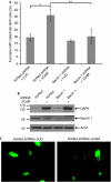
Figure 1
Figure 1
Functional interaction between DAPK and beclin 1. (A) HEK293 cells were transfected with DAPK ΔCaM or with a control vector (pcDNA3-luciferase, LUC), together with shRNAs targeting beclin 1 or HcRed, and with GFP–LC3 plasmid. After 72 h, cells were counted and lysates were prepared. (A) The percentage of cells with punctate GFP–LC3 fluorescence per total GFP–LC3-positive cells was quantified. Data presented are the mean±s.d. from a triplicate of 100 transfected cells. The asterisks denote a significance level of P=0.001. (B) Western blot analysis was performed using the indicated antibodies. (C) Representative GFP–LC3 staining of cells transfected with the control shRNA (HcRed) together with pcDNA3-luciferase or DAPKΔCaM. CaM, calmodulin; DAPK, death-associated protein kinase; GFP, green fluorescent protein; HEK, human embryonic kidney; shRNA, short hairpin RNA.
Figure 2
Figure 2
Beclin 1 is a new substrate of DAPK. (A) Flag-tagged DAPK (100 ng) was incubated with GST (900 ng) or GST–beclin-1 (750 ng) in the presence of Ca2+, calmodulin and [γ-33P]ATP for 30 min or 60 min. Phosphorylated proteins were visualized by X-ray film exposure, and GST/GST–beclin-1 levels were visualized by Ponceau S staining. The autophosphorylation of DAPK indicates that its catalytic activity was intact in all samples. (B) Flag-tagged DAPK (60 ng) was incubated with Flag-tagged beclin 1 (250 ng), which was purified from HEK293T cells, and a kinase assay was performed for 60 min. Where indicated (+LiCl), beclin-1-bound beads were first washed stringently in 0.5 M LiCl and 0.5 M KCl. Phosphorylated proteins were visualized by X-ray film exposure, and the levels of beclin 1 were visualized by Western blot analysis using beclin 1 antibodies. DAPK, death-associated protein kinase; GST, glutathione S-transferase; HEK, human embryonic kidney.
Figure 3
Figure 3
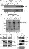
Physical interaction between DAPK and beclin 1. (Aa) Protein extracts from COS7 cells, HEK293T cells, or no extracts were added to bacterially produced GST or GST–beclin-1. The pulled-down proteins, as well as the total cell extracts, were blotted with the indicated antibodies. (Ab) Ponceau S staining of GST and GST–beclin-1 to which the extracts were added. (B) HEK293 cells were co-transfected with Flag-tagged beclin 1 or with Flag-tagged beclin 1 lacking the Bcl-2-binding domain (ΔBD) together with Bcl-XL and HA-tagged DAPK. Beclin 1 was immunoprecipitated using Flag antibodies, and the co-immunoprecipitated proteins, as well as the total cell extracts, were blotted with DAPK, Bcl-XL and beclin 1 antibodies. (C) HEK293 cells were transfected with Flag–beclin-1 or Flag–GFP, and the Flag-tagged proteins were immunoprecipitated using Flag antibodies, and eluted with an excess of Flag peptides. The blot was reacted with DAPK antibodies or with Flag antibodies at different time exposures. DAPK, death-associated protein kinase; GFP, green fluorescent protein; GST, glutathione S-transferase; HA, haemagglutinin; HEK, human embryonic kidney.
Figure 4
Figure 4
DAPK phosphorylates beclin 1 on Thr 119 located within its BH3 domain. (A) A bacterially purified catalytic domain of DAPK was incubated for 15 min at 30°C with increasing concentrations (5–10 nmol) of a peptide corresponding to the BH3 domain of beclin 1 (aa 108–127) and with the same peptide in which Thr 119 was substituted by alanine. An in vitro kinase assay was performed, and the reactions were applied to Whatman filters. Total levels of TCA-insoluble counts were measured and plotted against substrate concentration. Data are the mean±s.d. of three experiments. (B) A model of the interaction between the BH3 domain of beclin 1, and the hydrophobic pocket of Bcl-XL. The cyan arrow points to the groove in which phosphorylated Ser 113 can be accommodated. The black arrow points to the site that might be disrupted by phosphorylation of Thr 119. The magenta arrow points to the positively charged groove in which phosphorylated Ser 127 can be accommodated. (C) Flag-tagged DAPK (60 ng) was incubated with GST–WT beclin 1 or with GST–T119A beclin 1 (1000 ng) in the presence of Ca2+, calmodulin and ATP for 30 min. GST-beclin 1 levels were visualized by Ponceau S staining, and phosphorylation on Thr 119 was detected by a phosphoThr 119 antibody (Western blot). (D) HEK293 cells were co-transfected with Flag-tagged beclin 1 with or without ΔCaM DAPK. After 24 h, beclin 1 was immunoprecipitated from cells, using Flag antibodies, and the immunoprecipitates were reacted with phosphoThr 119 antibodies. The Ponceau staining shows equal amounts of immunoprecipitated beclin 1. The cell extract blots were reacted with haemagglutinin antibodies or with beclin 1 antibodies. CaM, calmodulin; DAPK, death-associated protein kinase; GST, glutathione S-transferase; HEK, human embryonic kidney; TCA, trichloro-acetic acid.
Figure 5
Figure 5
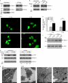
DAPK promotes the dissociation of beclin 1 from Bcl-XL. (A) HEK293 cells were transfected with Flag-tagged beclin 1 and Bcl-XL with or without haemagglutinin-tagged DAPK. Beclin 1 was immunoprecipitated using Flag antibodies, and the co-immunoprecipitated proteins, as well as the total cell extracts, were blotted using the indicated antibodies. (B) HEK293 cells were co-transfected with Flag-tagged T119A or T119E beclin 1 mutants and Bcl-XL. Beclin 1 was immunoprecipitated using Flag antibodies, and the co-immunoprecipitated proteins, as well as the total cell extracts, were blotted using the indicated antibodies. (C) HEK293 cells were transfected with 5 or 10 μg T119A or T119E beclin 1 mutants together with GFP–LC3 plasmid. After 24 h, cells were counted and lysates were prepared. (a) Representative GFP–LC3 staining. (b) The percentage of cells with punctate GFP–LC3 fluorescence per total GFP–LC3-positive cells was quantified. Data presented are the mean±s.d. from a triplicate experiment with 100 transfected cells. The asterisks denote significance level: *P=0.01; **P=0.005. (c) Western blot analysis was performed using the indicated antibodies. (D) HEK293 cells were transfected with Bcl-XL, Flag-tagged beclin 1 (WT) or Flag-tagged T119A beclin 1 mutant with or without haemagglutinin-tagged activated DAPK (ΔCaM). Beclin 1 was immunoprecipitated using Flag antibodies, and the co-immunoprecipitated proteins, as well as the total cell extracts, were blotted using the indicated antibodies. Levels of Bcl-XL were quantified using NIH image software, and the ratio between immunoprecipitated and expressed Bcl-XL was calculated. (E) Transmission electron micrographs of HEK293 cells 24 h after they were transfected with Bcl-XL, Flag-tagged beclin 1 and ΔCaM or pcDNA3-luciferase as a control. The images in (c) and (d) were taken at higher magnifications of the ΔCaM treatment (see the scale bars). ‘AV' indicates autophagic vacuoles. CaM, calmodulin; DAPK, death-associated protein kinase; GFP, green fluorescent protein; GST, glutathione S-transferase; HEK, human embryonic kidney; NIH, National Institutes of Health.

Similar articles

Cited by

References

    1. Berman HM, Westbrook J, Feng Z, Gilliland G, Bhat TN, Weissig H, Shindyalov IN, Bourne PE (2000) The Protein Data Bank. Nucleic Acids Res 28: 235–242 - PMC - PubMed
    1. Berry DL, Baehrecke EH (2007) Growth arrest and autophagy are required for salivary gland cell degradation in Drosophila. Cell 131: 1137–1148 - PMC - PubMed
    1. Bialik S, Kimchi A (2006) The death-associated protein kinases: structure, function, and beyond. Annu Rev Biochem 75: 189–210 - PubMed
    1. Cao Y, Klionsky DJ (2007) Physiological functions of Atg6/Beclin 1: a unique autophagy-related protein. Cell Res 17: 839–849 - PubMed
    1. Cohen O, Feinstein E, Kimchi A (1997) DAP-kinase is a Ca2+/calmodulin-dependent, cytoskeletal-associated protein kinase, with cell death-inducing functions that depend on its catalytic activity. EMBO J 16: 998–1008 - PMC - PubMed

Publication types

MeSH terms