. 2009 Jan 23;136(2):284-95.
doi: 10.1016/j.cell.2008.11.037.

Age-dependent deterioration of nuclear pore complexes causes a loss of nuclear integrity in postmitotic cells

Affiliations

Age-dependent deterioration of nuclear pore complexes causes a loss of nuclear integrity in postmitotic cells

Maximiliano A D'Angelo et al. Cell. .

Abstract

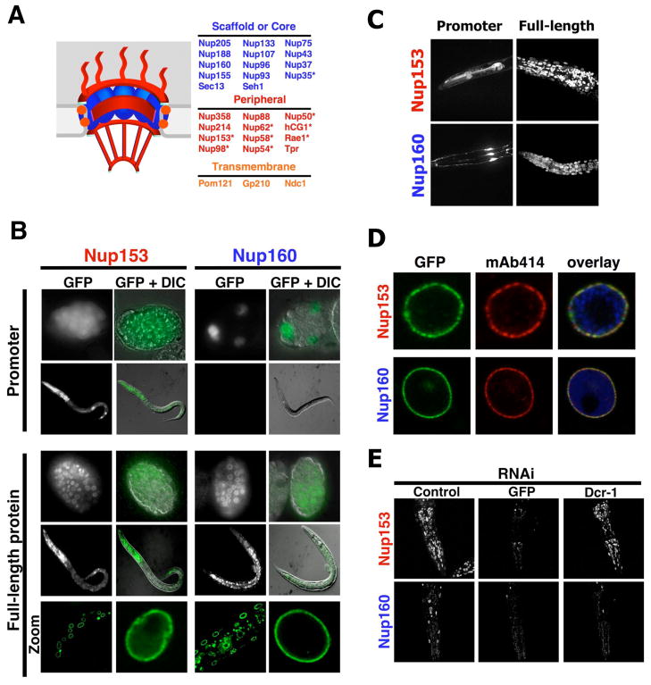
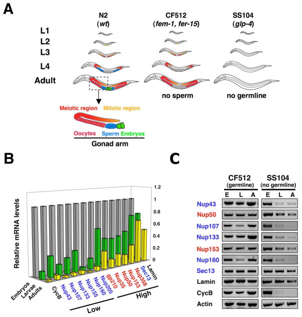
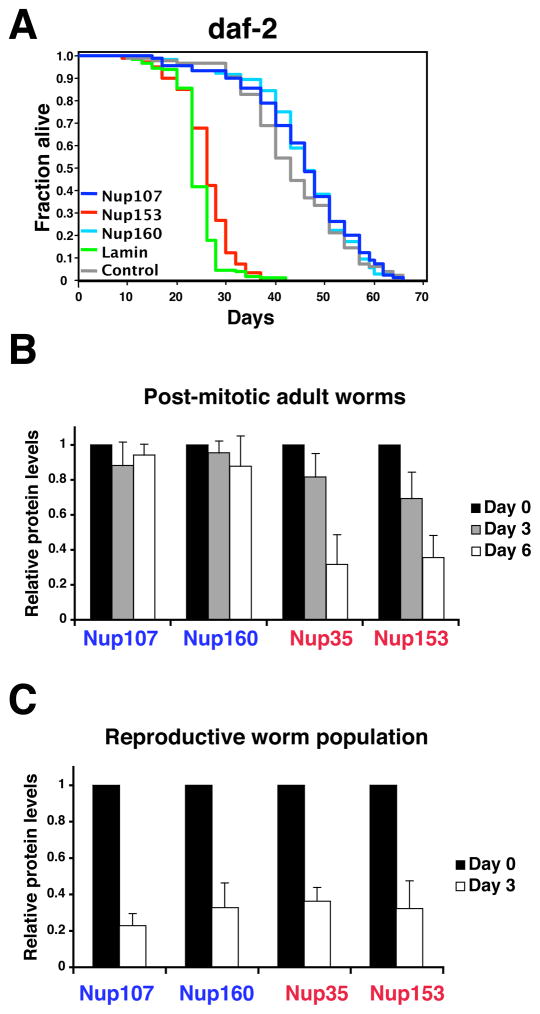
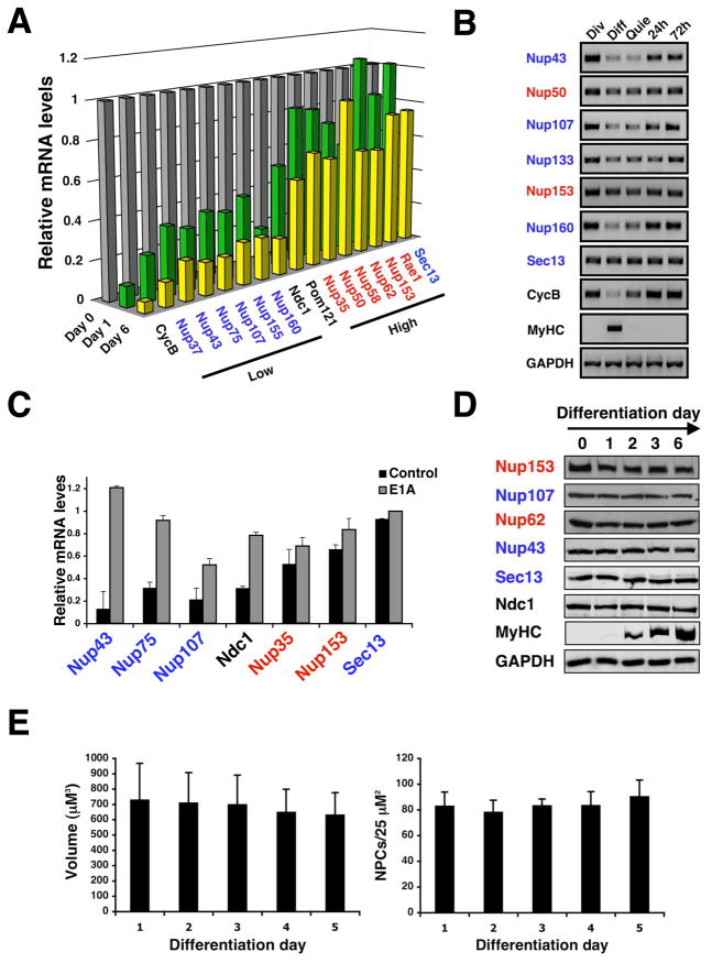
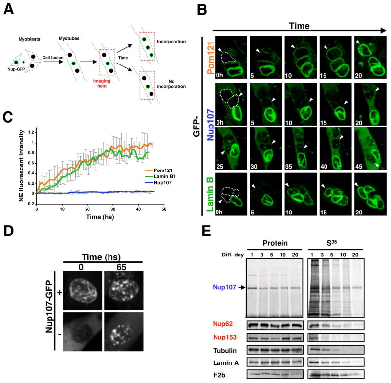
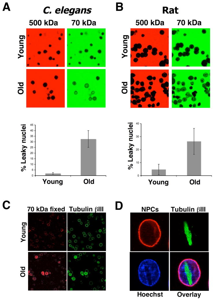
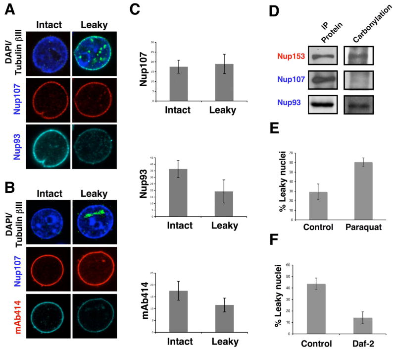
In dividing cells, nuclear pore complexes (NPCs) disassemble during mitosis and reassemble into the newly forming nuclei. However, the fate of nuclear pores in postmitotic cells is unknown. Here, we show that NPCs, unlike other nuclear structures, do not turn over in differentiated cells. While a subset of NPC components, like Nup153 and Nup50, are continuously exchanged, scaffold nucleoporins, like the Nup107/160 complex, are extremely long-lived and remain incorporated in the nuclear membrane during the entire cellular life span. Besides the lack of nucleoporin expression and NPC turnover, we discovered an age-related deterioration of NPCs, leading to an increase in nuclear permeability and the leaking of cytoplasmic proteins into the nucleus. Our finding that nuclear "leakiness" is dramatically accelerated during aging and that a subset of nucleoporins is oxidatively damaged in old cells suggests that the accumulation of damage at the NPC might be a crucial aging event.


Figures

Figure 1
Figure 1. ceNup160 scaffold nucleoporin shows life-long stability
(A). Scheme of the nuclear pore complex structure and composition. Asterisks denote dynamic nucleoporins. (B) C. elegans N2 wild type strain was injected with a vector expressing GFP under the control of either the ceNup153 promoter or the ceNup160 promoter (Promoter) or with vectors expressing ceNup153-GFP or ceNup160-GFP under their endogenous promoters (full-length protein). Expression of the reporter protein was analyzed by fluorescence microscopy and GFP signal was merged with differential interference contrast images (DIC). Correct localization of ceNup153-GFP and ceNup160-GFP fusion proteins to the NE was analyzed by confocal microscopy (Zoom). (C) The activity of ceNup153 and ceNup160 promoters and the localization of full-length proteins in the head of adult worms were analyzed by confocal microscopy. Image shows the maximal projection of 30 z-stacks. (D) Nuclei were purified from ceNup160-GFP and ceNup153-GFP transgenic worms and NPC insertion of the GFP-tagged nucleoporins (green) was confirmed by colocalization with the NPC antibody mAb414 (red). Chromatin is shown in blue. (E) ceNup153-GFP and ceNup160-GFP expressing worms were subjected to GFP RNAi until no fluorescent signal was detected. RNAi against C. elegans dicer (DCR-1) was used to release adult worms from the GFP RNAi. Adults were fed DCR-1 RNAi for 6 days before the GFP signal was analyzed. Dashed lines outline worms heads.
Figure 2
Figure 2. Expression of scaffold nucleoporins is down-regulated during C. elegans development
(A). Scheme of C. elegans strains used in this work (based on (Crittenden et al., 2006)). N2 is wild type strain, CF512 (fem-1, fer-15) has thermo-sensitive mutations that inhibit male germline production at 25°C, SS104 (glp-4) has a mutation that inhibits male and female germline production at 25°C, being adults worms at the restrictive temperature entirely post-mitotic. (B) Total RNA was extracted from the embryo, larva and adult developmental stages of C. elegans (SS104 strain). Expression levels were analyzed by RT-PCR (n=4) and normalized to embryonic levels. Cyclin B (CycB) was used as a marker for cell cycle exit. Standard deviation is below 20%. (C) Nucleoporin expression levels in embryos (E), larvae (L) and adults (A) worms of C. elegans sterile strains CF5120 and SS104 were analyzed by RT-PCR (n=4).
Figure 3
Figure 3. Expression of scaffold nucleoporins is not required during adulthood
(A). daf-2(e1370) mutant worms on day 1 of adulthood were fed bacteria expressing empty vector control (gray line), ceNup153 RNAi (red line), ceNup160 RNAi (light blue line) ceNup107 RNAi (blue line) or ceLamin RNAi (green line). Life span was assessed as described (Apfeld and Kenyon, 1998). Statistical analyses can be found in Table S1. (B) Total protein was extracted from sterile (25°C) adults SS104 worms subject to the indicated RNAis for 3 or 6 days and protein levels were assayed by western blot analysis. Values represent average ± SD from 3 independent experiments. (C) Total protein was extracted from a reproductive population (15°C) of SS104 worms subjecteded to the indicated RNAis for 3 days and protein levels were assayed by western blot. Values represent average ± SD from 3 independent experiments.
Figure 4
Figure 4. Down-regulation of scaffold nuceloporin expression is conserved in mammals
(A). Total RNA was extracted from dividing C2C12 myoblasts (day 0) or differentiated myotubes (day 3 and 6). Nucleoporin expression levels were analyzed by Q-PCR (n=3). Cyclin B was used as a marker for cell cycle exit. In all cases standard deviation is below 20%. (B) Total RNA was extracted from dividing (Div), differentiated (Diff) and quiescent (Quie) C2C12 cells. Nucleoporin expression levels were analyzed by RT-PCR. Cyclin B (CycB) and myosin heavy chain (MyHC) were used and controls for cell cycle exit and differentiation respectively. (C) Dividing C2C12 myoblasts were infected with retrovirus carrying a control vector or a vector expressing the E1A protein fused to the estrogen receptor. Cells were induced to differentiate for 3–4 days and E1A was activated with 4OH-tamoxifen for 24 hours. Total RNA was extracted and nucleoporin levels were analyzed by RT-PCR (n=3). (D) Total protein was extracted from dividing myoblasts (day 0) or differentiated myotubes (day 1–6). Nucleoporin protein levels during C2C12 differentiation were analyzed by western blotting. Myosin heavy chain (MyHC) was used as a differentiation control. (E) C2C12 cells expressing 3GFP-NLS were induced to differentiate. Dividing myoblasts (Day 0) and differentiated myotubes (Day 1–5) were fixed and NPCs were stained using anti-Nup153 antibody. Nuclear volume and NPC density were analyzed by confocal microscopy and quantified.
Figure 5
Figure 5. Nup107 scaffold nucleoporin does not exchange once inserted into NPCs
(A). Scheme of the imaging setup used to analyze the incorporation of GFP-nucleoporins or Lamin B1-GFP fusion proteins into the NE of differentiated myotubes. (B) Proliferating myoblasts were transfected with Pom121-GFP, GFP-Nup107 or Lamin B1-GFP expressing vectors, diluted with untransfected cells and induced to differentiate for 3 days in low serum containing media. Fields containing GFP positive and negative nuclei were selected and imaged using a spinning disk confocal microscope for at least 50 hs at 1 h intervals. Dotted lines show the position of GFP negative nuclei at time 0 and arrowheads are used to follow the GFP negative nuclei during time. (C) Incorporation of Pom121-GFP, GFP-Nup107 or Lamin B1-GFP at the NE of GFP negative nuclei was quantified using Image J. (D) Proliferating myoblasts were transfected with a GFP-Nup107 expressing vector, diluted with untransfected cells and induced to differentiate for 3 days. Fields containing nuclei that came from transfected cells (+) or untransfected cells (−) were selected and imaged using a spinning disk confocal. Images show the formation of Nup107 aggregates after 65 hs of overexpression. (E) Dividing C2C12 myoblasts were incubated with a S35-Methonine/S35-Cysteine mix for 24 hs (Pulse) and then switched to differentiating media (Chase). Total cell lysates were prepared from the indicated time points. Proteins were immunoprecipitated using specific antibodies, separated by SDS-PAGE and transfered to nitrocellulose membranes. The presence of S35-labeled proteins was analyzed using a phosphoimager and protein levels were determined by western blot.
Figure 6
Figure 6. C. elegans and mammals show an age-related increase of nuclear permeability
(A). Nuclei were isolated from brains of young (3 months) and old (28 months) rats Purified nuclei were incubated with a green 70kDa and a red 500kDa fluorescent dextrans. Nuclear permeability was analyzed by confocal microscopy. Images show each dextran in their original color. The percentage of nuclei that showed influx of the 70kDa dextran was determined using Photoshop CS3 Extended. (B). Nuclei were prepared from C. elegans SS104 worms at different times of adulthood. Nuclear permeability was analyzed and quantified as described in (A) (C) Young and old rat nuclei were incubated with the 70kDa fluorescent dextran, fixed and stained with an antibody against tubulin bIII. (F) Brain nuclei from old rats were fixed and stained with the mAb414 antibody (NPCs) and an anti- tubulin bIII.
Figure 7
Figure 7. Nuclei with increase permeability show deteriorated NPCs
(A). Nuclei were isolated from brains of old (28 months) rats, fixed and stained with antibodies against tubulin bIII and the scaffold nucleoporins Nup107 and Nup133. The staining of intact and leaky nuclei (the latter identified by the intranuclear accumulation of intranuclear tubulin bIII) was compared. (B) Intact and leaky nuclei isolated from old rat brains were stained with tubulin bIII, Nup107 and the mAb414 antibody. Intact and leaky nuclei were compared as described in (A). (C) The fluorescent intensity of the nuclei stained in (A) and (B) was quantified using Image J. (D) Total protein extracts were prepared from brains of old rats and nucleoporins were immunoprecipitated (IP) using specific antibodies. The IP proteins were treated with 2,4-dinitrophenylhydrazine to derivatize carbonyl groups to 2.4-dinitrophenylhydrazone (DNP-hydrazone) The DNP-derivatized proteins were detected by western blot using an anti-DNP antibody. (E) C. elegans SS104 worms on day 1 of adulthood were transfer to plates containing bacteria and buffer (Control) or Paraquat (0.5mM). On day 6 nuclei were isolated from worms and nuclear permeability was analyzed using the 70 and 500 kDa dextrans assay and quantified using Photoshop CS3 Extended. (F) SS104 adult day 1 worms were grown in bacteria expressing empty vector (Control) or daf-2 RNAi (Daf-2) for 8 days. Nuclei were isolated from worms and nuclear permeability was analyzed as in (D).

Comment in

  • Old nuclei spring new leaks.
    Kotwaliwale CV, Dernburg AF. Kotwaliwale CV, et al. Cell. 2009 Jan 23;136(2):211-2. doi: 10.1016/j.cell.2009.01.004. Cell. 2009. PMID: 19167324

Similar articles

Cited by

References

    1. Aguilaniu H, Gustafsson L, Rigoulet M, Nystrom T. Asymmetric inheritance of oxidatively damaged proteins during cytokinesis. Science. 2003;299:1751–1753. - PubMed
    1. Alber F, Dokudovskaya S, Veenhoff LM, Zhang W, Kipper J, Devos D, Suprapto A, Karni-Schmidt O, Williams R, Chait BT, et al. The molecular architecture of the nuclear pore complex. Nature. 2007;450:695–701. - PubMed
    1. Antonin W, Franz C, Haselmann U, Antony C, Mattaj IW. The Integral Membrane Nucleoporin pom121 Functionally Links Nuclear Pore Complex Assembly and Nuclear Envelope Formation. Mol Cell. 2005;17:83–92. - PubMed
    1. Apfeld J, Kenyon C. Cell nonautonomy of C. elegans daf-2 function in the regulation of diapause and life span. Cell. 1998;95:199–210. - PubMed
    1. Beanan MJ, Strome S. Characterization of a germ-line proliferation mutation in C. elegans. Development. 1992;116:755–766. - PubMed

Publication types

Substances

LinkOut - more resources