NOTCH3 expression is induced in mural cells through an autoregulatory loop that requires endothelial-expressed JAGGED1
- PMID: 19150886
- PMCID: PMC2747310
- DOI: 10.1161/CIRCRESAHA.108.184846
NOTCH3 expression is induced in mural cells through an autoregulatory loop that requires endothelial-expressed JAGGED1
Abstract
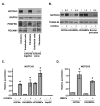
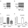
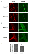
Endothelial cells and mural cells (smooth muscle cells, pericytes, or fibroblasts) are known to communicate with one another. Their interactions not only serve to support fully functional blood vessels but also can regulate vessel assembly and differentiation or maturation. In an effort to better understand the molecular components of this heterotypic interaction, we used a 3D model of angiogenesis and screened for genes, which were modulated by coculturing of these 2 different cell types. In doing so, we discovered that NOTCH3 is one gene whose expression is robustly induced in mural cells by coculturing with endothelial cells. Knockdown by small interfering RNA revealed that NOTCH3 is necessary for endothelial-dependent mural cell differentiation, whereas overexpression of NOTCH3 is sufficient to promote smooth muscle gene expression. Moreover, NOTCH3 contributes to the proangiogenic abilities of mural cells cocultured with endothelial cells. Interestingly, we found that the expression of NOTCH3 is dependent on Notch signaling, because the gamma-secretase inhibitor DAPT blocked its upregulation. Furthermore, in mural cells, a dominant-negative Mastermind-like1 construct inhibited NOTCH3 expression, and endothelial-expressed JAGGED1 was required for its induction. Additionally, we demonstrated that NOTCH3 could promote its own expression and that of JAGGED1 in mural cells. Taken together, these data provide a mechanism by which endothelial cells induce the differentiation of mural cells through activation and induction of NOTCH3. These findings also suggest that NOTCH3 has the capacity to maintain a differentiated phenotype through a positive-feedback loop that includes both autoregulation and JAGGED1 expression.
Figures


Comment in
-
Building a vessel wall with notch signaling.Circ Res. 2009 Feb 27;104(4):419-21. doi: 10.1161/CIRCRESAHA.109.194233. Circ Res. 2009. PMID: 19246684 Free PMC article. No abstract available.
Similar articles
-
Endothelial cells downregulate apolipoprotein D expression in mural cells through paracrine secretion and Notch signaling.Am J Physiol Heart Circ Physiol. 2011 Sep;301(3):H784-93. doi: 10.1152/ajpheart.00116.2011. Epub 2011 Jun 24. Am J Physiol Heart Circ Physiol. 2011. PMID: 21705670 Free PMC article.
-
Regulation of vascular smooth muscle cell phenotype in three-dimensional coculture system by Jagged1-selective Notch3 signaling.Tissue Eng Part A. 2014 Apr;20(7-8):1175-87. doi: 10.1089/ten.TEA.2013.0268. Epub 2014 Feb 10. Tissue Eng Part A. 2014. PMID: 24138322 Free PMC article.
-
Building a vessel wall with notch signaling.Circ Res. 2009 Feb 27;104(4):419-21. doi: 10.1161/CIRCRESAHA.109.194233. Circ Res. 2009. PMID: 19246684 Free PMC article. No abstract available.
-
Blockade of Jagged/Notch pathway abrogates transforming growth factor β2-induced epithelial-mesenchymal transition in human retinal pigment epithelium cells.Curr Mol Med. 2014 May;14(4):523-34. doi: 10.2174/1566524014666140331230411. Curr Mol Med. 2014. PMID: 24694299 Review.
-
Notch signaling regulates tumor angiogenesis by diverse mechanisms.Oncogene. 2008 Sep 1;27(38):5132-7. doi: 10.1038/onc.2008.227. Oncogene. 2008. PMID: 18758482 Free PMC article. Review.
Cited by
-
Temporal and embryonic lineage-dependent regulation of human vascular SMC development by NOTCH3.Stem Cells Dev. 2015 Apr 1;24(7):846-56. doi: 10.1089/scd.2014.0520. Epub 2015 Feb 25. Stem Cells Dev. 2015. PMID: 25539150 Free PMC article.
-
BRD4 Regulates Breast Cancer Dissemination through Jagged1/Notch1 Signaling.Cancer Res. 2016 Nov 15;76(22):6555-6567. doi: 10.1158/0008-5472.CAN-16-0559. Epub 2016 Sep 20. Cancer Res. 2016. PMID: 27651315 Free PMC article.
-
Pericyte recruitment during vasculogenic tube assembly stimulates endothelial basement membrane matrix formation.Blood. 2009 Dec 3;114(24):5091-101. doi: 10.1182/blood-2009-05-222364. Epub 2009 Oct 12. Blood. 2009. PMID: 19822899 Free PMC article.
-
Notch signalling in ischaemia-induced angiogenesis.Biochem Soc Trans. 2009 Dec;37(Pt 6):1221-7. doi: 10.1042/BST0371221. Biochem Soc Trans. 2009. PMID: 19909251 Free PMC article. Review.
-
Zebrafish Vascular Mural Cell Biology: Recent Advances, Development, and Functions.Life (Basel). 2021 Oct 3;11(10):1041. doi: 10.3390/life11101041. Life (Basel). 2021. PMID: 34685412 Free PMC article. Review.
References
-
- Armulik A, Abramsson A, Betsholtz C. Endothelial/pericyte interactions. Circ Res. 2005;97:512–523. - PubMed
-
- Jain RK. Molecular regulation of vessel maturation. Nat Med. 2003;9:685–693. - PubMed
-
- Montesano R, Pepper MS, Orci L. Paracrine induction of angiogenesis in vitro by Swiss 3T3 fibroblasts. J Cell Sci. 1993;105:1013–1024. - PubMed
Publication types
MeSH terms
Substances
Grants and funding
LinkOut - more resources
Full Text Sources
Other Literature Sources
Miscellaneous

