. 2009;4(1):107-15.
doi: 10.1038/nprot.2008.215. Epub 2009 Jan 8.

A methodology for the combined in situ analyses of the precursor and mature forms of microRNAs and correlation with their putative targets

Affiliations

A methodology for the combined in situ analyses of the precursor and mature forms of microRNAs and correlation with their putative targets

Gerard J Nuovo et al. Nat Protoc. 2009.

Abstract

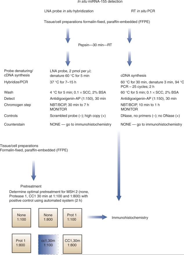
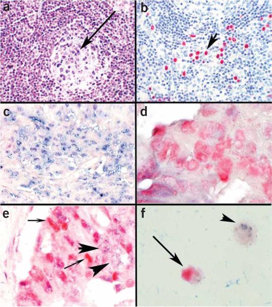
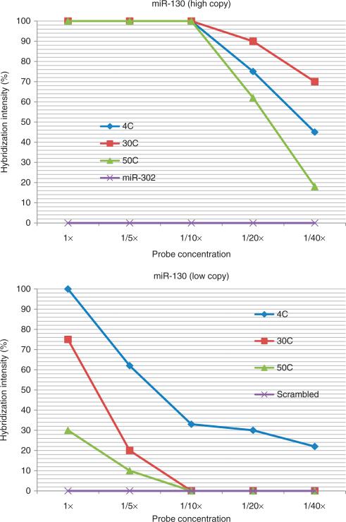
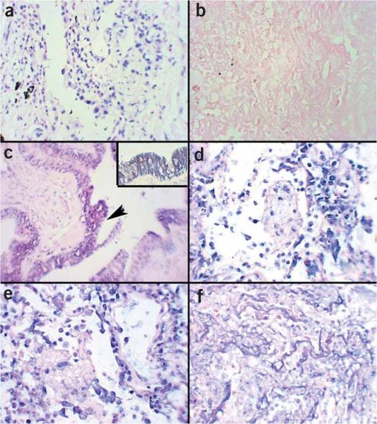
There are relatively few protocols described for the in situ detection of microRNA (miRNA) and they often use cryostat sections, signal amplification and hybridization or washes of 50-60 degrees C. This protocol describes in situ miRNA detection that can be done in paraffin-embedded, formalin-fixed tissue. Detection of the miRNA precursors can be done by RT in situ PCR, which can theoretically detect one copy per cell. The key variable for the RT in situ PCR protocol is optimal protease digestion, which is then followed by overnight DNase digestion and target specific incorporation of the reported nucleotide into the amplified cDNA. Detection of mature miRNAs is achieved by in situ hybridization with locked nucleic acid probes. This part of the protocol involves a brief protease digestion, followed by an overnight hybridization, short low stringency wash and detection of the labeled probe. The key variables for this method include probe concentration and stringency conditions. Each miRNA in situ method takes 1 d. The final step of the protocol involves colabeling by immunohistochemistry for the putative target of the miRNA, which is done after the in situ hybridization step and takes a few hours.


Figures

Figure 1
Figure 1
A flowchart schematic for the detection of miRNA (mature and precursors) and putative protein targets by colabeling. A simplified protocol for miRNA detection by in situ hybridization with an LNA probe and RT in situ PCR is presented. The LNA probe most likely detects the mature miRNA, whereas the RT in situ PCR detects only the precursor molecules. After either in situ test, one can do colabeling with immunohistochemistry for the putative protein target, assuming that the optimal condition for the latter includes either protease digestion or cell conditioning. One needs to determine the optimal pretreatment for the antigen. The six boxes represent either no pretreatment (None) with dilute (1:800) or concentrated (1:100) primary antibody, protease 1 digestion (Prot 1) or cell conditioning for 30 min (cc1,30) at the two different concentrations. Figure 2c–e shows a representative example of results obtained using the protocol with the detection of miR-155 in colon cancer and colabeling with a possible target, MSH 2.
Figure 2
Figure 2
Determination of putative targets of miRNAs by colocalization analysis with immunohistochemistry. The miRNA miR-155 is markedly upregulated in lymphoproliferative diseases, such as AIDS, and is also increased in colon cancer in which there is often dysregulation of the DNA repair gene MSH 2. (a) miR-155 is present in the atypical lymphocytes but not the endothelial cells as determined by in situ hybridization with an LNA probe; the arrow points to the germinal center where several blue-staining cells indicative of miR-155 expression are seen. Many more miR-155 cells are seen in the interfollicular zone. (b) By analyzing serial sections with immunohistochemistry using an antibody directed against HHV8 and a red chromogen, it was simple to demonstrate that there were over 25 times more miR-155-positive cells than HHV8-positive cells, showing that HHV-8 per se was not responsible for upregulating miR-155; the arrow points to several HHV-8 red staining cells. (c) We performed colocalization experiments with miR-155 and MSH 2, using cell conditioning for 30 min for the MSH2 protein. Note that in cancer cell groups strongly positive for miR-155 (blue, in situ hybridization with an LNA probe), MSH 2 (red, immunohistochemistry) is not evident. (d) Conversely, in colon cancer cell groups where the neoplastic cells expressed MSH 2 (red, immunohistochemistry), miR-155 (blue, in situ hybridization with an LNA probe) was not evident. (e) In some groups of cancer cells, both miR-155 and MSH 2 were present. The colocalization experiments in these groups showed that the cells making MSH 2 (small arrows, red—immunohistochemistry) did not contain miR-155 (large arrows, blue—in situ hybridization with an LNA probe) and vice versa. These data suggest that miR-155 may be downregulating MSH 2. (f) Colocalization experiments with cell lines can also be used to get useful information on whether a given miRNA may be regulating a specific protein. miR-16 downregulates the protein bcl-2, which is an important anti-apoptotic factor in several lymphomas. Colocalization experiments showed that miR-16-positive cells (small arrow, blue—in situ hybridization with an LNA probe) were bcl-2 negative, and the bcl-2-positive cells (large arrow, red—immunohistochemistry, ) were miR-16 negative, lending supporting evidence to a direct role for miR-16 in bcl-2 regulation. Panels a–c are at ×400 and panels d–f are at ×1,000 original magnification.
Figure 3
Figure 3
Optimizing experiments for miRNA detection by in situ hybridization with an LNA probe: importance of miRNA copy number, probe concentration and temperature of the post-hybridization wash. The top panel depicts a series of experiments in which placenta were tested for miR-130 with an LNA probe. The concentration of the probe ranged from 2 pmol μl−1 (1×) to 0.05 pmol μl−1 (1/40×). The temperature of the post-hybridization wash was set at 4, 30 or 50 °C. We also tested miR-302 by in situ hybridization as a negative control, as this miRNA is not expressed in the placenta; the post-hybridization wash was at 4 °C. The data points represent the mean of duplicate analyses done blinded to the reaction conditions; the standard error of the mean was no more than 10% of the mean value. In comparison, the bottom panel depicts an equivalent series of experiments with miR-130 but with brain tissues. miR-130 is present in a copy number in brain that is about 93% less than in placenta. The data with the scrambled probe (negative control) are included; the post-hybridization wash was done at 4 °C. Note the strong relationship between probe concentration, stringency of the post-hybridization wash and the relative miRNA copy number.
Figure 4
Figure 4
LNA-based in situ detection of miRNAs: keys to successful hybridization. The placenta contains high-copy miR-130. In situ hybridization for miR-130 shows an intense signal (brown, NBT/BCIP) with a probe concentration of 0.4 pmol μl−1 if the post-hybridization wash is at (a) 4 °C or (b) 50 °C. The inset shows no signal with the scrambled probe under the same conditions of either panel a or b. In comparison, the copy number of miR-130 is 93% less in the brain compared with the placenta. Panel c shows a signal in the brain at a probe concentration of 0.05 pmol μl−1 if the post-hybridization wash is at 4 °C but no signal if the post-hybridization wash is at 50 °C, (d) even if the probe concentration is increased to 0.8 pmol ml−1. The inset in panel d shows that, under similar conditions, a strong signal is seen in mouse embryo if the stem cell marker miR-302 probe is used at a concentration of 0.4 pmol μl−1. By combining RT in situ PCR for the precursor with LNA in situ hybridization, one can determine that benign tissues (such as the leiomyoma in panel e) can show miR-221 precursors (blue signal) and not the mature form (inset—LNA probe where no signal was evident); note the nuclear localization of the miRNA precursor. (f) When the mature form of miR-221 is evident, as is typical in this invasive leiomyosarcoma, the miR-221 precursor (RTISPCR) and mature miR-221 signal from the LNA probe both tend to localize to the cytoplasm (each panel is at ×400 original magnification).
Figure 5
Figure 5
RT in situ PCR for precursor miRNA detection: keys to successful amplification. The key to troubleshooting with RT in situ PCR for miRNA precursor detection is to analyze the positive (no DNase) and negative (DNase, irrevelant or scrambled primers control) before examining the test section. (a) The PCR in situ positive control must show an intense nuclear-based signal that is completely eliminated in b the negative control for the results for c the miRNA precursors to be accurate. The test is for miRNA-126, which is present in the pulmonary epithelium; note how the signal is evident only in these cells (arrow) and not the surrounding tissue, which is an important histological-based internal control; the inset shows the cytoplasmic signal that localizes to the bronchiole epithelium. Note what happens if the protease digestion time is too short; a signal is seen in the negative control (DNase, no primers—panel d) and (e) many different cell types besides the bronchiole epithelium are positive in the test. Also note how the colorimetric reaction occurs in the nucleus as compared with the cytoplasmic pattern when the controls work properly (panel c). Finally, note that when the protease digestion time is too long, the cell morphology is no longer recognizable and only the more resistant basement membranes are seen; thus, no signal is evident (panel f). Each panel is at 400× except the inset, which is at 1,000× original magnification.

Similar articles

Cited by

References

    1. Nuovo GJ. In situ detection of precursor and mature microRNAs in paraffin embedded, formalin fixed tissues and cell preparations. Methods. 2008;44:39–46. - PubMed
    1. Lee EJ, et al. Classification of microRNA processing patterns in tissues, cell lines, and tumors. RNA. 2008;14:35–42. - PMC - PubMed
    1. Calin GA, Croce CM. MicroRNA signatures in human cancers. Nat. Rev. Cancer. 2006;6:857–866. - PubMed
    1. Garzon R, Fabbri M, Cimmino A, Calin GA, Croce CM. MicroRNA expression and function in cancer. Trends Mol. Med. 2006;12:580–587. - PubMed
    1. Yanaihara N, et al. Unique microRNA molecular profiles in lung cancer diagnosis and prognosis. Cancer Cell. 2006;9:189–198. - PubMed

Publication types

MeSH terms