. 2009 Mar 13;284(11):6661-73.
doi: 10.1074/jbc.M806735200. Epub 2009 Jan 8.

An upstream open reading frame regulates translation of GADD34 during cellular stresses that induce eIF2alpha phosphorylation

Affiliations

An upstream open reading frame regulates translation of GADD34 during cellular stresses that induce eIF2alpha phosphorylation

Yun-Young Lee et al. J Biol Chem. .

Abstract

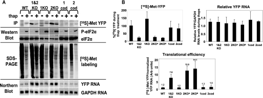
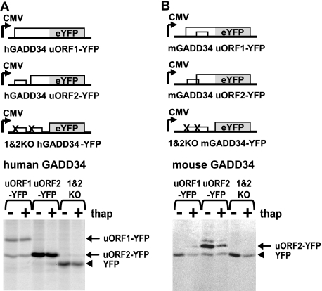
Cellular stress such as endoplasmic reticulum stress, hypoxia, and viral infection activates an integrated stress response, which includes the phosphorylation of the eukaryotic initiation factor 2alpha (eIF2alpha) to inhibit overall protein synthesis. Paradoxically, this leads to translation of a subset of mRNAs, like transcription factor ATF4, which in turn induces transcription of downstream stress-induced genes such as growth arrest DNA-inducible gene 34 (GADD34). GADD34 interacts with protein phosphatase 1 to dephosphorylate eIF2alpha, resulting in a negative feedback loop to recover protein synthesis and allow translation of stress-induced transcripts. Here, we show that GADD34 is not only transcriptionally induced but also translationally regulated to ensure maximal expression during eIF2alpha phosphorylation. GADD34 mRNAs are preferentially associated with polysomes during eIF2alpha phosphorylation, which is mediated by its 5'-untranslated region (5'UTR). The human GADD34 5'UTR contains two non-overlapping upstream open reading frames (uORFs), whereas the mouse version contains two overlapping and out of frame uORFs. Using 5'UTR GADD34 reporter constructs, we show that the downstream uORF mediates repression of basal translation and directs translation during eIF2alpha phosphorylation. Furthermore, we show that the upstream uORF is poorly translated and that a proportion of scanning ribosomes bypasses the upstream uORF to recognize the downstream uORF. These findings suggest that GADD34 translation is regulated by a unique 5'UTR uORF mechanism to ensure proper GADD34 expression during eIF2alpha phosphorylation. This mechanism may serve as a model for understanding how other 5'UTR uORF-containing mRNAs are regulated during cellular stress.


Figures

FIGURE 1.
FIGURE 1.
Polysomal association of GADD34 mRNA during eIF2α phosphorylation in human HepG2 and mouse Hepa cells. HepG2 cells (A) or Hepa (1–6C) cells (B) were treated with 1 μm thapsigargin or 2 mm DTT for the indicated times and subjected to [35S]methionine/cysteine pulse-labeling. Radiolabel incorporation into newly synthesized protein was measured by trichloroacetic acid precipitation and normalized to that in untreated cells (100%) (A) or by autoradiogram (B) of SDS-PAGE analysis. The phosphorylation status of eIF2α and the total eIF2α in cell lysates were monitored by Western blot analysis using a phospho-specific antibody to phosphorylated eIF2α (top) and an antibody that recognizes the C-terminal region of eIF2α (bottom), respectively. C, sedimentation profiles at absorbance 254 nm of HepG2 lysates untreated (left) or treated with 1 μm thapsigargin for 30 min (right). Cell lysates were fractionated by a 10–50% (w/v) sucrose gradient centrifugation. The top to bottom of the gradient is represented from left to right, respectively. The sedimentation of the 40 S, 60 S, 80 S fractions and polysomes are indicated. D and E, polysomal Northern blot analysis of RNA in fractions from HepG2 (D) and Hepa (E) cell lysates after 1 μm 30 min thapsigargin (thap), 2 mm 30 min DTT, or untreated (unt) treatments as indicated to the right. The distribution of mRNAs is indicated to the left by Northern blotting of polysomal RNA. Fractions from top to bottom of the gradient are represented from left to right, respectively. Fractions containing 80 S and polysomes are indicated at the bottom. Quantitation of the Northern blots in D and E are shown in F and G, respectively. The amount of radioactive probe specific to the indicated mRNA in each fraction is indicated as a percentage of total radioactivity in all fractions within each sucrose gradient (% radioactivity).
FIGURE 2.
FIGURE 2.
GADD34 expression in HepG2 cells during thapsigargin and actinomycin D treatment. A, Northern blots of RNA and (C) immunoblots of lysates from HepG2 cells alone (unt) or treated with 5 μg/ml actinomycin D (actD), 1 μm thapsigargin (thap), or thapsigargin/actinomycin D (actD/thap) for the indicated times. A, radiolabeled probes specific to GADD34, ATF4, CHOP, and GAPDH mRNAs (indicated to the right) were quantitated (B) using phosphorimaging analysis and normalized to the amount of RNA at the 3-h untreated time point. C, GADD34, ATF4, phosphorylated eIF2α, total eIF2, and CHOP were detected by antibodies as indicated to the right. Representative Western blots of at least three independent experiments are shown. For GADD34, two independent experiments are shown, which shows reproducible induction of GADD34 protein levels under thapsigargin alone and thapsigargin and actinomycin D treatment. The arrow indicates the migration of GADD34 protein, and the asterisk indicates nonspecific proteins recognized by the GADD34 antibody. D, quantitation of a representative GADD34 immunoblot. The amount of GADD34 protein was normalized to the amount of GADD34 at the 3-h untreated time point (Odyssey-Licor). E, polysomal Northern blot analysis of RNA in fractions from untreated HepG2 cells (unt) or from cells after a 30-min treatment with 1 μm thapsigargin (thap), 5 μg/ml actinomycin D (actD), or 1 μm thapsigargin (actD/thap) as indicated to the right. The distribution of GADD34 and GAPDH mRNAs is shown by Northern blot analysis using radiolabeled DNA probes. Fractions from top to bottom of the gradient are represented from left to right, respectively. Locations of 80 S and polysomes across the gradient are indicated at the bottom. Quantitation of the amount of radioactivity within each fraction is shown in supplemental Fig. S1.
FIGURE 3.
FIGURE 3.
Schematic of uORFs within select mammalian GADD34 5′UTRs. Representative cDNAs encoding GADD34-related sequences in GenBank™ are shown, including human (NM_014330), rat (NM_133546), hamster (L28147), and mouse (NM_008654) GADD34 5′UTRs. The AUG codon of the GADD34 ORF is shown. The length of the 5′UTR and the space between uORFs are indicated above (nt, nucleotides). Each box represents uORF1 and uORF2 and the size of the predicted translated uORF (aa, amino acid length). A close-up view of the overlap of the mouse GADD34 uORFs is shown below. The upstream uORF (uORF1) overlaps with the downstream uORF (uORF2) by 1 nucleotide. The stop codon of uORF1 is underlined, and the AUG of uORF2 is in gray.
FIGURE 4.
FIGURE 4.
The 5′UTR of human and mouse GADD34 mediates polysome association during thapsigargin treatment in HepG2 cells. Sucrose gradient density centrifugation and fractionation of lysates from stable cell lines expressing reporter YFP RNA fused with wild-type human GADD34 5′UTR (hGADD34-YFP) (A), mutant human GADD34 5′UTR (1&2KO hGADD34-YFP) (B), mouse GADD34 5′UTR (mGADD34-YFP) (C), or human ATF4 5′UTR (hATF4-YFP) (D) that were incubated in the absence (unt) or presence of 1 μm thapsigargin for 30 min (thap) were performed. The reporter RNAs were transcribed by the cytomegalovirus promoter (CMV). The distribution of specific RNAs across the gradient was detected by Northern blot analysis as indicated to the left. Fractions from top to bottom of the gradient are represented from left to right, respectively. Fractions that contain the 80 S and polysomes are indicated below the fractions. Quantitations of the amount of radioactivity within each fraction are shown in supplemental Fig. S2.
FIGURE 5.
FIGURE 5.
The uORFs of hGADD34 5′UTR mediate translation regulation during eIF2α phosphorylation. In A: Top row, immunoprecipitates of [35S]methionine-labeled YFP from lysates of cells expressing IGR IRES-YFP, hATF4-YFP, wild-type hGADD34-YFP, YFP alone, or mutant human GADD34 5′UTR fused with YFP where the AUG codon of the upstream uORF (1KO), downstream uORF (2KO), or both uORFs were mutated (1KO, 2KO, and 1&2KO, respectively) were either left untreated (U) or treated with 1 μm thapsigargin (T), 2 mm DTT (D), or 100 μg/ml arsenite for 45 min (A). Cells were pulse-labeled with [35S]methionine for 20 min prior to harvesting. Immunoprecipitates were separated by SDS-PAGE and exposed by phosphorimaging analysis. Second row, immunoblots of lysates using antibodies that recognize phospho-eIF2α or total eIF2α. Third row, lysates were subjected to SDS-PAGE analysis and exposed to autoradiography. Bottom row, in parallel, RNA from treated cells was subjected to Northern blot analysis using probes specific for YFP or GAPDH. B, quantitation of newly synthesized [35S]methionine-labeled YFP immunoprecipitates (top left) and levels of YFP RNA normalized to GAPDH (top right) as described in A. Each bar represents the percent of [35S]methionine-labeled YFP expressed or the YFP RNA levels during the indicated drug treatments as compared with that in untreated cells, which is set at 100%. For Northern blot analysis, YFP mRNA levels were normalized to GAPDH mRNA levels. Bottom, translational efficiency of reporter YFP RNA in unstressed cells was calculated by the amount of newly synthesized [35S]methionine YFP protein normalized to YFP/GAPDH mRNA. Above each bar shows the average translational efficiency normalized to the wild-type hGADD34-YFP RNA as 1. Shown are averages ± S.D. from at least three independent experiments.
FIGURE 6.
FIGURE 6.
The overlapping uORFs of the mouse 5′UTR mGADD34 mediate translational control during eIF2α phosphorylation. Top row, immunoprecipitates of [35S]methionine-labeled YFP from lysates of cells stably expressing wild-type or mutant mouse 5′UTR mGADD34-YFP reporter RNAs as indicated above under untreated or 1μm thapsigargin treatment for 45 min. 1&2KO, 1KO, and 2KO represent mutant mouse mGADD34 5′UTR reporter RNAs in which the AUG start codon of uORF1 (1KO), uORF2 (2KO), or both (1&2KO) uORFs was mutated. Mutations that inserted one (1cod) or two (2cod) codons between the AUG codon of the uORF2 and the stop codon of the upstream uORF1 were engineered into the 5′UTR mGADD34-YFP reporter RNA (see “Experimental Procedures” for sequence). Second row, immunoblots of lysates using antibodies that recognize phospho-eIF2α or total eIF2α. Third row, cells were pulse-labeled with [35S]methionine for 20 min prior to harvesting. Lysates were subjected to SDS-PAGE analysis and exposed to autoradiograph. Bottom row, in parallel, RNA from treated cells was subjected to Northern blot analysis using probes specific for YFP or GAPDH. B, quantitation of [35S]methionine-labeled YFP immunoprecipitates (top left) and of YFP RNA levels normalized to GAPDH (top right). Bottom, each bar represents the percentage of [35S]methionine-labeled YFP expressed or YFP RNA levels during the thapsigargin treatment as compared with that in untreated cells. Translational efficiency of reporter YFP RNA in unstressed cells was calculated by the amount of newly synthesized [35S]methionine precipitates normalized to YFP/GAPDH mRNA levels. Above each bar shows the average translational efficiency normalized to the wild-type mGADD34-YFP RNA. Shown are averages ± S.D. from at least three independent experiments.
FIGURE 7.
FIGURE 7.
The downstream uORF2 of hGADD34 and mGADD34 5′UTRs are translated efficiently in HepG2 cells. Schematics of human (A) or mouse (B) GADD34 5′UTR-YFP reporter RNAs stably expressed in HepG2 cells. The upstream (uORF1) or downstream (uORF2) uORFs were engineered such that the reading frames of the uORF were fused with the reporter YFP ORF. For the human hGADD34 uORF1-YFP RNA, the uORF2 is in-frame with uORF1 and the YFP ORF, thus both uORF1-YFP and uORF2-YFP fusion protein products can be monitored. Below, immunoprecipitates of newly synthesized 35S-labeled YFP protein from lysates of cells in the presence or absence of 1 μm thapsigargin. Reactions were resolved on an SDS-PAGE and exposed by phosphorimaging analysis. uORF1-YFP, uORF2-YFP, and YFP ORF proteins are indicated to the right.

Similar articles

Cited by

References

    1. Ron, D., and Harding, H. P. (2007) in Translational Control in Biology and Medicine (Mathews, M. B., Sonenberg, N., and Hershey, J., eds) pp. 345–368, Cold Spring Harbor Laboratory Press, Cold Spring Harbor, NY
    1. Dever, T. E., Dar, A. C., and Sicheri, F. (2007) in Translational Control in Biology and Medicine (Mathews, M. B., Sonenberg, N., and Hershey, J., eds) pp. 319–344, Cold Spring Harbor Laboratory Press, Cold Spring Harbor, NY
    1. Kostura, M., and Mathews, M. B. (1989) Mol. Cell. Biol. 9 1576–1586 - PMC - PubMed
    1. Han, A. P., Yu, C., Lu, L., Fujiwara, Y., Browne, C., Chin, G., Fleming, M., Leboulch, P., Orkin, S. H., and Chen, J. J. (2001) EMBO J. 20 6909–6918 - PMC - PubMed
    1. Chen, J. J., Throop, M. S., Gehrke, L., Kuo, I., Pal, J. K., Brodsky, M., and London, I. M. (1991) Proc. Natl. Acad. Sci. U. S. A 88 7729–7733 - PMC - PubMed

Publication types

MeSH terms

LinkOut - more resources