

Cutting edge: Ikaros is a regulator of Th2 cell differentiation
- PMID: 19124715
- PMCID: PMC2718557
- DOI: 10.4049/jimmunol.182.2.741
Cutting edge: Ikaros is a regulator of Th2 cell differentiation
Abstract
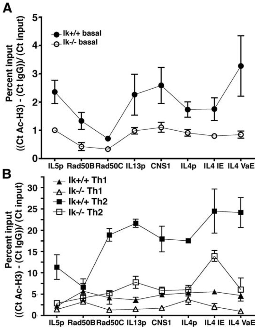
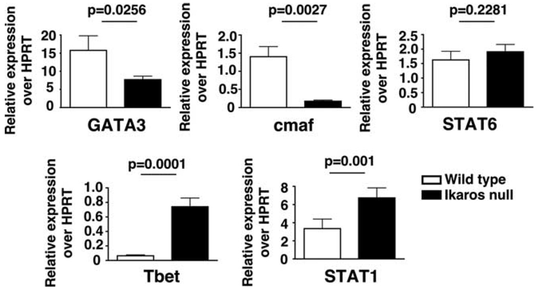
Ikaros, a hematopoietic transcription factor, has well defined effects on early lymphocyte development in the bone marrow and thymus. In this study we demonstrate that Ikaros is a positive regulator of Th2 cytokine gene expression in peripheral T cells. CD4+ T cells from naive Ikaros(null) mice cultured under Th2-skewing conditions express the Th1 cytokine IFN-gamma and have reduced IL-4, IL-5, and IL-13 expression. Ikaros directly associates with several Th2 locus regulatory regions in naive CD4+ T cells. The decreased ability to express Th2 cytokines in Ikaros(null)T cells corresponds with histone 3 hypoacetylation across the Th2 cytokine locus as well as decreased GATA3 and cMaf and increased T-bet and STAT1 expression. These data support a model whereby Ikaros directly activates Th2 gene expression by promoting local chromatin accessibility during CD4+ T cell differentiation and also acts indirectly to regulate expression of Th2- and Th1-specific transcription factors.
Conflict of interest statement
The authors have no financial conflict of interest.
Figures


Similar articles
-
Ikaros is a regulator of Il10 expression in CD4+ T cells.J Immunol. 2009 Nov 1;183(9):5518-25. doi: 10.4049/jimmunol.0901284. Epub 2009 Oct 14. J Immunol. 2009. PMID: 19828627 Free PMC article.
-
Ikaros silences T-bet expression and interferon-gamma production during T helper 2 differentiation.J Biol Chem. 2010 Jan 22;285(4):2545-53. doi: 10.1074/jbc.M109.038794. Epub 2009 Nov 18. J Biol Chem. 2010. PMID: 19923223 Free PMC article.
-
A distinct region of the murine IFN-gamma promoter is hypomethylated from early T cell development through mature naive and Th1 cell differentiation, but is hypermethylated in Th2 cells.J Immunol. 2004 Dec 15;173(12):7377-84. doi: 10.4049/jimmunol.173.12.7377. J Immunol. 2004. PMID: 15585862
-
Chromatin remodeling and T helper subset differentiation.IUBMB Life. 2000 Jun;49(6):473-8. doi: 10.1080/15216540050166990. IUBMB Life. 2000. PMID: 11032239 Review.
-
An updated view on transcription factor GATA3-mediated regulation of Th1 and Th2 cell differentiation.Int Immunol. 2011 Jul;23(7):415-20. doi: 10.1093/intimm/dxr029. Epub 2011 Jun 1. Int Immunol. 2011. PMID: 21632975 Free PMC article. Review.
Cited by
-
Differentiation of effector CD4 T cell populations (*).Annu Rev Immunol. 2010;28:445-89. doi: 10.1146/annurev-immunol-030409-101212. Annu Rev Immunol. 2010. PMID: 20192806 Free PMC article. Review.
-
Insulin-InsR signaling drives multipotent progenitor differentiation toward lymphoid lineages.J Exp Med. 2015 Dec 14;212(13):2305-21. doi: 10.1084/jem.20150618. Epub 2015 Nov 16. J Exp Med. 2015. PMID: 26573296 Free PMC article.
-
Ikaros is a regulator of Il10 expression in CD4+ T cells.J Immunol. 2009 Nov 1;183(9):5518-25. doi: 10.4049/jimmunol.0901284. Epub 2009 Oct 14. J Immunol. 2009. PMID: 19828627 Free PMC article.
-
Transcriptional regulation of Th2 cell differentiation.Immunol Cell Biol. 2010 Mar-Apr;88(3):244-9. doi: 10.1038/icb.2009.114. Epub 2010 Jan 12. Immunol Cell Biol. 2010. PMID: 20065998 Free PMC article. Review.
-
Ikaros silences T-bet expression and interferon-gamma production during T helper 2 differentiation.J Biol Chem. 2010 Jan 22;285(4):2545-53. doi: 10.1074/jbc.M109.038794. Epub 2009 Nov 18. J Biol Chem. 2010. PMID: 19923223 Free PMC article.
References
-
- Reiner SL. Development in motion: helper T cells at work. Cell. 2007;129:33–36. - PubMed
-
- Ansel KM, Lee DU, Rao A. An epigenetic view of helper T cell differentiation. Nat. Immunol. 2003;4:616–623. - PubMed
-
- Ansel KM, Greenwald RJ, Agarwal S, Bassing CH, Monticelli S, Interlandi J, Djuretic IM, Lee DU, Sharpe AH, Alt FW, Rao A. Deletion of a conserved Il4 silencer impairs T helper type 1-mediated immunity. Nat. Immunol. 2004;5:1251–1259. - PubMed
-
- Lee DU, Agarwal S, Rao A. Th2 lineage commitment and efficient IL-4 production involves extended demethylation of the IL-4 gene. Immunity. 2002;16:649–660. - PubMed
-
- Lee GR, Kim ST, Spilianakis CG, Fields PE, Flavell RA. T helper cell differentiation: regulation by cis elements and epigenetics. Immunity. 2006;24:369–379. - PubMed
Publication types
MeSH terms
Substances
Grants and funding
LinkOut - more resources
Full Text Sources
Molecular Biology Databases
Research Materials
Miscellaneous