. 2009 Jan 12;184(1):31-44.
doi: 10.1083/jcb.200811035. Epub 2009 Jan 5.

Lamin A/C-mediated neuromuscular junction defects in Emery-Dreifuss muscular dystrophy

Affiliations

Lamin A/C-mediated neuromuscular junction defects in Emery-Dreifuss muscular dystrophy

Alexandre Méjat et al. J Cell Biol. .

Abstract

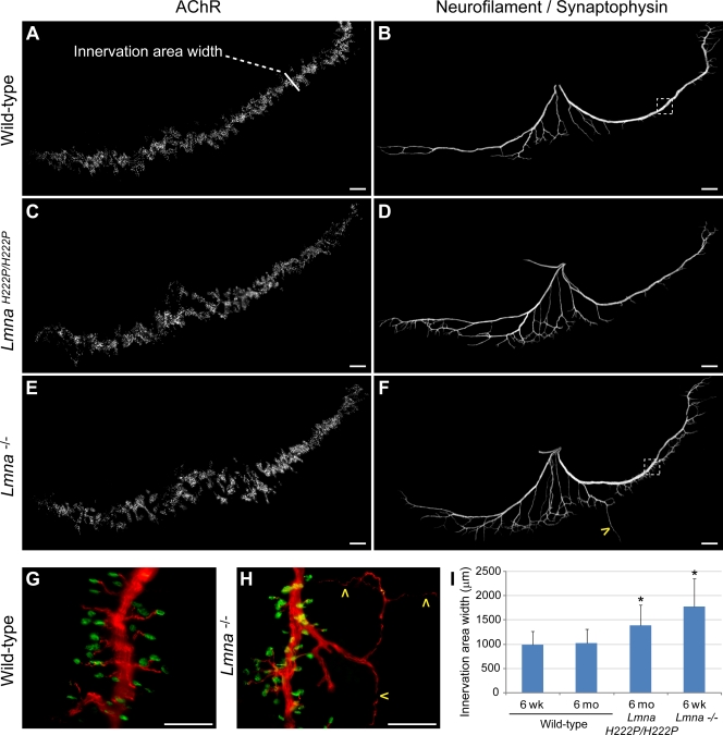
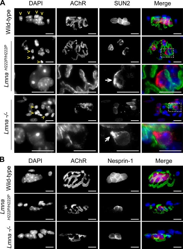
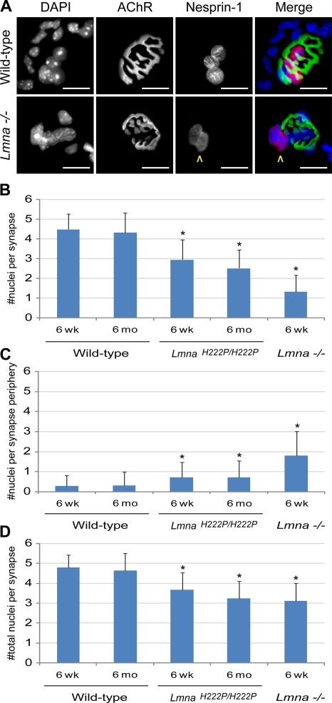
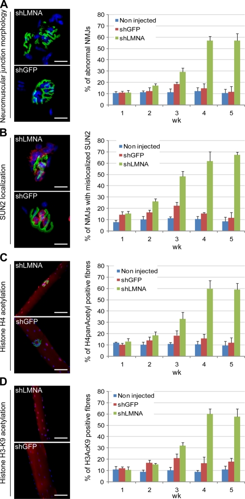
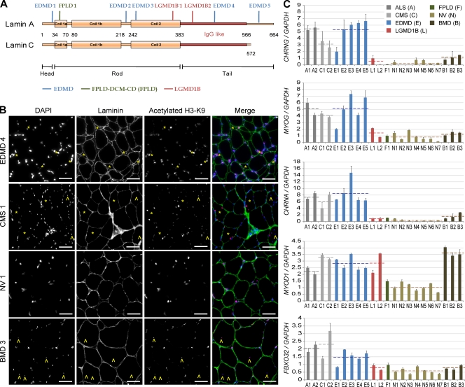
The LMNA gene encodes lamins A and C, two intermediate filament-type proteins that are important determinants of interphase nuclear architecture. Mutations in LMNA lead to a wide spectrum of human diseases including autosomal dominant Emery-Dreifuss muscular dystrophy (AD-EDMD), which affects skeletal and cardiac muscle. The cellular mechanisms by which mutations in LMNA cause disease have been elusive. Here, we demonstrate that defects in neuromuscular junctions (NMJs) are part of the disease mechanism in AD-EDMD. Two AD-EDMD mouse models show innervation defects including misexpression of electrical activity-dependent genes and altered epigenetic chromatin modifications. Synaptic nuclei are not properly recruited to the NMJ because of mislocalization of nuclear envelope components. AD-EDMD patients with LMNA mutations show the same cellular defects as the AD-EDMD mouse models. These results suggest that lamin A/C-mediated NMJ defects contribute to the AD-EDMD disease phenotype and provide insights into the cellular and molecular mechanisms for the muscle-specific phenotype of AD-EDMD.


Figures

Figure 1.
Figure 1.
Innervation patterns in wild-type and AD-EDMD mouse models. Whole-mount diaphragms of 6-mo-old wild-type (A and B), 6-mo-old LmnaH222P/H222P (C and D), and 6-wk-old Lmna−/− (E and F) mice were stained with Bgt to delineate the NMJs (AChR; A, C, and E), and neurofilament p150 and synaptophysin were used to stain the motor axon (B, D, and F). A representative right hemidiaphragm of each phenotype is shown. LmnaH222P/H222P and Lmna−/− diaphragms showed markedly broader synapse bands (A, C, and E) and highly ramified phrenic nerves (B, D, and F). (G and H) Enlargements of indicated areas (boxes in B and F) double-stained with neurofilament/synaptophysin (red) and Bgt (green) showing intense nerve sprouting from both the main nerve axis and secondary branches in Lmna−/− diaphragm (arrowheads). (I) Quantitation of postsynaptic innervation area width. Values shown are mean ± SD of at least 200 measurements per diaphragm from three mice per genotype. *, P < 0.001 relative to wild-type littermate mice. Bars, 500 μm.
Figure 2.
Figure 2.
Abnormal NMJs in muscle from wild-type and AD-EDMD mouse models. (A–C) Representative synapse morphologies from wild-type, LmnaH222P/H222P, and Lmna−/− mice isolated fibers from tibialis anterior stained with DAPI and Bgt. (A) Synapses from 6-mo-old wild-type mice showed characteristic “pretzel-like” AChR networks, and between four and eight nuclei directly aggregated beneath the NMJ (white arrowheads). (B) Synapses from 6-mo-old LmnaH222P/H222P were frequently fragmented with muscle nuclei located at the periphery of the AChR network (yellow arrowhead). (C) AChR staining in 6-wk-old Lmna−/− muscles was generally dramatically disorganized, with barely discernable branches, whereas few to no nuclei were observed underneath or at the periphery (yellow arrowheads) of the NMJ. (D) The percentage of abnormal NMJs observed in 6-wk-old and 6-mo-old wild-type, LmnaH222P/H222P, and Lmna−/− mice muscles. Values are mean ± SD of at least 100 NMJ from three different animals in each genotype. Bars, 10 μm.
Figure 3.
Figure 3.
Mislocalization of nuclear envelope components in synaptic nuclei of AD-EDMD mouse models. Isolated muscle fibers from 6-mo-old wild-type, 6-mo-old LmnaH222P/H222P, and 6-wk-old Lmna−/− mice tibialis anterior muscles were stained with DAPI, Bgt, and SUN2 (A) or Nesprin-1 antibodies (B). SUN2 and Nesprin-1 were expressed in all muscle nuclei but with a lower staining in mutant synaptic nuclei (A and B). Boxes indicate the area enlarged in the panel below. Compared with wild type, SUN2 appeared heterogeneous and frequently aggregated on one side of synaptic nuclei in AD-EDMD mouse models (A, white arrows). Nesprin-1 staining was reduced in LmnaH222P/H222P and Lmna−/− synaptic nuclei and partly relocalized (B). In all cases, Schwann cell nuclei (yellow arrowheads) were negative for both Nesprin-1 and SUN2. Bars, 10 μm.
Figure 4.
Figure 4.
Altered recruitment of the muscle synaptic nuclei. (A) Isolated muscle fibers were stained with DAPI, Bgt, and Nesprin-1 antibodies to unequivocally identify synaptic myonuclei (yellow arrowheads). Bars, 10 μm. (B–D) Nesprin-1–positive myonuclei were quantified according to their relative position to the Bgt staining. Percentage of synaptic (B), perisynaptic (C), and total (D) nuclei per muscle synapse of 6-wk-old and 6-mo-old wild-type, 6-wk-old and 6-mo-old LmnaH222P/H222P, and 6-wk-old Lmna−/− mice are shown. Values are mean ± SD of at least 100 NMJs from three different animals in each genotype. *, P < 0.001 relative to littermate wild type.
Figure 5.
Figure 5.
Hallmarks of denervation in AD-EDMD mouse models. (A) mRNA expression levels of Chrng, Chrna, Myog, MyoD1, Fbxo32, and Hdac9 were evaluated in wild-type, LmnaH222P/H222P, and Lmna−/− mice gastrocnemius muscles by quantitative RT-PCR (A). Mutant muscles showed higher expression levels of electrical activity–dependent genes (Chrng, Chrna, Myog, and MyoD1) and atrophy-associated genes (Fbxo32). In contrast, mutant muscles expressed less Hdac9 than the wild type. All gene expression levels were normalized to Gapdh. Values are mean ± SD from six muscles (two muscles from three mice) per genotype. *, P < 0.001 relative to the wild type. Expression levels in each sample were evaluated in at least two independent quantitative RT-PCR experiments. (B) Wild-type and Lmna mutant isolated muscle fibers from tibialis anterior muscle were stained with DAPI, Bgt, and acetylated histone H3-K9 or acetylated histone H4 antibodies. A high proportion of LmnaH222P/H222P and Lmna−/− fibers displayed increased histone acetylation levels. The percentage of histone hyperacetylated muscle fibers are indicated for the different genotypes. Values are means ± SD from at least 100 fibers from three different mice per genotype. Bars, 25 μm.
Figure 6.
Figure 6.
Transient A-type lamin knockdown in wild-type tibialis anterior. Wild-type tibialis anterior muscles injected with AAV6 expressing shRNAs against LMNA (shLMNA) or GFP (shGFP) were assessed at the indicated time after AAV injection for several phenotypes observed in LmnaH222P/H222P and Lmna−/− muscles: NMJ morphology (A), SUN2 localization (B), and H3 and H4 histone acetylation levels (C and D). For each assay, a representative NMJ or muscle fiber from shLMNA- or shGFP-injected muscle 4 wk after injection is provided with the quantification of the number of altered NMJ per fiber at each time point. Hyperacetylated nuclei visible on shGFP (C) correspond to nuclei from nonmuscle interstitial cells and were used as positive controls for the staining. Values are means ± SD from at least 100 fibers from three different mice per criteria.
Figure 7.
Figure 7.
Hallmarks of denervation in human patients. (A) Schematic representation of A-type lamin proteins. The position of the different mutations studied is indicated along the length of the protein. (B) Muscle biopsies from AD-EDMD, BMD, and CMS patients as well as normal volunteer (NV) were stained with DAPI (blue), α2-laminin antibody (green), and acetylated-histone H3-lysine 9 (K9) antibody (red). Atrophic muscle fibers (asterisks) showed a very high level of acetylation and no grouping of atrophic fibers. Muscle fibers of normal size occasionally showed hyperacetylated nuclei in AD-EDMD muscles. In contrast, hyperacetylated nuclei were only observed in nonmuscle interstitial cells in NV muscles. Although many centrally located nuclei could be observed in BMD muscles (arrowheads), most nuclei were in a subsarcolemmal position in AD-EDMD and CMS patients and in NV muscles. Bars, 30 μm. (C) Expression levels of CHRNG, CHRNA, MYOG, MYOD1, and FBXO32 were evaluated by quantitative RT-PCR on human biopsies. Patients affected by AD-EDMD were compared with patients affected by LGMD1B, FLPD-DCM-CD, ALS, CMS, BMD, and NV. Compared with NV, EDMD patients showed high, though similar to denervation-related diseases (ALS and CMS), levels of CHRNG, CHRNA, MYOG, and FBXO32. In contrast, expression levels of patients affected by the nondenervation-related disease BMD were similar to NV. Patients affected by other laminopathies (LGMD1B and FPLD-DCM-CD) showed low, though similar to normal volunteers, levels of CHRNG, CHRNA, and MYOG. All patients affected by neuromuscular diseases (ALS, CMS, EDMD, LGMD1B, and BMD) showed a high level of MYOD1 compared with NV but this could be caused by two independent mechanisms: misregulation in the muscle fibers (ALS, CMS, and potentially EDMD) and/or regeneration by the activated satellite cells (BMD). All gene expression levels were normalized to the GAPDH expression level. Values provided for each patient are means ± SD from at least three independent experiments. Broken lines indicate mean values of a group of patients.

Similar articles

Cited by

References

    1. Apel, E.D., R.M. Lewis, R.M. Grady, and J.R. Sanes. 2000. Syne-1, a dystrophin- and Klarsicht-related protein associated with synaptic nuclei at the neuromuscular junction. J. Biol. Chem. 275:31986–31995. - PubMed
    1. Arimura, T., A. Helbling-Leclerc, C. Massart, S. Varnous, F. Niel, E. Lacene, Y. Fromes, M. Toussaint, A.M. Mura, D.I. Keller, et al. 2005. Mouse model carrying H222P-Lmna mutation develops muscular dystrophy and dilated cardiomyopathy similar to human striated muscle laminopathies. Hum. Mol. Genet. 14:155–169. - PubMed
    1. Bakay, M., Z. Wang, G. Melcon, L. Schiltz, J. Xuan, P. Zhao, V. Sartorelli, J. Seo, E. Pegoraro, C. Angelini, et al. 2006. Nuclear envelope dystrophies show a transcriptional fingerprint suggesting disruption of Rb-MyoD pathways in muscle regeneration. Brain. 129:996–1013. - PubMed
    1. Bhagavati, S., A. Ghatpande, S.A. Shafiq, and B. Leung. 1996. In situ hybridization analysis for expression of myogenic regulatory factors in regenerating muscle of mdx mouse. J. Neuropathol. Exp. Neurol. 55:509–514. - PubMed
    1. Broers, J.L., E.A. Peeters, H.J. Kuijpers, J. Endert, C.V. Bouten, C.W. Oomens, F.P. Baaijens, and F.C. Ramaekers. 2004. Decreased mechanical stiffness in LMNA−/− cells is caused by defective nucleo-cytoskeletal integrity: implications for the development of laminopathies. Hum. Mol. Genet. 13:2567–2580. - PubMed

Publication types

MeSH terms