. 2008 Dec 29;183(7):1287-98.
doi: 10.1083/jcb.200807023.

The microtubule-binding protein CLIP-170 coordinates mDia1 and actin reorganization during CR3-mediated phagocytosis

Affiliations

The microtubule-binding protein CLIP-170 coordinates mDia1 and actin reorganization during CR3-mediated phagocytosis

Elodie Lewkowicz et al. J Cell Biol. .

Abstract

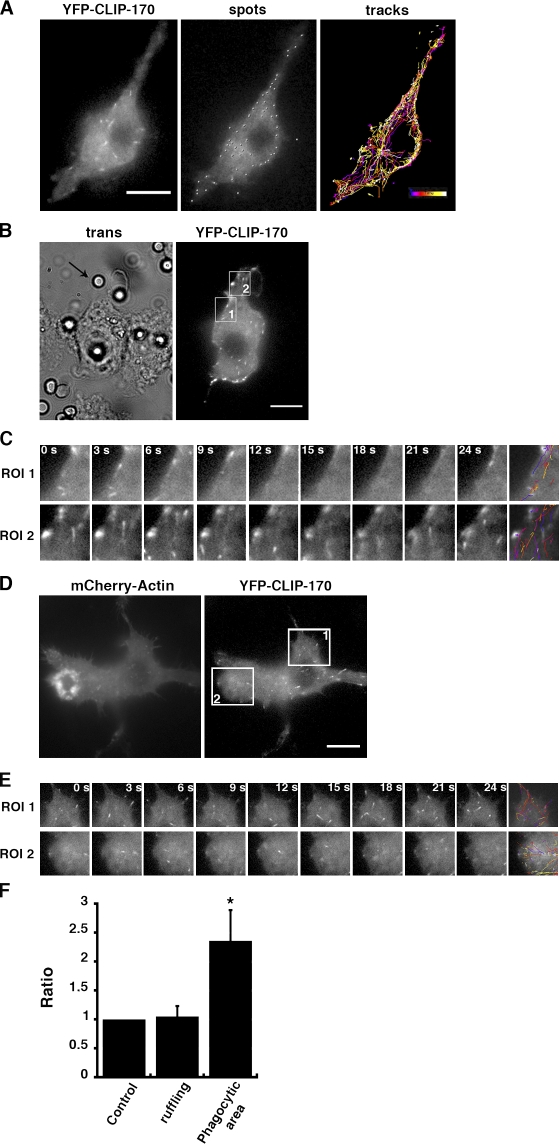
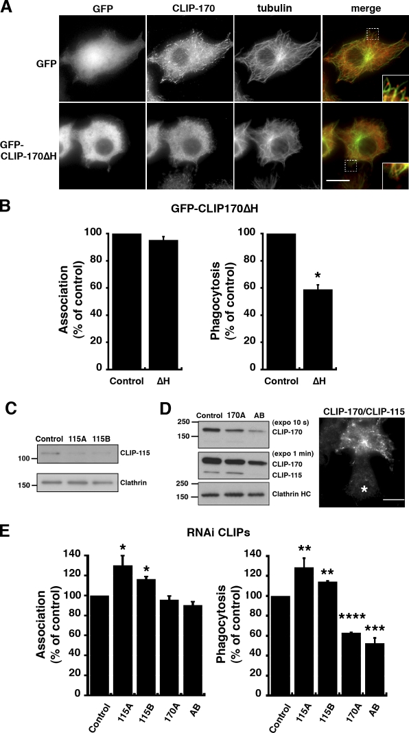
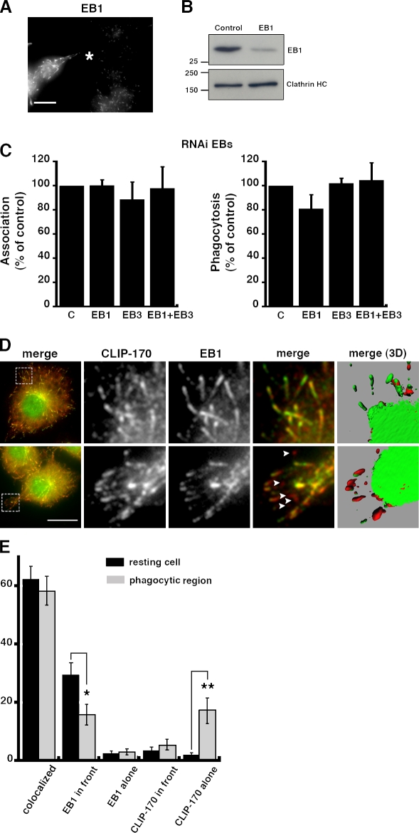
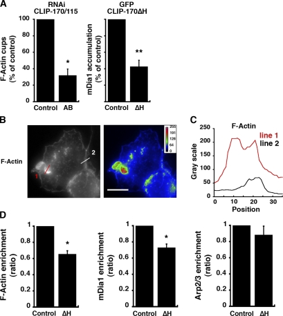
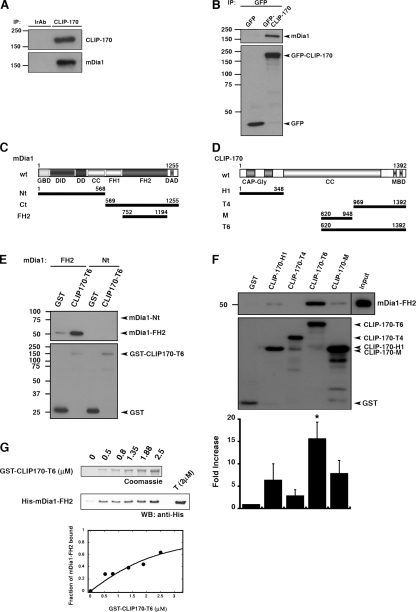
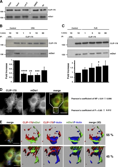
Microtubule dynamics are modulated by regulatory proteins that bind to their plus ends (+TIPs [plus end tracking proteins]), such as cytoplasmic linker protein 170 (CLIP-170) or end-binding protein 1 (EB1). We investigated the role of +TIPs during phagocytosis in macrophages. Using RNA interference and dominant-negative approaches, we show that CLIP-170 is specifically required for efficient phagocytosis triggered by alphaMbeta2 integrin/complement receptor activation. This property is not observed for EB1 and EB3. Accordingly, whereas CLIP-170 is dynamically enriched at the site of phagocytosis, EB1 is not. Furthermore, we observe that CLIP-170 controls the recruitment of the formin mDia1, an actin-nucleating protein, at the onset of phagocytosis and thereby controls actin polymerization events that are essential for phagocytosis. CLIP-170 directly interacts with the formin homology 2 domain of mDia1. The interaction between CLIP-170 and mDia1 is negatively regulated during alphaMbeta2-mediated phagocytosis. Our results unravel a new microtubule/actin cooperation that involves CLIP-170 and mDia1 and that functions downstream of alphaMbeta2 integrins.


Figures

Figure 1.
Figure 1.
CLIP-170 is recruited at sites of phagocytosis. (A) Dynamics of CLIP-170–positive comets at steady-state in macrophages. Cells were transfected with pYFP–CLIP-170 and 24 h later time-lapse imaging was performed (left). Tracking of CLIP-170–positive structures was performed using Imaris 5.7 software (spots corresponding to CLIP-170–positive structures [middle] and tracks followed during 150 s [right]). Colored bar, time scale. Bar, 10 μm. (B) Dynamics of CLIP-170 in phagocytosing macrophages. RAW264.7 macrophages were incubated at 37°C with C3bi-SRBCs and observed by wide-field fluorescence microscopy. Transmission (left) and fluorescence (right) images were recorded every second during 150 s. Transmission acquisition shows phagocytosis of an opsonized SRBC (left, arrow). The cell was divided in two regions of interest (ROI1 and ROI2). ROI1 corresponds to a nonphagocytosing area, whereas ROI2 is a phagocytosing area. Bar, 10 μm. (C) Time stack of images acquired in ROI1 and ROI2 as described in B. The last image shows the tracks followed during 150 s. (D) Dynamics of CLIP-170 in RAW264.7 macrophages treated with macrophage colony-stimulating factor to induce ruffling and spreading. Cells were transfected with pYFP–CLIP-170 and pmCherry-actin and 24 h later time-lapse imaging was performed as in A. mCherry-actin (left) and YFP–CLIP-170 (right) images were recorded every second during 150 s. The cell was divided into an area with actin activity (ROI2) and a resting area (ROI1). Bar, 10 μm. (E) Time stack of images acquired in ROI1 and ROI2 as described in D. Selected images are shown. The last image shows the tracks followed during 150 s. (F) Cells were analyzed as described in B and D. The number of comets per surface unit was calculated for regions of interest in different cells and the ratio between phagocytic and nonphagocytic (control) areas or between areas with actin activity (ruffling) and resting areas (control) was measured. The means ± SEM of 2,026 comets (in 7 phagocytosing cells) and the means ± SEM of 13,285 comets (in 11 cells with ruffles) are plotted. *, P < 0.05.
Figure 2.
Figure 2.
CLIP-170 is important for CR3-mediated phagocytosis. (A) RAW264.7 cells were transfected with pEGFP–CLIP-170ΔH, encoding a dominant-negative mutant of CLIP-170 (bottom) or with pEGFP as a control (top). After 24 h, macrophages were labeled with the anti–CLIP-170/CLIP-115 antibodies (#2221 serum) and with anti-tubulin antibodies, and then analyzed by wide-field fluorescence microscopy. Insets with magnified images are shown on the right. Bar, 10 μm. (B) A dominant-negative form of CLIP-170 inhibits CR3-mediated phagocytosis. Macrophages were transfected with pEGFP–CLIP-170ΔH or with pEGFP as a control. After 24 h, macrophages were allowed to phagocytose C3bi-SRBCs for 3 and 60 min at 37°C, and then fixed and stained with Cy3–anti-rabbit IgG antibodies to detect SRBCs. Efficiencies of association and phagocytosis were scored in 50 cells expressing GFP–CLIP-170ΔH and 50 GFP-expressing control cells. Results are expressed as a percentage of control cells. The means ± SEM of at least three independent experiments are plotted. *, P < 0.0001. (C) RAW264.7 macrophages were transfected with pSUPER-115A, pSUPER-115B, or pSUPER-G (directed against giantin) as a control. After 48 h, lysates were prepared and Western blotting was performed with anti–CLIP-170/115 antibodies (top) or with anti-clathrin (bottom) as a loading control. (D) RAW264.7 macrophages were transfected with pSUPER-170A (directed against CLIP-170), pSUPER-AB (directed against CLIP-170 and CLIP-115), or pSUPER-G (directed against giantin) as a control. After 48 h, lysates were prepared and Western blotting was performed with anti–CLIP-170/115 antibodies (10 s [top] or 1 min [middle] of film exposure) or with anti-clathrin (bottom). In addition, cells were fixed, permeabilized, and labeled with anti–CLIP-170/115 antibodies followed by Cy3–anti-rabbit IgG antibodies. Cells were analyzed by wide-field fluorescence microscopy (right). The image shows a cell depleted of CLIP-170/115 with pSUPER-AB (asterisk). Bar, 10 μm. (E) Depletion of CLIP-170 but not CLIP-115 inhibits phagocytosis. Macrophages were transfected as described in C and D, and then allowed to phagocytose C3bi-SRBCs for 3 and 60 min at 37°C and fixed and stained with Cy2–anti-rabbit IgG antibodies and, after permeabilization, with anti–CLIP-170 or anti–CLIP-115 antibodies. The efficiencies of association and phagocytosis were calculated for 50 CLIP-115–depleted cells (115A and 115B), 50 CLIP-170–depleted cells (170A), 50 CLIP-170/115–depleted cells (AB), and 50 control cells. Results are expressed as a percentage of control cells. The means ± SEM of three independent experiments are plotted. *, P < 0.05; **, P < 0.005; ***, P < 0.001; ****, P < 0.0001.
Figure 3.
Figure 3.
EB1 is not recruited in phagocytic cups and neither EB1 nor EB3 are required for phagocytosis. (A) RAW264.7 macrophages were transfected with siRNA directed against EB1 and after 24 h were analyzed by immunofluorescence with anti-EB1 antibodies followed by Cy3–anti-mouse IgG. Asterisk shows an EB1-depleted cell. Bar, 10 μm. (B) RAW264.7 macrophages were transfected with siRNA directed against EB1 or GFP as a control, and then lysed and analyzed by Western blotting with anti-EB1 antibodies and then with anti-clathrin as a loading control. (C) RAW264.7 macrophages were transfected with siRNA directed against EB1, EB3, or EB1 and EB3 or GFP as a control, and then allowed to phagocytose C3bi-SRBCs for 3 and 60 min. The efficiencies of association and phagocytosis were scored in 50 EB1-, EB3-, or EB1+EB3-depleted and 50 control cells (cells depleted for GFP). Results are expressed as a percentage of control cells. The means ± SEM of three independent experiments are plotted. (D) Differential recruitment of CLIP-170 and EB1 during phagocytosis. Macrophages were preactivated with PMA during 15 min (top) or preactivated and then allowed to internalize C3bi-SRBCs for 10 min (bottom). They were then analyzed by immunofluorescence with anti–CLIP-170/115 and anti-EB1 antibodies followed by Cy3–anti-rabbit IgG and Cy2–anti-mouse IgG, respectively. Bar, 10 μm. Insets show a peripheral region of a nonphagocytosing cell (top) or a phagocytosing area (bottom). Arrowheads indicate comets labeled with CLIP-170 only. 3D reconstructions of the stacks of merged images are presented in the right panels. (E) Positioning of CLIP-170/EB1 comets. Comets of CLIP-170 and EB1 were compared and the number of comets with colocalized EB1 and CLIP-170, EB1 in front of CLIP-170, EB1 alone, CLIP-170 in front of EB1, or CLIP-170 alone were counted in resting cells (black bars) and in phagocytic cups from phagocytosing cells (gray bars). Results are expressed as a percentage of the total comets counted. The means ± SEM of 586 comets (in 37 cells) from three independent experiments are plotted. *, P < 0.05; **, P < 0.005.
Figure 4.
Figure 4.
CLIP-170 function is important for actin polymerization and mDia1 recruitment. (A) RAW264.7 macrophages were transfected with pSUPER-AB (directed against CLIP-170 and CLIP-115 [left]) or pEGFP–CLIP-170ΔH (right) and then allowed to phagocytose C3bi-SRBCs for 10 min at 37°C. The cells were fixed and stained with AMCA– or Cy2–anti-rabbit IgG to detect the external particles, Alexa 488– or Alexa 350–phalloidin to stain F-actin, and anti-mDia1 or anti–CLIP-170 antibodies followed by Cy3–anti-mouse IgG. 50 CLIP-170–inhibited cells and 50 control cells were scored for the presence or absence of F-actin or mDia1 accumulation around bound particles. Results are expressed as a percentage of control cells. Means ± SEM of three independent experiments are plotted. *, P < 0.001; **, P < 0.005. (B) RAW264.7 macrophages were allowed to phagocytose C3bi-SRBCs for 10 min at 37°C, fixed, permeabilized, and incubated with Alexa 546–phalloidin. Images are shown using the Grayscale (left) or Rainbow2 (right) look-up tables. Values on the color scales in the right corner of the right panel indicate the fluorescence intensities. Bar, 10 μm. (C) The profiles of F-actin fluorescence intensities along the line drawn at the phagocytic site (line 1) and in the cell body (line 2) are shown. (D) Macrophages were transfected with pEGFP–CLIP-170ΔH or pEGFP as a control and then phagocytosis was performed as in B. mDia1 was detected with specific antibodies and the Arp2/3 complex was stained with anti-p16 antibodies, each followed by Cy3-F(ab′)2 anti-mouse IgG antibodies. The fluorescence intensities measured in the phagocytic cups were background subtracted and expressed as a percentage of the intensities in the cell body. Data obtained for F-actin (left), mDia1 (middle), and Arp2/3 (right) were plotted. Data are the mean ± SEM of three independent experiments. 15 CLIP-170ΔH–expressing and 15 GFP-expressing control cells were analyzed in each experiment. *, P < 0.005. Enrichment of Arp2/3 was not significantly impaired (P > 0.1).
Figure 5.
Figure 5.
CLIP-170 interacts with the FH2 domain of mDia1 directly. (A) Endogenous mDia1 and CLIP-170 coimmunoprecipitate. RAW264.7 macrophages were lysed and immunoprecipitated with an anti–CLIP-170 antibody (clone F-3) or with an irrelevant antibody as a control (anti-myc). The samples were loaded on 7.5% SDS-PAGE and revealed by Western blotting with anti-mDia1 antibodies and after stripping with anti–CLIP-170/CLIP-115 antibodies (top). (B) GFP–CLIP-170 interacts with endogenous mDia1. HeLa cells transiently expressing GFP–CLIP-170 or GFP as a control were lysed and incubated with anti-GFP. The precipitates were analyzed for mDia1 by Western blotting. After stripping, the membranes were detected with anti-GFP antibodies (bottom). (C) Schematic representation of the His-tagged mDia1 constructs. (D) Schematic representation of the GST-fused CLIP-170 constructs. (E) CLIP-170 interacts directly with the FH2 domain of mDia1. GST or GST–CLIP-170–T6 (400 nM) bound to glutathione sepharose beads were incubated with recombinant His-tagged mDia1 constructs (500 nM). Bound material was subjected to Western blotting with anti-His antibodies and after stripping with anti-GST antibodies. The results are representative of 4–10 independent experiments with three different preparations of the GST construct, two of His-mDia1-FH2 and one of His-mDia1-Nter. (F) A C-terminal domain of CLIP-170 interacts directly with mDia1-FH2. GST or the indicated GST–CLIP-170 constructs (400 nM) bound to glutathione sepharose beads were incubated with recombinant His-FH2 (500 nM) and bound material was subjected to Western blotting with anti-His antibodies. Input represents 10% of the amount of His-FH2 added in the assay. After stripping, membranes were detected with anti-GST antibodies. The intensities of the bands were calculated using ImageJ and background subtracted. A ratio was calculated for each GST-CLIP construct as compared with GST (bottom). Results are the means ± SEM of four to eight independent experiments with three different preparations of the GST constructs. Binding of GST–CLIP-170–T6 to mDia1-FH2 was significantly higher than binding of GST (*, P < 0.005), whereas binding of other constructs was not (P > 0.05). (G) His-tagged mDia1 FH2 domain (2 μM) pull-down assays using GST–CLIP-170–T6 at the concentrations indicated. Total input (T) and bound fractions were probed with anti-His antibodies. The results were quantified to obtain the binding curve. The best fit of the curve gave a Kd of 0.8 μM.
Figure 6.
Figure 6.
CLIP-170 and mDia1 interaction does not depend on actin or microtubules and is regulated during phagocytosis. (A) Cells were treated with 10 μM taxol, 0.1 μM latrunculin A, or 0.5 μM nocodazole during 45 min and then lysed. Lysates were analyzed as in Fig. 5 A. Data are representative of four independent experiments. (B and C) The mDia1–CLIP-170 complex is regulated during CR3- (B) but not FcR-mediated (C) phagocytosis. RAW264.7 macrophages were incubated with nonopsonized (control), C3bi-, or IgG-SRBCs for various times at 37°C. Lysates were incubated with monoclonal anti–CLIP-170 antibodies or an irrelevant antibody (IrAb). Western blots were performed using anti-mDia1 antibodies, and then with anti–CLIP-170/CLIP-115 antibodies (#2221 serum). The intensities of the bands were analyzed as in Fig. 5 F except that the ratio was calculated for each time point as compared with the control. Data are representative of three to four independent experiments. *, P < 0.05; **, P < 0.005; ***, P < 0.001; ****, P < 0.0005. (D) Recruitment of CLIP-170 and mDia1 in the phagocytic cup. Macrophages were allowed to internalize C3bi-SRBCs for 10 min. They were then fixed and analyzed by immunofluorescence with anti–CLIP-170/CLIP-115 and anti-mDia1 antibodies followed by Cy3–anti-rabbit IgG, Cy2–anti-mouse IgG, and Alexa 350–phalloidin to stain F-actin. Analysis of colocalization was performed on one plane of a stack acquired with a 0.2-μm step using the JaCop plugin of ImageJ. Colocalization was assessed with Pearson's coefficient. CLIP-170 and mDia1 were significantly more colocalized in nonphagocytosing regions of macrophages (Pearson's coefficient 0.91 ± 0.006) than in phagocytosing regions (Pearson's coefficient 0.849 ± 0.012; P < 0.0001). Data are representative of three independent experiments with 27 different cells in total. (E) RAW264.7 macrophages were analyzed as described in D. A z plane acquired with a 0.2-μm step is shown (left). Bar, 10 μm. Insets show a phagocytic cup presented as 3D reconstructions of the stacks of images. (top) CLIP-170, mDia1, and F-actin were closely interconnected in the phagocytic cup (55% of the cases out of 29 cells analyzed in total). (bottom) CLIP-170 was found on the cytosolic side of the cup or “below” the cup, whereas mDia1 and F-actin were intimately associated (45% of the cases out of 29 cells analyzed in total). Data are representative of three independent experiments.

Similar articles

Cited by

  • mDia1-3 in mammalian filopodia.
    Goh WI, Ahmed S. Goh WI, et al. Commun Integr Biol. 2012 Jul 1;5(4):340-4. doi: 10.4161/cib.20214. Commun Integr Biol. 2012. PMID: 23060957 Free PMC article.
  • Unleashing formins to remodel the actin and microtubule cytoskeletons.
    Chesarone MA, DuPage AG, Goode BL. Chesarone MA, et al. Nat Rev Mol Cell Biol. 2010 Jan;11(1):62-74. doi: 10.1038/nrm2816. Epub 2009 Dec 9. Nat Rev Mol Cell Biol. 2010. PMID: 19997130 Review.
  • Microtubules regulate brush border formation.
    Tonucci FM, Ferretti A, Almada E, Cribb P, Vena R, Hidalgo F, Favre C, Tyska MJ, Kaverina I, Larocca MC. Tonucci FM, et al. J Cell Physiol. 2018 Feb;233(2):1468-1480. doi: 10.1002/jcp.26033. Epub 2017 Aug 4. J Cell Physiol. 2018. PMID: 28548701 Free PMC article.
  • Phagocytic Integrins: Activation and Signaling.
    Torres-Gomez A, Cabañas C, Lafuente EM. Torres-Gomez A, et al. Front Immunol. 2020 Apr 30;11:738. doi: 10.3389/fimmu.2020.00738. eCollection 2020. Front Immunol. 2020. PMID: 32425937 Free PMC article. Review.
  • Formins and microtubules.
    Bartolini F, Gundersen GG. Bartolini F, et al. Biochim Biophys Acta. 2010 Feb;1803(2):164-73. doi: 10.1016/j.bbamcr.2009.07.006. Epub 2009 Jul 23. Biochim Biophys Acta. 2010. PMID: 19631698 Free PMC article. Review.

References

    1. Aderem, A., and D.M. Underhill. 1999. Mechanisms of phagocytosis in macrophages. Annu. Rev. Immunol. 17:593–623. - PubMed
    1. Akhmanova, A., and M.O. Steinmetz. 2008. Tracking the ends: a dynamic protein network controls the fate of microtubule tips. Nat. Rev. Mol. Cell Biol. 9:309–322. - PubMed
    1. Allen, L.A., and A. Aderem. 1996. Molecular definition of distinct cytoskeletal structures involved in complement- and Fc receptor–mediated phagocytosis in macrophages. J. Exp. Med. 184:627–637. - PMC - PubMed
    1. Araki, N., T. Hatae, A. Furukawa, and J.A. Swanson. 2003. Phosphoinositide-3-kinase-independent contractile activities associated with Fcgamma-receptor-mediated phagocytosis and macropinocytosis in macrophages. J. Cell Sci. 116:247–257. - PubMed
    1. Bartolini, F., J.B. Moseley, J. Schmoranzer, L. Cassimeris, B.L. Goode, and G.G. Gundersen. 2008. The formin mDia2 stabilizes microtubules independently of its actin nucleation activity. J. Cell Biol. 181:523–536. - PMC - PubMed

Publication types