. 2008 Dec 26;32(6):791-802.
doi: 10.1016/j.molcel.2008.10.028.

Structural and functional analysis of the E. coli NusB-S10 transcription antitermination complex

Affiliations

Structural and functional analysis of the E. coli NusB-S10 transcription antitermination complex

Xiao Luo et al. Mol Cell. .

Abstract

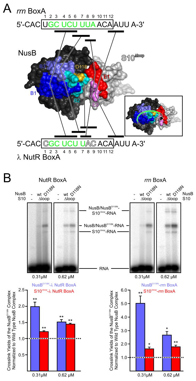
Protein S10 is a component of the 30S ribosomal subunit and participates together with NusB protein in processive transcription antitermination. The molecular mechanisms by which S10 can act as a translation or a transcription factor are not understood. We used complementation assays and recombineering to delineate regions of S10 dispensable for antitermination, and determined the crystal structure of a transcriptionally active NusB-S10 complex. In this complex, S10 adopts the same fold as in the 30S subunit and is blocked from simultaneous association with the ribosome. Mass spectrometric mapping of UV-induced crosslinks revealed that the NusB-S10 complex presents an intermolecular, composite, and contiguous binding surface for RNAs containing BoxA antitermination signals. Furthermore, S10 overproduction complemented a nusB null phenotype. These data demonstrate that S10 and NusB together form a BoxA-binding module, that NusB facilitates entry of S10 into the transcription machinery, and that S10 represents a central hub in processive antitermination.


Figures

Figure 1
Figure 1. Analysis of the S10Δloop Mutant
(A) Co-purification of GST-S10 or GST-S10Δloop and mutants with His6-NusB and mutants. Groups of three lanes show the soluble extract from co-overexpression experiments (first lane), the wash (second lane) and the elution (third lane) from glutathione beads. Co-expressed proteins are indicated above the group of lanes. M - molecular mass marker. (B) Double filter-binding assays of a NusB-S10 complex to an rrn BoxA-containing 19mer RNA. Upper panel - nitrocellulose layer representing bound RNA. Lower panel - nylon filter representing unbound RNA. The upper lanes correspond to the full-length complex, the lower lanes to the NusB-S10Δloop complex. Numbers indicate protein concentrations in μM. (C) Gel analysis of nusE<>kan recombinants. Kanamycin resistant cells from a single colony were analyzed by PCR for configuration of the targeted chromosomal nusE region. Lane 1 -DNA marker (Invitrogen). Lanes 2 and 3 - PCR products from recombinant cells that contained pBADnusE. Lanes 4 and 5 - PCR products from recombinant cells that contained pBADnusEΔloop initially selected either with (lanes 2 and 4) or without (lanes 3 and 5) 0.2 % arabinose. Lane 6 -PCR product control of wt nusE from the bacterial chromosome. Note that a haploid nusE<>kan knockout can be made only when pBADnusE is induced by arabinose, i.e. when wt nusE is expressed from the plasmid (lane 2).
Figure 2
Figure 2. Structure of the NusB-S10Δloop Complex
(A) Ribbon plot of the E. coli NusB-S10Δloop complex. NusB - blue, S10Δloop - red. Secondary structure elements and termini are labeled. The red sphere marks the site at which the ribosome-binding loop of S10 has been replaced by a single serine. I, II –interaction regions on the flank of the first three helix bundle (I) and on a tip of the second three helix bundle (II) of NusB. Inset 1 -NMR structure of ecoNusB (PDB ID 1EY1; (Altieri et al., 2000)) after global superpositioning on the NusB molecule of the present complex. Inset 2 - Structure of S10 from the E. coli 30S subunit (PDB ID 2AVY; (Schuwirth et al., 2005)) after global superpositioning on the S10Δloop molecule of the present complex. (B) Electrostatic surface potentials mapped on the surfaces of NusB (left) and S10Δloop (right) showing a view on the interfaces of both molecules. Blue - positive charge, red - negative charge. Protomers were rotated 90° relative to panel (A) as indicated. (C–F) Details of the NusB-S10Δloop interaction. Interacting residues and secondary structure elements are labeled. Residues of interest are colored by atom type: carbon - as the respective molecules; oxygen - red; nitrogen - blue. Cyan spheres - water molecules. Dashed lines -hydrogen bonds or salt bridges. Views relative to Figure 2A are indicated.
Figure 3
Figure 3. Aspects of the NusB-S10Δloop Interaction
(A) Stereo ribbon plot showing induced fit adjustment of the loop between helices α4 and α5 in NusB. An NMR structure of E. coli NusB (gray; PDB ID 1EY1; (Altieri et al., 2000)) was superimposed on the NusB subunit of the present NusB-S10Δloop complex (blue and red, respectively). The view relative to Figure 2A is indicated. Glu75 of NusB changes its position upon complex formation (arrow) in order to engage in a salt bridge with Arg16 of S10Δloop. Relevant residues are in sticks and colored by atom type as before. (B) Comparison of S10Δloop (red) binding to NusB (blue) and S10 binding to the remainder of the 30S subunit (rRNA - gold; r-proteins - gray). The orientation relative to Figure 2A is indicated. (C) Comparison of the present NusB-S10Δloop complex (blue and red, left) with the M. tuberculosis NusB dimer (blue and cyan, right; PDB ID 1EYV; (Gopal et al., 2000)). The blue NusB molecules of both complexes are in the same orientation.
Figure 4
Figure 4. BoxA RNA Binding
(A) Mapping of crosslinked peptides on the surface of the NusB-S10Δloop complex. The view is from the top of Figure 2A. NusB - dark gray, S10 - light gray. Crosslinked peptides of NusB (B1, B2, B3; see Table S3 for peptide sequences) are dark blue, cyan and steel blue, respectively. Crosslinked peptides of S10 (E1 and E3) are red and violet, respectively. Asp118 - gold. RNAs encompassing the rrn and λ BoxA elements and used for crosslinking are given above and below the structure, respectively. Boxed regions with residue numbers indicate the core BoxA elements. Residues in green of rrn BoxA RNA and λ BoxA RNA have previously been implicated in recruitment of NusB and S10 to antitermination complexes by mutational analysis (Mogridge et al., 1998). Outlined residues differ in λ BoxA compared to rrn BoxA. Black bars designate crosslinked regions of the RNAs. They are connected by lines to the peptides, to which they have been crosslinked (Table S3). Inset 1 illustrates the deduced topology of the NusB-S10-BoxA RNA complexes. (B) Top: Representative crosslinking of λ NutR BoxA RNA (left two panels) or rrn BoxA RNA (right two panels) to NusB-S10Δloop or NusB101-S10Δloop (NusBAsp118Asn-S10Δloop). Two concentrations of protein complex (0.31 μM and 0.62 μM) were crosslinked, resolved on SDS gels and visualized by autoradiography. In each panel, RNA alone is in the left lane, NusB-S10Δloop complex in the central lane and NusB101-S10Δloop complex in the right lane. Bottom: Quantification of crosslink yields. Values are the crosslink yields of the protein components of the NusB101-S10Δloop samples, relative to the crosslink yields of the corresponding components of the NusB-S10Δloop samples. The crosslink yields of the components of the NusB-S10Δloop samples were set at 100 % (dashed lines). Values represent the mean of three independent experiments +/− the standard errors of the mean. Asterisks - p 0.032; double asterisks - p 0.020.
Figure 5
Figure 5. NusB-S10Δloop,Ala86Asp Complex
(A) Comparison of the NusB-S10Δloop complex (left) with the NusB-S10Δloop,Ala86Asp complex (right). Gray meshes - final 2Fo-Fc electron densities covering residue 86 and neighboring residues of the S10Δloop molecules, contoured at the 1σ level. Insets - closeup views of the residue 86 regions. The orientation relative to Figure 2A is indicated. (B) Comparison of the electrostatic surface potentials of the complexes. Blue - positive charge, red - negative charge. Left - NusB-S10Δloop complex. Right - NusB-S10Δloop,Ala86Asp complex. The positions of residue 86 are circled. The orientations are the same as in panel A.

Comment in

Similar articles

Cited by

References

    1. Altieri AS, Mazzulla MJ, Horita DA, Heath Coats R, Wingfield PT, Das A, Court DL, Byrd AR. The structure of the transcriptional antiterminator NusB from Escherichia coli. Nat Struct Biol. 2000;7:470–474. - PMC - PubMed
    1. Belogurov GA, Vassylyeva MN, Svetlov V, Klyuyev S, Grishin NV, Vassylyev DG, Artsimovitch I. Structural basis for converting a general transcription factor into an operon-specific virulence regulator. Mol Cell. 2007;26:117–129. - PMC - PubMed
    1. Berg KL, Squires C, Squires CL. Ribosomal RNA operon anti-termination. Function of leader and spacer region box B-box A sequences and their conservation in diverse micro-organisms. J Mol Biol. 1989;209:345–358. - PubMed
    1. Beuth B, Pennell S, Arnvig KB, Martin SR, Taylor IA. Structure of a Mycobacterium tuberculosis NusA-RNA complex. EMBO J. 2005;24:3576–3587. - PMC - PubMed
    1. Bonin I, Robelek R, Benecke H, Urlaub H, Bacher A, Richter G, Wahl MC. Crystal structures of the antitermination factor NusB from Thermotoga maritima and implications for RNA binding. Biochem J. 2004;383:419–428. - PMC - PubMed

Publication types

MeSH terms

LinkOut - more resources