. 2009 Feb 6;284(6):3728-38.
doi: 10.1074/jbc.M808788200. Epub 2008 Dec 15.

Induction of microRNA-221 by platelet-derived growth factor signaling is critical for modulation of vascular smooth muscle phenotype

Affiliations

Induction of microRNA-221 by platelet-derived growth factor signaling is critical for modulation of vascular smooth muscle phenotype

Brandi N Davis et al. J Biol Chem. .

Abstract

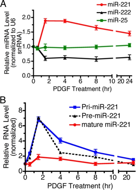
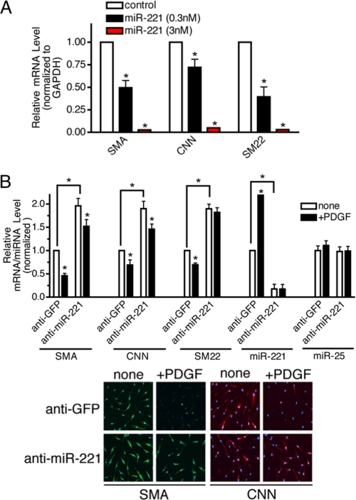
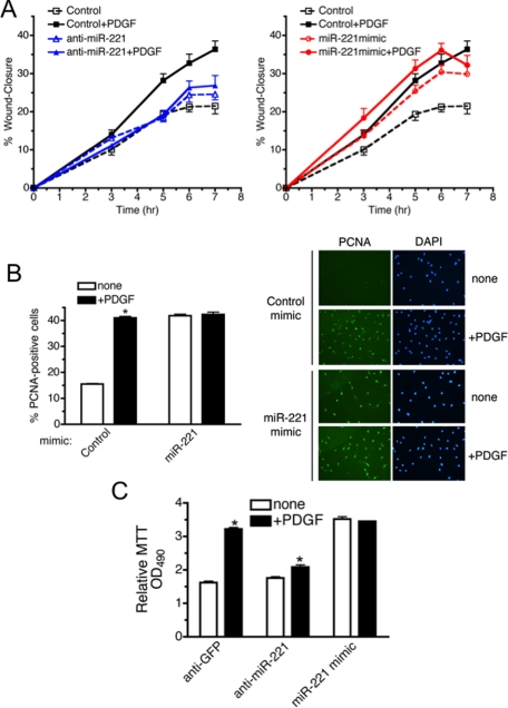
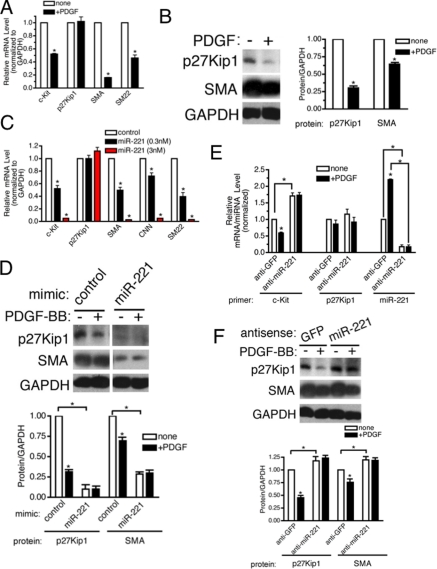
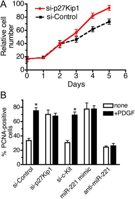
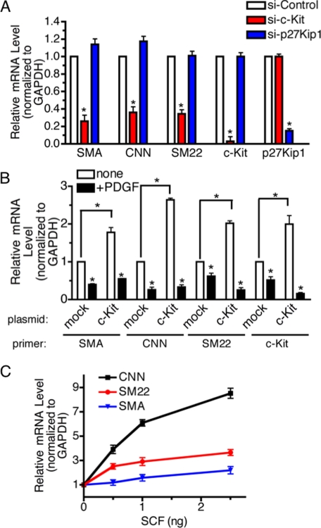
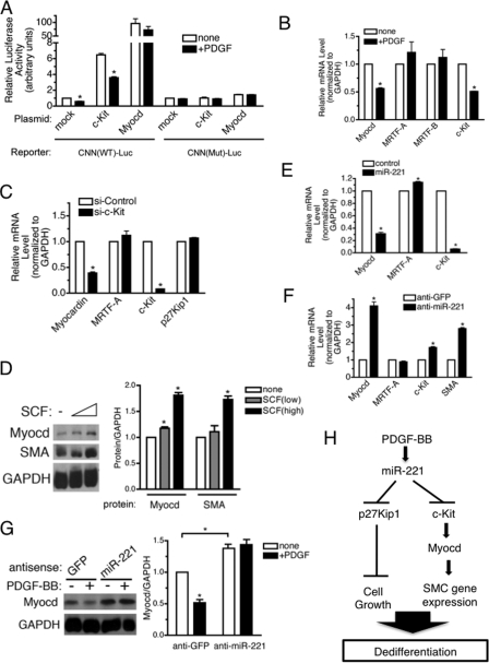
The platelet-derived growth factor (PDGF) signaling pathway is a critical regulator of animal development and homeostasis. Activation of the PDGF pathway leads to neointimal proliferative responses to artery injury; it promotes a switch of vascular smooth muscle cells (vSMC) to a less contractile phenotype by inhibiting the SMC-specific gene expression and increasing the rate of proliferation and migration. The molecular mechanism for these pleiotropic effects of PDGFs has not been fully described. Here, we identify the microRNA-221 (miR-221), a small noncoding RNA, as a modulator of the phenotypic change of vSMCs in response to PDGF signaling. We demonstrate that miR-221 is transcriptionally induced upon PDGF treatment in primary vSMCs, leading to down-regulation of the targets c-Kit and p27Kip1. Down-regulation of p27Kip1 by miR-221 is critical for PDGF-mediated induction of cell proliferation. Additionally, decreased c-Kit causes inhibition of SMC-specific contractile gene transcription by reducing the expression of Myocardin (Myocd), a potent SMC-specific nuclear coactivator. Our study demonstrates that PDGF signaling, by modulating the expression of miR-221, regulates two critical determinants of the vSMC phenotype; they are SMC gene expression and cell proliferation.


Figures

FIGURE 1.
FIGURE 1.
miR-221 is regulated by the PDGF-BB signaling pathway. A, time-course expression of miR-221 (red), miR-222 (black), and miR-25 (green) normalized to U6 small nuclear RNA was examined by qRT-PCR in PASMC stimulated with 20 ng/ml PDGF-BB for 0.5–24 h as indicated. Changes of miR-25 upon PDGF-BB treatment was not statistically significant. B, time-course analysis of the relative expression of miR-221 transcripts (Pri-miR-221), Pre-miR-221 or mature miR-221 normalized to GAPDH (for Pri-miR-221 or Pre-miR-221), or U6 small nuclear RNA (for mature miR-221) in PASMC treated with PDGF-BB (20 ng/ml) for 0.5–24 h as indicated. The average of three experiments, each performed in triplicate with S.E., are presented.
FIGURE 2.
FIGURE 2.
miR-221 is critical for the PDGF-dependent inhibition of SMC gene markers. A, PASMC were transfected with negative control mimic or two different doses of miR-221 mimic (0.3 or 3 nm). Total RNA was harvested 48 h after transfection. The level of expression of SMC genes (SMA, CNN, and SM22) was examined by qRT-PCR analysis. B, PASMC were transfected with antisense oligonucleotides (106 nm) to miR-221 (anti-mIR-221) or GFP (anti-GFP) as control. Cells were then treated with PDGF-BB (20 ng/ml) for 48 h and subjected to qRT-PCR analysis or immunofluorescence staining of SMC markers (SMA, CNN, and SM22). The effect and specificity of anti-miR-221 on miR-221 expression was evaluated by qRT-PCR analysis of miR-221 and miR-25 (control) presented as a ratio to the U6 small nuclear RNA level. SMA and CNN protein expression was examined by FITC-conjugated anti-SMA antibody (green) and Cy3-conjugated anti-CNN antibody (red), respectively. Nuclei were stained with DAPI (blue).
FIGURE 3.
FIGURE 3.
miR-221 is critical for the PDGF-dependent promotion of cell migration and growth. A, PASMC transfected with anti-GFP (Control), anti-miR-221 (left panel), or 0.3 nm miR-221 mimic (right panel) were subjected to the scratch wound assay in the presence or absence of 20 ng/ml PDGF-BB. Results are the mean ± S.E. of triplicate measurements of three independent experiments. B, PASMC transfected with miR-221 mimic (0.3 nm) or control mimic followed by PDGF-BB treatment for 24 h were stained with FITC-conjugated antibody against proliferation marker PCNA and DAPI. >150 cells were counted per condition, and the percentage of PCNA-positive cells is presented. Results are the mean ± S.E. for triplicate assays of three independent experiments. C, PASMC transfected with anti-miR-221, anti-GFP (control), or miR-221 mimic (0.3 nm) followed by PDGF-BB treatment for 24 h were subjected to MTT cell proliferation assay. Results are indicated as the absorbance readings at 490 nm. Results are the mean ± S.E. for triplicate assays of three independent experiments.
FIGURE 4.
FIGURE 4.
miR-221 mediates reduced expression of c-kit and p27Kip1. A, PASMC were treated with 20 ng/ml PDGF-BB for 24 h followed by qRT-PCR analysis of two known targets of miR-221, c-Kit, and p27Kip1 and two SMC marker genes, SMA and SM22. B, immunoblotting with anti-p27Kip1, anti-SMA, and anti-GAPDH antibody (loading control) was performed in parallel with qRT-PCR analysis (right panel). Proteins bands were quantitated by densitometry, and relative amounts of proteins normalized to GAPDH are presented. C, PASMC were transfected with negative control miRNA or two different doses of miR-221 mimic (0.3 or 3 nm). Total RNA was harvested 48 h after transfection, and the level of expression of c-kit, p27Kip1, and SMC genes (SMA, CNN, and SM22) was examined by qRT-PCR analysis. D, immunoblot analysis of p27Kip1, SMA, and GAPDH using cell lysates of PASMC treated equally to B. Proteins bands were quantitated by densitometry, and relative amounts of proteins normalized to GAPDH are presented. E, PASMC were transfected with antisense oligonucleotides (106 nm) to miR-221 (anti-miR-221) or GFP (anti-GFP) as control. Cells were then treated with PDGF-BB (20 ng/ml) for 48 h and subjected to qRT-PCR analysis of c-kit and p27Kip1. The effect of anti-miR-221 was evaluated by qRT-PCR analysis of miR-221 normalized to U6 small nuclear RNA. F, immunoblot analysis of p27Kip1, SMA, or GAPDH using cell lysates of PASMC treated as described in E. Proteins bands were quantitated by densitometry, and relative amounts of proteins normalized to GAPDH are presented.
FIGURE 5.
FIGURE 5.
p27Kip1 is responsible for PDGF-mediated promotion of cell growth. A, after transfection of si-Control or si-p27Kip1 in PASMC, 5 × 10⁵ cells were seeded, and proliferation was determined by counting cell number over 5 days. Transfection efficiency of siRNA was at least 95% as determined by FITC-labeled siRNA (data not shown). B, PASMC transfected with si-p27Kip1, si-c-Kit, si-Control, miR-221 mimic (0.3 nm), or anti-miR-221 followed by PDGF-BB treatment for 24 h. Cells were stained with FITC-conjugated anti-PCNA antibody and DAPI. A total of >150 cells were counted per condition, and the percentages of PCNA-positive cells are presented.
FIGURE 6.
FIGURE 6.
Targeted inhibition of c-kit mediates PDGF-dependent suppression of SMC genes. A, PASMC were transfected with non-targeting control siRNA (si-Control) or siRNA for c-kit (si-c-Kit), or p27Kip1 (si-p27Kip1). Forty-eight hours after transfection, total RNAs were extracted and subjected to qRT-PCR analysis to examine mRNA levels of SMA, CNN, SM22, c-kit, or p27Kip1 normalized to GAPDH. The average of three experiments, each performed in triplicate with S.E., are presented. B, PASMC were transfected with empty vector (mock) or human c-Kit expression plasmid followed by PDGF-BB treatment for 24 h and qRT-PCR analysis of SMC genes (SMA, CNN, and SM22) and human c-kit. C, PASMC were treated with 0.5, 1.0, or 2.5 ng of SCF for 24 h followed by qRT-PCR analysis to examine mRNA levels of SMA, CNN, or SM22.
FIGURE 7.
FIGURE 7.
c-Kit pathway induces SMC gene expression through induction of a critical transcription factor Myocd. A, wild type (CNN(WT)-Luc) or mutant (CNN(Mut)-Luc) CNN gene promoter-luciferase reporter construct was transiently transfected into PASMC. Cells were treated with or without PDGF-BB for 20 h followed by luciferase assay. Luciferase activities were normalized to the β-galactosidase activities. B, PASMC were treated with 20 ng/ml PDGF-BB for 24 h followed by RNA extraction and qRT-PCR analysis of mRNA encoding Myocd family proteins (Myocd, MRTF-A, and MRTF-B) or c-kit. C, PASMC were transfected with 4 nm non-targeting control siRNA (si-Control) or siRNA for c-kit (si-c-Kit). Forty-eight hours after transfection, total RNAs were extracted and subjected to qRT-PCR analysis to examine mRNA levels of Myocd, MRTF-A, c-kit, or p27Kip1 normalized to GAPDH. D, PASMC were treated with 2.5 or 5 ng of SCF for 8 h followed by immunoblot analysis to examine protein levels of Myocd, SMA, and GAPDH (loading control). Proteins bands were quantitated by densitometry, and relative amounts of proteins normalized to GAPDH are presented. E, PASMC were transfected with control mimic, or miR-221 mimic RNA was harvested 48 h after transfection and subjected to qRT-PCR analysis of mRNA encoding Myocd family proteins (Myocd and MRTF-A) or c-kit normalized to GAPDH. F, PASMC were transfected with anti-GFP (control) or anti-miR-221 mimic followed by qRT-PCR analysis of mRNA encoding Myocd family proteins (Myocd and MRTF-A), c-kit or SMA normalized to GAPDH. G, PASMC transfected with anti-GFP (control) or anti-miR-221 mimic were treated with 20 ng/ml PDGF-BB for 48 h. Total cell lysates were subjected to immunoblotting with anti-Myocd and anti-GAPDH antibody (loading control). Proteins bands were quantitated by densitometry, and relative amounts of proteins normalized to GAPDH are presented. H, schematic diagram of pleiotropic PDGF action mediated by induction of miR-221.

Similar articles

Cited by

References

    1. Andrae, J., Gallini, R., and Betsholtz, C. (2008) Genes Dev. 22 1276-1312 - PMC - PubMed
    1. Owens, G. K., Kumar, M. S., and Wamhoff, B. R. (2004) Physiol. Rev. 84 767-801 - PubMed
    1. Kim, V. N. (2005) Nat. Rev. Mol. Cell Biol. 6 376-385 - PubMed
    1. Niwa, R., and Slack, F. J. (2007) Curr. Opin. Genet. Dev. 17 145-150 - PubMed
    1. Hammond, S. M. (2006) Cancer Chemother. Pharmacol. 58 63-68 - PubMed

Publication types

MeSH terms

LinkOut - more resources