. 2008 Dec 22;205(13):3145-58.
doi: 10.1084/jem.20081661. Epub 2008 Dec 15.

Interleukin (IL)-1 promotes allogeneic T cell intimal infiltration and IL-17 production in a model of human artery rejection

Affiliations

Interleukin (IL)-1 promotes allogeneic T cell intimal infiltration and IL-17 production in a model of human artery rejection

Deepak A Rao et al. J Exp Med. .

Abstract

Interleukin (IL) 1alpha produced by human endothelial cells (ECs), in response to tumor necrosis factor (TNF) or to co-culture with allogeneic T cells in a TNF-dependent manner, can augment the release of cytokines from alloreactive memory T cells in vitro. In a human-mouse chimeric model of artery allograft rejection, ECs lining the transplanted human arteries express IL-1alpha, and blocking IL-1 reduces the extent of human T cell infiltration into the artery intima and selectively inhibits IL-17 production by infiltrating T cells. In human skin grafts implanted on immunodeficient mice, administration of IL-17 is sufficient to induce mild inflammation. In cultured cells, IL-17 acts preferentially on vascular smooth muscle cells rather than ECs to enhance production of proinflammatory mediators, including IL-6, CXCL8, and CCL20. Neutralization of IL-17 does not reduce T cell infiltration into allogeneic human artery grafts, but markedly reduces IL-6, CXCL8, and CCL20 expression and selectively inhibits CCR6(+) T cell accumulation in rejecting arteries. We conclude that graft-derived IL-1 can promote T cell intimal recruitment and IL-17 production during human artery allograft rejection, and suggest that targeting IL-1 in the perioperative transplant period may modulate host alloreactivity.


Figures
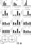
Figure 1.
Figure 1.
Human ECs use cell-associated IL-1α to promote alloreactive memory CD4+ T cell IL-17 production. (A) ECs or SMCs were treated with 10 ng/ml TNF for the indicated times, and cell lysates and supernatants were assayed for IL-1α by ELISA. (B) MHC class II–positive ECs were co-cultured with allogeneic CD4+ T cells for the indicated times, and then ECs were purified and assayed for IL-1α in EC lysates and supernatants by ELISA. For A and B, data from one of three experiments with similar results are shown. (C) MHC class II–positive ECs were co-cultured with CD4+ T cells for 48 h with a neutralizing anti-TNF antibody or isotype control, and IL-1α in purified EC lysates was measured by ELISA. Means ± SEM (n = 3 experiments) are shown. (D) Untreated or TNF-pretreated ECs were co-cultured with allogeneic memory CD4+ T cells with 5 μg/ml of a neutralizing anti–IL-1α antibody or isotype control. Culture medium was assayed for IFN-γ and IL-17 by ELISA at 24 h. (E) CD4+ T cells from primary co-cultures were restimulated with fresh ECs, and the culture medium was assayed for IFN-γ, IL-17, and IL-5 by ELISA at 24 h. Means ± SEM of fold changes relative to the untreated, isotype control group from seven (D) or three (E) experiments are shown. The ranges of values in the control group were 1–33 pg/ml (IFN-γ) and 1–10 pg/ml (IL-17) in D, and 301–645 pg/ml (IFN-γ), 5–128 pg/ml (IL-17), and 8–428 pg/ml (IL-5) in E. (F) Purified memory CD4+ T cells were co-cultured with CD32-transduced, anti-CD3 mAb–coated ECs that were either pretreated with TNF or left untreated, and then T cells were restimulated with PMA plus ionomycin and analyzed by ICS. A neutralizing antibody to IL-1α or isotype control was added to the primary co-culture as indicated. Means ± SEM of fold changes relative to the untreated, isotype control group from six different donors are shown. (G) CFSE-labeled memory CD4+ T cells co-cultured with allogeneic ECs for 14 d were restimulated with PMA and ionomycin and analyzed by ICS. Representative dot plots of CFSE dilution and IL-17 expression in CD45+-gated lymphocytes are shown (numbers indicate percentages). (H) Scatter plot of the fraction of CFSE-low, IL-17+ cells in individual microtiter co-cultures. Pooled data from two experiments with similar results are shown. Horizontal bars indicate means. *, P < 0.05; #, P < 0.01; π, P < 0.001.
Figure 2.
Figure 2.
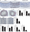
IL-1 blockade reduces T cell–mediated injury and IL-17 production in human artery allografts. (A) Immunohistochemical detection of human endothelium (CD31) and IL-1α in a human artery aortic interposition graft. Staining with an isotype-matched primary antibody is shown as a control. Bars, 25 μm. (B) 5-μm transverse sections were stained with EVG, and the total vessel area (area bounded by the external elastic lamina and the lumen) and the intimal area (area bounded by the internal elastic lamina and the lumen) were quantified using ImageJ software. Bars, 100 μm. (C) Immunohistochemical identification and quantification of human CD45RO+ T cells in graft intimas. Bars, 25 μm. (D) RT-PCR analysis of CD3ε (normalized to GAPDH), IFN-γ, and IL-17 (normalized to CD3ε), and the ratio of IL-17/IFN-γ in artery grafts undergoing T cell–mediated rejection. For B–D, means ± SEM (n = 10 per group) are shown. *, P < 0.05.
Figure 3.
Figure 3.
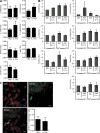
IL-1 blockade does not alter memory CD4+ T cell expression of Th17-associated markers. (A) RT-PCR analysis of T cell genes normalized to CD3ε in artery grafts undergoing T cell–mediated rejection treated with IL-1Ra or PBS. Means ± SEM (n = 10 per group) are shown. (B) RT-PCR analysis of T cells activated by co-culture with CD32-transduced ECs overlayed with anti-CD3 mAb. ECs pretreated with TNF or left untreated were overlayed with anti-CD3 mAb and co-cultured with purified, memory CD4+ T cells with an anti–IL-1α antibody for 3 d. T cells were then rested with 3 d and RNA was isolated. Gene expression was normalized to CD3ε, and means ± SEM of fold changes relative to the untreated, isotype control group from six different donors are shown. (C) Immunofluorescence microscopy of intimal T cells stained with anti-CD3 (red) and anti-CD161 (green). The arrowhead points to a cell positive for both CD161 and CD3. Quantification of CD161+/CD3+ double-positive cells is expressed as a fraction of total CD3+ T cells in the intimas of artery grafts treated with IL-1Ra or PBS. Means ± SEM (n = 7 per group) are shown. Bars, 10 μm.
Figure 4.
Figure 4.
IL-17 induces acute inflammatory responses in human skin grafts. Well-healed skin grafts on CB.17 SCID/beige mice were harvested 6 h after injection of 1 μg IL-17 or PBS. (A) Immunohistochemical quantification of Gr-1+ mouse neutrophils in human skin grafts. Bars, 100 μm. (B) Quantitative RT-PCR analysis of IL-6 and CCL20 expression normalized to GAPDH in injected grafts. (C) Immunohistochemical identification of human E-selectin+ vessels (arrow) in skin grafts. Bars, 25 μm. Data are pooled from two independent experiments with similar results, and means ± SEM (n = 6 per group) are shown in A and B. *, P < 0.05; #, P < 0.01.
Figure 5.
Figure 5.
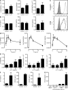
IL-17 induces inflammatory gene expression in cultured human SMCs. (A) ECs were treated with 100 ng/ml IL-17 for 6 h, and target gene expression was measured by quantitative RT-PCR. Data shown are means ± SD of triplicate samples from one of two independent experiments with similar results. (B) ECs were treated with 100 ng/ml IL-17, and the culture medium was assayed for IL-6, CXCL8, and CCL20 by ELISA. Means ± SEM (n = 3 experiments) are shown. (C) Measurement of surface E-selectin and ICAM-1 expression on ECs treated for 6 h with 100 ng/ml IL-17 (dashed line), 10 ng/ml TNF (dotted line), or vehicle control (continuous line) by flow cytometry. Shaded histograms represent staining with an isotype-matched control antibody. Representative data are from one of three independent experiments with similar results. (D and E) SMCs were treated with 100 ng/ml IL-17 for the indicated times (D) or with IL-17 at the indicated concentrations for 6 h (E), and target gene expression was measured by quantitative RT-PCR. (F) SMCs were treated with 100 ng/ml IL-17 for 24 h, and the culture medium was assayed for IL-6, CXCL8, and CCL20 by ELISA. (G) SMCs were treated with the IL-17 and TNF alone or in combination, and the culture medium was assayed for CCL20 by ELISA. Means ± SEM from four (E and G) or six (D and F) experiments are shown.
Figure 6.
Figure 6.
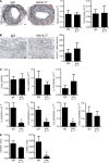
IL-17 blockade reduces inflammatory responses during allogeneic T cell–mediated rejection of human artery grafts. Human PBMCs were adoptively transferred into mice bearing human artery interposition grafts, and mice were treated three times weekly with a neutralizing anti–IL-17 antibody or an isotype control. (A) 5-μm transverse sections were EVG stained, and the vessel area and the intimal area were quantified using ImageJ software. Bars, 100 μm. (B) Immunohistochemical identification and quantification of human CD45RO+ T cells in graft intimas. Bars, 25 μm. (C) RT-PCR analysis of cytokine and chemokine expression (normalized to GAPDH) in grafts from mice treated with an anti–IL-17 antibody or an isotype control. (D) RT-PCR analysis of chemokine receptor expression (normalized to CD3ε) in grafts from mice treated with an anti–IL-17 antibody or an isotype control. Means ± SEM are shown (IgG, n = 6; anti–IL-17, n = 5). *, P < 0.05; #, P < 0.01.

Similar articles

Cited by

References

    1. Libby, P., and J.S. Pober. 2001. Chronic rejection. Immunity. 14:387–397. - PubMed
    1. Nickeleit, V., E.C. Vamvakas, M. Pascual, B.J. Poletti, and R.B. Colvin. 1998. The prognostic significance of specific arterial lesions in acute renal allograft rejection. J. Am. Soc. Nephrol. 9:1301–1308. - PubMed
    1. Racusen, L.C., K. Solez, R.B. Colvin, S.M. Bonsib, M.C. Castro, T. Cavallo, B.P. Croker, A.J. Demetris, C.B. Drachenberg, A.B. Fogo, et al. 1999. The Banff 97 working classification of renal allograft pathology. Kidney Int. 55:713–723. - PubMed
    1. Trpkov, K., P. Campbell, F. Pazderka, S. Cockfield, K. Solez, and P.F. Halloran. 1996. Pathologic features of acute renal allograft rejection associated with donor-specific antibody: analysis using the Banff grading schema. Transplantation. 61:1586–1592. - PubMed
    1. Choi, J., D.R. Enis, K.P. Koh, S.L. Shiao, and J.S. Pober. 2004. T lymphocyte-endothelial cell interactions. Annu. Rev. Immunol. 22:683–709. - PubMed

Publication types

MeSH terms