. 2009 Jan 7;28(1):69-80.
doi: 10.1038/emboj.2008.254. Epub 2008 Dec 4.

Identification, structure, and functional requirement of the Mediator submodule Med7N/31

Affiliations

Identification, structure, and functional requirement of the Mediator submodule Med7N/31

Tobias Koschubs et al. EMBO J. .

Abstract

Mediator is a modular multiprotein complex required for regulated transcription by RNA polymerase (Pol) II. Here, we show that the middle module of the Mediator core contains a submodule of unique structure and function that comprises the N-terminal part of subunit Med7 (Med7N) and the highly conserved subunit Med31 (Soh1). The Med7N/31 submodule shows a conserved novel fold, with two proline-rich stretches in Med7N wrapping around the right-handed four-helix bundle of Med31. In vitro, Med7N/31 is required for activated transcription and can act in trans when added exogenously. In vivo, Med7N/31 has a predominantly positive function on the expression of a specific subset of genes, including genes involved in methionine metabolism and iron transport. Comparative phenotyping and transcriptome profiling identify specific and overlapping functions of different Mediator submodules.


Figures
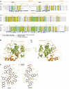
Figure 1
Figure 1
Structure of the Med7N/31 Mediator subcomplex. (A) Multiple sequence alignment and structural conservation of Med7 and Med31 from Saccharomyces cerevisiae (S.c.), Schizosaccharomyces pombe (S.p.), Caenorhabditis elegans (C.e.), Drosophila melanogaster (D.m.), Homo sapiens (H.s.) and Arabidopsis thaliana (A.t.). Secondary structure elements are indicated above the sequences (spirals, α- and 310(η)-helices; arrows, β-strands; lines, ordered but without secondary structure; dashed lines, disordered regions). Invariant and conserved residues are highlighted in green and yellow, respectively. Surface accessibility is indicated below the sequences (blue, exposed; white, buried). Cleavage sites revealed by limited proteolysis with trypsin or chymotrypsin are indicated with black arrows. The loop between Med7N helices α1 and η2 (residues 41–55) was disordered. Sequence alignments were done with MUSCLE (Edgar, 2004) and figures were prepared with ESPript (Gouet et al, 1999). (B) Ribbon model representation of the Med7N/31 crystal structure. Two views are shown that are related by a 180° rotation around the vertical axis. Med7 is depicted in orange and Med31 in green. Secondary structure elements are labelled according to (A). This and other figures were prepared with PyMol (DeLano, 2002). (C) Ligplot view (Wallace et al, 1995) of the interactions of the Med7N polyproline stretches pPS1 and pPS2 with Med31.
Figure 2
Figure 2
Analysis of the Med7N/31 structure. (A) Surface conservation of the Med7N/31 subcomplex. Invariant and conserved residues are highlighted in green and yellow, respectively. The surface is semitransparent to show the underlying residues in a stick model. Two views are shown that are related by a 180° rotation around the vertical axis. (B) Surface charge distribution. Red, blue, and white areas indicate negative, positive, and neutral charges, respectively. (C) The Med7N polyproline stretches adopt a CTD-like conformation. Superimposition of Med7N pPS1 and pPS2 (orange) with the Y(SEP)PT(SEP)PS peptide from the human Pin1 structure (1F8A, light purple). (D) Comparison of pPS1 in the crystal structures of Med7N/31 and Med7N/31 with pPS1 mutated to a CTD heptad repeat (mutant 6; Supplementary Table II).
Figure 3
Figure 3
Functional analysis of Med7N/31 in vivo and in vitro. (A) Yeast complementation assays. Plasmids encoding full-length MED7 or N-terminal truncations of MED7 were cloned into the SmaI/XbaI restriction sites of a pAL vector or through SalI/XbaI into a pRS315 vector. Individual plasmids were transformed into the MED7 shuffle strain (Supplementary Table III) and streaked onto 5-FOA-containing plates to shuffle out the MED7 encoding URA3 plasmid. Yeast cells carrying only MED7C (102–222) are viable, whereas the Med7 N-terminal part including part of the linker (residues 1–101) cannot rescue cell growth. (B) Mutant strains exhibit a slow-growth phenotype. Note that growth curves are on a logarithmic scale. (C) Med7N/31 is required for Gal4–VP16-activated transcription in vitro (lane 1). Transcription can be rescued by the addition of TAP-purified Mediator (0.2 pmol, lane 2) or recombinant Med7N/31 (rMed7N/31, 2–200 pmol, lanes 3–5). Addition of both rMed7N/31 (2–200 pmol) and recombinant TFIIS (rTFIIS, 20 pmol, lanes 6–8) enhances the signal. (D) A Med7N/31Δ nuclear extract that was rescued by the addition of recombinant Med7N/31 (lane 2, 200 pmol) was additionally stimulated by the addition of TFIIS (lane 5, 60 pmol). TFIIS alone can partially compensate for loss of Med7N/31 (lanes 3 and 4, 20–60 pmol). This stimulation was not observed with a med18Δ nuclear extract that was rescued by the addition of recombinant Med18/20 (rMed18/20, lanes 7–10, 20–60 pmol). (E) TFIIS addition stimulates basal and Gal4–VP16-activated transcription of wild-type (WT) nuclear extracts (lanes 2 and 3, and 5 and 6, respectively, 20–60 pmol). Recombinant Med7/31 or Med18/20 did not stimulate WT nuclear extracts. (F) Deletion of Med7N/31 or its subunits does not lead to loss of additional Mediator subunits. C-terminally TAP-tagged Med18 was purified from med31Δ/Med18-TAP, med7NΔ/Med18-TAP, and med7N/31Δ/Med18-TAP strains as described (Puig et al, 2001; Larivière et al, 2008). Copurifying proteins were separated on a 12% NuPAGE gel (Invitrogen), and bands were stained with Coomassie blue. The identity of all Mediator subunits except Med31 was confirmed by mass spectrometry (Supplementary Table V). Contaminants such as ribosomal proteins or degradation products of Mediator subunits were especially detected at lower molecular weights and do partially overlap with smaller subunits such as Med7C.
Figure 4
Figure 4
Transcriptome profiling analysis and phenotypic correlation. (A) Cluster diagram (Euclidean distance) of genes exhibiting significantly altered mRNA levels (at least 1.7-fold, vertical axis) for different Mediator subunit deletion strains and TFIIS (horizontal axis). Changes in mRNA levels compared with the wild-type strain are depicted in red (up), green (down), or black (no change). (B) Pearson's correlation matrix for expression profiles of the Mediator middle subunit deletion strains med31Δ, med7NΔ, med7N/31Δ, the head subunit med8CΔ, med18Δ, med20Δ, the tail subunit deletion strains med2Δ, med3Δ, and the deletion profile of the general transcription factor dst1Δ strain. (C) Venn diagram showing the overlap between the investigated med31Δ, med7NΔ, and med7N/31Δ strains. Although these three strains exhibit a very high overlap, the overlap with the dst1Δ strain was not regarded as statistically significant. (D) Number of significantly altered genes of all four investigated deletion strains, split into up- and downregulated genes. (E) Selected phenotyping analysis results. Using 10-fold serial dilutions of yeast strains spotted onto selective agar plates, we screened for phenotypes, occurring under certain growth conditions. All assays were compared with a control plate on YPD, synthetic complete (SC), or SD media plates. As typical examples, the results from the growth inhibition assay on SD (-met) plates, the siderophore uptake assay (detected using bathophenanthroline disulphonic acid (BPDS)), and the growth inhibition on paramomycin assay are depicted.
Figure 5
Figure 5
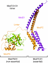
Structural overview of the trimeric Med7/21/31 complex architecture. Structures of the non-essential Med7N/31 submodule (this study) and the previously described essential Med7C/21 subcomplex are drawn to scale.

Similar articles

Cited by

References

    1. Adzhubei AA, Sternberg MJ (1993) Left-handed polyproline II helices commonly occur in globular proteins. J Mol Biol 229: 472–493 - PubMed
    1. Alexa A, Rahnenfuhrer J, Lengauer T (2006) Improved scoring of functional groups from gene expression data by decorrelating GO graph structure. Bioinformatics 22: 1600–1607 - PubMed
    1. Askree SH, Yehuda T, Smolikov S, Gurevich R, Hawk J, Coker C, Krauskopf A, Kupiec M, McEachern MJ (2004) A genome-wide screen for Saccharomyces cerevisiae deletion mutants that affect telomere length. Proc Natl Acad Sci USA 101: 8658–8663 - PMC - PubMed
    1. Asturias FJ, Jiang YW, Myers LC, Gustafsson CM, Kornberg RD (1999) Conserved structures of Mediator and RNA polymerase II holoenzyme. Science 283: 985–987 - PubMed
    1. Bäckström S, Elfving N, Nilsson R, Wingsle G, Björklund S (2007) Purification of a plant Mediator from Arabidopsis thaliana identifies PFT1 as the Med25 subunit. Mol Cell 26: 717–729 - PubMed

Publication types

MeSH terms