. 2008 Dec 2:8:57.
doi: 10.1186/1471-230X-8-57.

Wnt-reporter expression pattern in the mouse intestine during homeostasis

Affiliations

Wnt-reporter expression pattern in the mouse intestine during homeostasis

Paige S Davies et al. BMC Gastroenterol. .

Abstract

Background: The canonical Wnt signaling pathway is a known regulator of cell proliferation during development and maintenance of the intestinal epithelium. Perturbations in this pathway lead to aberrant epithelial proliferation and intestinal cancer. In the mature intestine, proliferation is confined to the relatively quiescent stem cells and the rapidly cycling transient-amplifying cells in the intestinal crypts. Although the Wnt signal is believed to regulate all proliferating intestinal cells, surprisingly, this has not been thoroughly demonstrated. This important determination has implications on intestinal function, especially during epithelial expansion and regeneration, and warrants an extensive characterization of Wnt-activated cells.

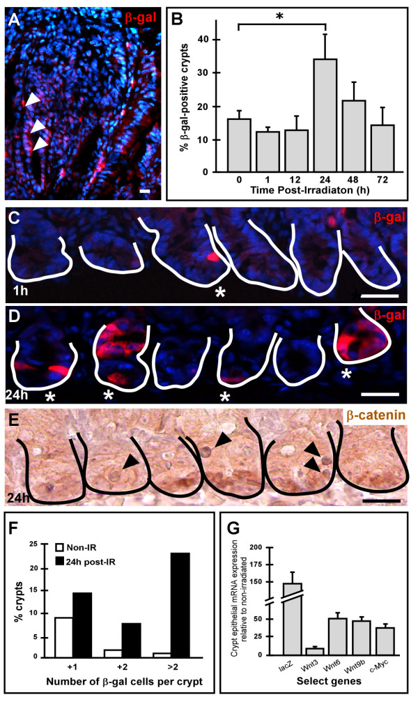
Methods: To identify intestinal epithelial cells that actively receive a Wnt signal, we analyzed intestinal Wnt-reporter expression patterns in two different mouse lines using immunohistochemistry, enzymatic activity, in situ hybridization and qRT-PCR, then corroborated results with reporter-independent analyses. Wnt-receiving cells were further characterized for co-expression of proliferation markers, putative stem cell markers and cellular differentiation markers using an immunohistochemical approach. Finally, to demonstrate that Wnt-reporter mice have utility in detecting perturbations in intestinal Wnt signaling, the reporter response to gamma-irradiation was examined.

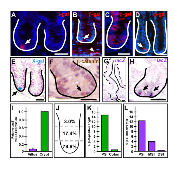
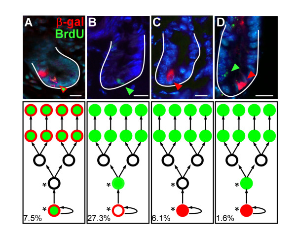
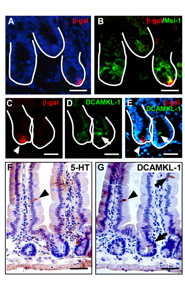
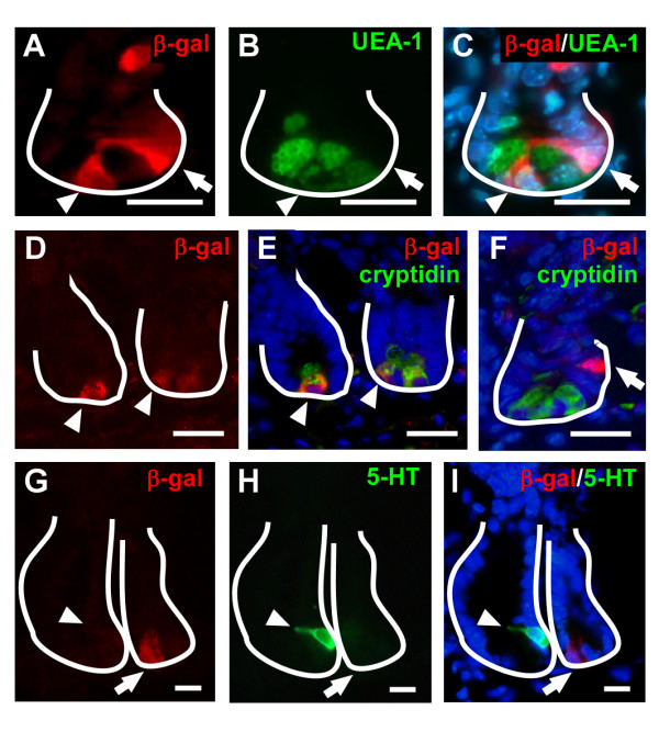
Results: Wnt-activated cells were primarily restricted to the base of the small intestinal and colonic crypts, and were highest in numbers in the proximal small intestine, decreasing in frequency in a gradient toward the large intestine. Interestingly, the majority of the Wnt-reporter-expressing cells did not overlap with the transient-amplifying cell population. Further, while Wnt-activated cells expressed the putative stem cell marker Musashi-1, they did not co-express DCAMKL-1 or cell differentiation markers. Finally, gamma-irradiation stimulated an increase in Wnt-activated intestinal crypt cells.

Conclusion: We show, for the first time, detailed characterization of the intestine from Wnt-reporter mice. Further, our data show that the majority of Wnt-receiving cells reside in the stem cell niche of the crypt base and do not extend into the proliferative transient-amplifying cell population. We also show that the Wnt-reporter mice can be used to detect changes in intestinal epithelial Wnt signaling upon physiologic injury. Our findings have an important impact on understanding the regulation of the intestinal stem cell hierarchy during homeostasis and in disease states.


Figures

Figure 1
Figure 1
Adult mouse expression pattern of Wnt-receiving epithelial cells. (A,C,D) Cryopreserved adult TOPGAL mouse proximal small intestinal (PSI) or colonic and (B) BatGal mouse PSI tissue sections were stained with antibodies against β-galactosidase (β-gal, red) and counterstained with Hoechst dye (blue). (A,B) The majority of crypts in the PSI contained only one Wnt-activated cell or was devoid of positive cells (arrow). There were occasional mesenchymal cells positive for β-gal (arrowhead) in BatGal intestines (B). (C) Occasionally, β-gal-expressing cells were detected throughout the crypt epithelium and on adjacent villi. (D) Colonic crypts contained only rare single β-gal positive cells near the crypt base. (E) Wnt-receiving cells detected by enzymatic activity, X-gal staining (blue, arrow). (F) Adult wild-type mouse PSI was stained with antibodies against β-catenin (brown; arrow) to detect nuclear expression and counterstained with Hematoxylin. (G-I) Analyses of reporter RNA expression pattern and localization was determined by in situ hybridization (G,H; purple, arrow) and are consistent with the expression pattern in (A). (I) qRT-PCR for lacZ gene expression in isolated crypt or villus epithelial cells from TOPGAL PSI demonstrated expression in the crypts. (J) The crypt localization of β-gal-positive cells was highest in the lower third and decreased in numbers in the middle and upper third. (K) Crypts with Wnt-receiving cells in TOPGAL intestinal sections were higher in the PSI (15.2%) and decreased down the length of the intestine to 0.8% in the colon. (L) The number of β-gal-positive villi also reflected a decreasing gradient with the highest numbers in the PSI (12.3%), less in the the middle small intestine (MSI; 4.0%) and the least in the distal small intestine (DSI; 0.2%). Solid white or black line demarks the epithelial-mesenchymal boundary. Dashed line outlines the apical epithelial border. Bar = 25 μm.
Figure 2
Figure 2
Wnt-activated cells represent progenitor cells within the intestinal crypt. (A-C) Cryopreserved intestinal tissue sections from TOPGAL adult mice co-stained with antibodies to β-gal (red) and Ki67 (green) then counter-stained with Hoechst (blue). Arrow indicates a cell with β-gal staining and arrowhead designates a cell co-staining for both markers. (D-G) Co-localization of BrdU (green) and β-gal (red) expression in crypt and villus epithelial cells from adult TOPGAL mice injected with BrdU 2 days prior to sacrifice. Green arrow denotes β-gal-positive cells at the lagging edge of migrating BrdU-positive cells up the villus. Red bracket indicates β-gal-positive villus epithelium. White asterisk marks β-gal and BrdU double-positive crypt cells. (F-G) Higher magnification of crypt regions in D-E. Solid white line demarks the epithelial-mesenchymal boundary. Dashed white line outlines the apical epithelial border. Counter-stained with Hoechst dye (blue). Bar = 25 μm.
Figure 3
Figure 3
β-gal and BrdU co-staining scenarios. Wnt-reporter mouse intestines were injected with BrdU 2 days prior to analyses to assess the proliferative status of the β-gal positive crypt-based cells. (A) Approximately 7.5% of crypts contained a cell that was dual-labeled for β-gal and BrdU, reflecting cells that have been retained within the crypt (label-retaining cells) and that were Wnt-activated. (B) Approximately 27.3% of crypts contained a single BrdU-positive cell, possibly representing a "stem cell" that is not designated by the Wnt signaling pathway. This would be in line with the recently identified Bmi-1 positive stem cell. (C) 6.1% of crypts contained a single positive β-gal cell. This cell likely represents a cell that is activated by the Wnt signal after the effective BrdU labeling half-life in the animal. Finally, (D) a small percentage of crypts, 1.6%, contained a β-gal-positive cell and a BrdU-positive cell distinct from one another, likely representing a combination of the described scenarios. These scenarios are schematized in cartoon form beneath the corresponding fluorescent image that describes our perception of what each scenario may represent. In classical stem cell hierarchy, the lowest circle represents a progenitor cell residing near the base of the crypt and upper circles represent the progeny. Solid green circles represent BrdU-positive cells, solid red circles represent an activated Wnt cell, open red circles represent a cell that may have been Wnt-activated prior to BrdU labeling. These many different scenarios reflect the complex nature of the role of Wnt signaling on the stem cell hierarchy within the intestinal crypt.
Figure 4
Figure 4
Characterization of putative stem cell markers in Wnt-activated cells. (A-B) The putative stem cell marker, Musashi-1 (Msi-1; green) had broad expression within the crypt and co-localized with crypt β-gal-expressing cells (red). (C-E) β-gal-positive cells (red) do not co-localize with another putative stem cell marker, DCAMKL-1 (green). Solid white line marks the epithelial-mesenchymal boundary of the intestinal crypt. (F-G) DCAMKL-1 is expressed in a subset of enteroendocrine cells. Serial sections of mouse PSI were stained for serotonin (5-HT, an enteroendocrine marker; F) or DCAMKL-1 (G), a proposed intestinal stem cell marker. Arrowheads mark a single cell that co-labeled with both antibodies. Arrows mark DCAMKL-1-positive cells that do not express serotonin. Bar = 25 μm.
Figure 5
Figure 5
Characterization of epithelial differentiation markers in Wnt-activated cells. (A-C) Co-incubation of antibodies to β-gal (red) and UEA-1 (green), a lectin to mark Paneth and goblet cells, identifies distinct Wnt-activated cells (arrow) and overlapping expression (arrowhead). (D-F) Similar results are observed for the Paneth-cell-specific marker, cryptidin (green) when co-stained with β-gal (red). (G-I) Co-localization is not observed with dual staining of β-gal (red) and the enteroendocrine marker serotonin (5-HT; green). Solid white line marks the epithelial-mesenchymal boundary of the intestinal crypt. Bar = 25 μm.
Figure 6
Figure 6
Stimulation of Wnt signaling in intestinal epithelial cells. (A) An increased number of Wnt-activated cells are detected in an intestinal adenoma from a progeny of a BatGal and Min mouse mating. β-gal-positive cells are in red (arrows). Wnt signaling is stimulated in response to gamma-irradiation-induced injury. (B) Intestinal tissue sections from lethally irradiated TOPGAL mice harvested at various timepoints were stained with antibodies to β-gal (red) and quantified. At 24 h post-irradiation, the number of crypts harboring Wnt-receiving cells significantly increased (p = 0.004; asterisk) relative to non-irradiated controls. (C-D) Comparison of representative intestinal tissue sections from lethally irradiated TOPGAL mice stained with antibodies to β-gal (red) and counterstained with Hoechst dye (blue) at 1 h post-irradiation (C) and 24 h post-irradiation (D). (E) Wild type mice, 24 h post-irradiation, were examined with antibodies to β-catenin (brown) and counterstained with Hematoxylin (purple). Solid line marks the epithelial-mesenchymal boundary of the intestinal crypts. At 1 h post-irradiation, the number of β-gal-expressing cells was similar to the 0 h control, but increased in 24 h post-irradiated tissues. Asterisks denote β-gal or nuclear β-catenin positive crypts; black arrowheads denote apoptotic cells. Bar = 25 μm. (F) The number of β-gal-positive cells per crypt was scored in both non-irradiated (Non-IR) and 24 h post-irradiated (post-IR) intestines. The percentage of crypts with 1, 2 or greater than 2 β-gal-positive cells are shown. (G) qRT-PCR performed on mRNA from small intestinal crypt fractions of TOPGAL mice 24 h post-irradiation revealed an increase in the lacZ reporter gene compared to non-irradiated samples. In addition, three Wnt ligands known to be expressed in the intestinal epithelium (Wnt3, Wnt6, and Wnt9b) and a Wnt target gene (c-Myc) increased in response to the irradiation stimulus.

Similar articles

Cited by

References

    1. Korinek V, Barker N, Moerer P, van Donselaar E, Huls G, Peters PJ, Clevers H. Depletion of epithelial stem-cell compartments in the small intestine of mice lacking Tcf-4. Nat Genet. 1998;19:379–383. doi: 10.1038/1270. - DOI - PubMed
    1. Kuhnert F, Davis CR, Wang HT, Chu P, Lee M, Yuan J, Nusse R, Kuo CJ. Essential requirement for Wnt signaling in proliferation of adult small intestine and colon revealed by adenoviral expression of Dickkopf-1. Proc Natl Acad Sci USA. 2004;101:266–271. doi: 10.1073/pnas.2536800100. - DOI - PMC - PubMed
    1. Pinto D, Gregorieff A, Begthel H, Clevers H. Canonical Wnt signals are essential for homeostasis of the intestinal epithelium. Genes Dev. 2003;17:1709–1713. doi: 10.1101/gad.267103. - DOI - PMC - PubMed
    1. Booth C, Potten CS. Gut instincts: thoughts on intestinal epithelial stem cells. J Clin Invest. 2000;105:1493–1499. doi: 10.1172/JCI10229. - DOI - PMC - PubMed
    1. Kinzler KW, Vogelstein B. Lessons from hereditary colorectal cancer. Cell. 1996;87:159–170. doi: 10.1016/S0092-8674(00)81333-1. - DOI - PubMed

MeSH terms

LinkOut - more resources