. 2008 Dec;118(12):4036-48.
doi: 10.1172/JCI36264. Epub 2008 Nov 13.

Invariant NKT cells reduce the immunosuppressive activity of influenza A virus-induced myeloid-derived suppressor cells in mice and humans

Affiliations

Invariant NKT cells reduce the immunosuppressive activity of influenza A virus-induced myeloid-derived suppressor cells in mice and humans

Carmela De Santo et al. J Clin Invest. 2008 Dec.

Abstract

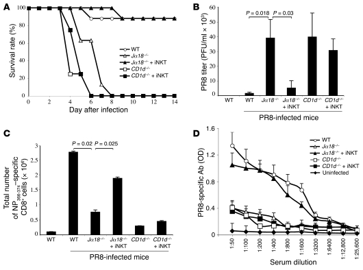
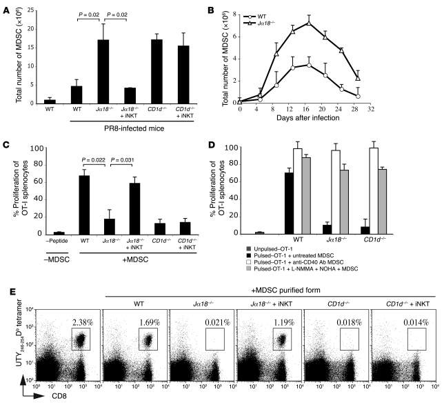
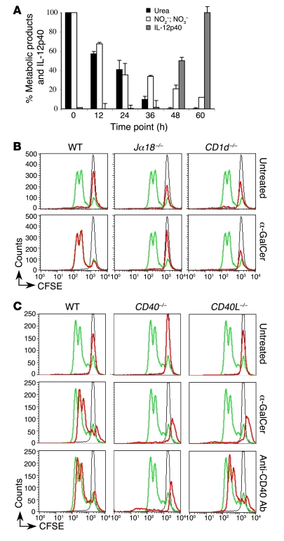
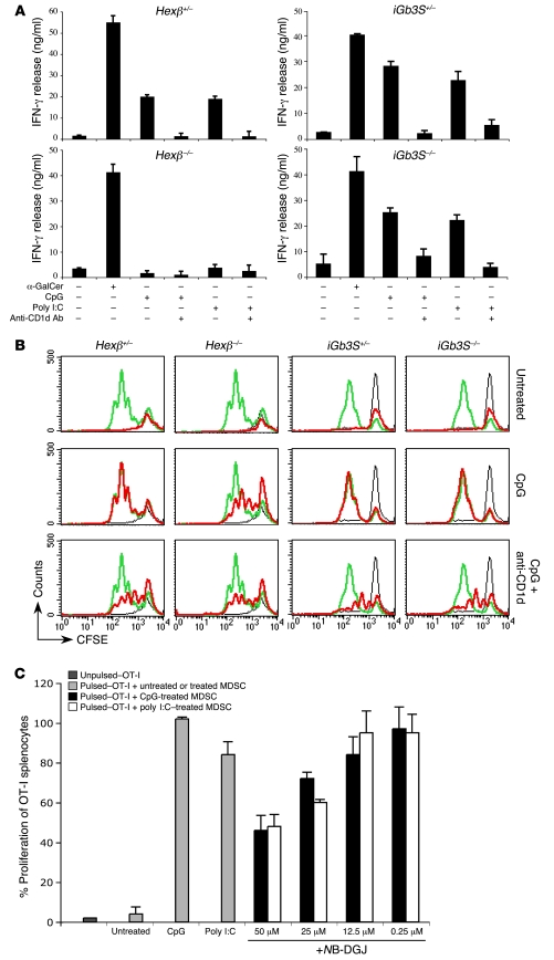
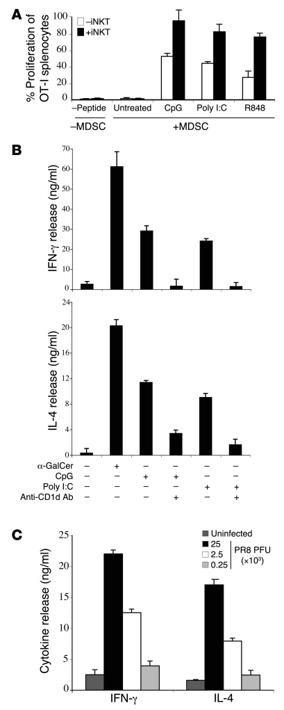
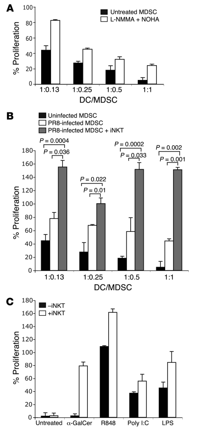
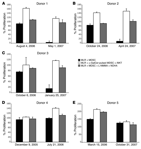
Infection with influenza A virus (IAV) presents a substantial threat to public health worldwide, with young, elderly, and immunodeficient individuals being particularly susceptible. Inflammatory responses play an important role in the fatal outcome of IAV infection, but the mechanism remains unclear. We demonstrate here that the absence of invariant NKT (iNKT) cells in mice during IAV infection resulted in the expansion of myeloid-derived suppressor cells (MDSCs), which suppressed IAV-specific immune responses through the expression of both arginase and NOS, resulting in high IAV titer and increased mortality. Adoptive transfer of iNKT cells abolished the suppressive activity of MDSCs, restored IAV-specific immune responses, reduced IAV titer, and increased survival rate. The crosstalk between iNKT and MDSCs was CD1d- and CD40-dependent. Furthermore, IAV infection and exposure to TLR agonists relieved the suppressive activity of MDSCs. Finally, we extended these results to humans by demonstrating the presence of myeloid cells with suppressive activity in the PBLs of individuals infected with IAV and showed that their suppressive activity is substantially reduced by iNKT cell activation. These findings identify what we believe to be a novel immunomodulatory role of iNKT cells, which we suggest could be harnessed to abolish the immunosuppressive activity of MDSCs during IAV infection.


Figures

Figure 1
Figure 1. Adoptive transfer of iNKT cells mediates protection from lethal doses of PR8.
WT, Jα18–/–, and CD1d–/– mice (n = 8/group) were injected intranasally with PR8 (3 × 10⁴ PFU/mouse). Liver-purified iNKT cells were adoptively transferred i.v. into Jα18–/– (Jα18–/– + iNKT) and CD1d–/– (CD1d–/– + iNKT) mice 1 day after infection. (A) Increased mortality of Jα18–/– mice following PR8 infection is prevented by the adoptive transfer of iNKT cells. Survival rate is shown as the percentage of live mice at different time points after the infection. Data are representative of 5 separate experiments. (B) PR8 titer in Jα18–/– mice is reduced by the adoptive transfer of iNKT cells. Lung homogenates from PR8-infected mice were assayed for number of PFU 6 days after infection. Results of statistical analyses, performed using Student’s t test, are shown. (C) Total number of NP366–374–specific CTLs in PR8-infected Jα18–/– mice is restored by the adoptive transfer of iNKT cells. The number of NP366–374–specific CTLs represents the average (±SD) of results obtained in n = 8 mice/group. Results of statistical analyses, performed using Student’s t test, are shown. (D) Anti-PR8 Ab titers in PR8-infected Jα18–/– mice are restored by the adoptive transfer of iNKT cells. The titer of anti-PR8–specific IgG was measured 6 days after infection.
Figure 2
Figure 2. Adoptive transfer of iNKT cells reduces PR8-induced MDSC expansion.
(A) iNKT cells adoptively transferred into PR8-infected Jα18–/– mice reduced the total number of lung MDSCs. PR8-infected WT, Jα18–/–, and CD1d–/– mice were injected with iNKT cells. Lung homogenates were stained with CD11b and Gr-1 Abs. (B) Injection of sublethal doses of PR8 (3 × 10² PFU) reduces the total numbers of lung MDSCs. (C) Adoptive transfer of iNKT cells reduces in vitro suppressive activity of PR8-induced MDSCs. MDSCs were purified from lungs of PR8-infected mice and cocultured with CFSE-labeled OT-I splenocytes. (D) The suppressive activity of MDSCs from PR8-infected mice is reduced by treatment with anti–CD40 agonist Ab and NOS2 and ARG1 inhibitors. Lung-purified MDSCs were left untreated (black bars) or treated in vitro with either anti–CD40 agonist Ab (white bars) or with NG-monomethyl-l-arginine (L-NMMA) and NOHA (light gray bars) and then added to OT-I splenocytes. Proliferation of unpulsed OT-I splenocytes in the absence of MDSCs is shown (dark gray bars). (E) Adoptive transfer of MDSCs from infected mice suppresses the expansion of UTY246–254–specific CD8+ T cell responses. UTY246–254 CTL responses were assessed in vaccinated mice injected with MDSCs from PR8-infected mice (see Methods). H-2Db/UTY246–254 tetramer (UTY246–254Db tetramer) versus CD8 dot plots are shown for gated propidium iodide–negative lymphocytes, and the percentage of cells staining positively with the tetramers is indicated. In AE, data represent the average ± SD of n = 5 mice/group, and results of statistical analyses, performed using Student’s t test, are shown.
Figure 3
Figure 3. The crosstalk between iNKT and MDSCs is CD1d and CD40 dependent.
(A) Loss of ARG1 and NOS2 activity in α-GalCer–pulsed MDSCs incubated with BM-derived iNKT cells. MDSCs were treated with α-GalCer in the presence of iNKT cells and collected at different time points. ARG1 and NOS2 activities were measured using a colorimetric assay to evaluate urea and nitrate/nitrite release, respectively (17, 18). An ELISA was used to evaluate IL-12p40 production. Values are shown as a percentage relative to time point 0. The maximum amount of urea released by untreated MDSCs (1 × 10⁶) was 91.7 μg. The maximum amount of NO2 and NO3 measured in the supernatant of untreated MDSCs was 120 μM. The maximum amount of IL-12p40 was 200 ng/ml. (B) Incubating α-GalCer–pulsed MDSCs with BM-derived iNKT cells abolishes their suppressive function. CFSE-labeled OT-I cell proliferation was analyzed in the presence (red) or absence (green) of MDSCs derived from WT, Jα18–/–, or CD1d–/– mice. MDSCs were left untreated or pulsed with α-GalCer. Proliferation of OT-I cells without SIINFEKL peptide is superimposed in all panels (black). (C) The effect of iNKT cells on α-GalCer–pulsed MDSCs is CD40 dependent. CFSE-labeled OT-I proliferation was analyzed in the presence (red) or absence (green) of MDSCs derived from WT, CD40–/–, or CD40L–/– mice. MDSCs were left untreated or pulsed with α-GalCer. To assess the role of the CD40 molecules, BM-derived MDSCs were treated with anti–CD40 agonist Ab for 48 hours. Proliferation of CFSE-labeled OT-I cells without SIINFEKL peptide is superimposed in all panels (black).
Figure 5
Figure 5. TLR-L–treated MDSCs derived from Hexβ–/– mice fail to stimulate iNKT cells.
(A) iNKT cells derived from WT mice fail to produce IFN-γ when incubated with TLR-L–matured MDSCs derived from Hexβ–/– mice. IFN-γ secretion by iNKT cells derived from WT mice, coincubated with TLR-L–matured MDSCs derived from Hexβ–/– or iGb3S–/– mice. Different protocols are indicated. (B) CpG-treated Hexβ–/– MDSCs fail to foster the crosstalk between iNKT cells and MDSCs. CFSE-labeled OT-I proliferation in the presence of TLR-L–treated MDSCs derived from either Hexβ–/– or iGb3S–/– mice is shown. BM-derived Hexβ–/– and iGb3S–/– MDSCs were treated with CpG in the presence or absence of blocking anti-CD1d Ab. Proliferation of OT-I cells in the presence (red) or absence (green) of MDSCs is shown. Proliferation without the SIINFEKL peptide (black) is superimposed in all panels. (C) Inhibition of GSL biosynthesis in TLR-L–matured MDSCs reduces iNKT cell activation. MDSCs derived from WT mice were incubated with either poly I:C or CpG and then treated with increasing concentrations of N-butyldeoxygalactonojirimycin (NB-DGJ). MDSCs were incubated with iNKT cells for 24 hours and tested for their effect on OT-I proliferation (see Methods). Addition of NB-DGJ to OT-I splenocytes in the absence of MDSCs did not affect OT-I proliferation (data not shown). Proliferation of unpulsed OT-I splenocytes in the absence of MDSCs is shown (dark gray). Treatment of MDSCs with TLR-L (light gray bars) relieves CFSE-labeled OT-I proliferation, as compared with the lack of OT-I proliferation observed after addition of untreated MDSCs. In contrast, combined treatment of MDSCs with either CpG (black bars) or poly I:C (white bars) plus increasing doses of NB-DGJ reduces OT-I proliferation.
Figure 4
Figure 4. Infection of MDSCs with PR8 fosters their ability to activate iNKT cells.
(A) TLR agonists rescue suppressive activity of MDSCs. BM-derived MDSCs were treated with TLR agonists in the presence or absence of liver-purified iNKT cells as described in Methods. CFSE-labeled OT-I proliferation was analyzed after addition of TLR agonist–treated MDSCs in the presence (black bars) or absence (white bars) of iNKT cells. Data represent the average ± SD of 3 replicates and are representative of 3 separate experiments. As negative control, proliferation of unpulsed CFSE-labeled OT-I splenocytes in the absence of MDSCs is shown. (B) IFN-γ and IL-4 release from iNKT cells incubated for 48 hours with CpG- or poly I:C–treated MDSCs, in the presence or absence of anti-CD1d blocking Ab. Data represent the average ± SD of 3 replicates and are representative of 3 separate experiments. (C) Amounts of IFN-γ and IL-4 released by iNKT cells incubated in the presence of MDSCs infected with increasing doses of PR8.
Figure 6
Figure 6. Inhibition of alloreactive T cell proliferation by human MDSCs can be rescued by iNKT cells.
(A) Healthy donors’ GM-CSF–differentiated MDSCs have MLR-suppressive activity, which can be blocked by the addition of NOHA and L-NMMA. The data are expressed as described in Methods. Addition of NOHA and L-NMMA to the MLR in the absence of MDSCs does not affect PBL proliferation (data not shown). The ratio of DCs to human GM-CSF–treated MDSCs is shown. (B) PR8 infection of healthy donors’ GM-CSF–treated MDSCs rescues MLR proliferation. Human GM-CSF–treated MDSCs were left untreated (black bars) or infected with PR8 and cocultured in the presence (gray bars) or absence (white bars) of iNKT cells (2.5 × 10⁴) for 24 hours. After irradiation, PR8-infected and uninfected MDSCs were added to the MLR. The data are expressed as described in Methods. The ratio of DCs to human MDSCs used is shown. Results of statistical analyses, performed using Student’s t test, are shown. (C) TLR-L incubation of healthy donors’ GM-CSF–differentiated MDSCs rescues T cell proliferation. Human GM-CSF–treated MDSCs were treated with LPS (10 μg/ml), R848 (5 μg/ml), poly I:C (10 μg/ml), or α-GalCer (100 ng/ml) and cocultured in the presence (white bars) or absence (black bars) of iNKT cells for 24 hours. The data are expressed as described in Methods. The ratio of DCs to human GM-CSF–treated MDSCs used was 1:1.
Figure 7
Figure 7. Inhibition of T cell proliferation by MDSCs purified from IAV-infected individuals.
CD11b+ cells were bead purified from PBLs derived from either IAV-infected individuals (AC) or healthy donors (D and E). All donors were bled twice. For donors 1, 2, and 3, the first blood sample was collected before the clinical symptom onset, while the second blood sample was collected within 30–60 days after the acute respiratory illness. Donors 4 and 5 did not have any respiratory illness within the time frame of the collection of the 2 blood samples. Irradiated purified CD11b+ cells were then added to allogenic PBLs and incubated with allogeneic irradiated DCs. Purified CD11b+ cells were either untreated (black bars) or treated with either α-GalCer (100 ng/ml) in the presence of iNKT cells at a MDSC/iNKT ratio of 1:0.25 (white bars) or L-NMMA and NOHA (gray bars). The data are expressed as described in Methods. Addition of either iNKT cells or L-NMMA and NOHA to the alloreactive PBLs in the absence of CD11b+ cells did not affect T cell proliferation (data not shown). The ratio of irradiated DCs to purified irradiated CD11b+ cells was 1:1.

Similar articles

Cited by

References

    1. Palese P. Influenza: old and new threats. Nat. Med. 2004;10:S82–S87. doi: 10.1038/nm1141. - DOI - PubMed
    1. Kobasa D., et al. Aberrant innate immune response in lethal infection of macaques with the 1918 influenza virus. Nature. 2007;445:319–323. doi: 10.1038/nature05495. - DOI - PubMed
    1. de Jong M.D., et al. Fatal outcome of human influenza A (H5N1) is associated with high viral load and hypercytokinemia. Nat. Med. 2006;12:1203–1207. doi: 10.1038/nm1477. - DOI - PMC - PubMed
    1. Doherty P.C., et al. Effector CD4+ and CD8+ T-cell mechanisms in the control of respiratory virus infections. Immunol. Rev. 1997;159:105–117. doi: 10.1111/j.1600-065X.1997.tb01010.x. - DOI - PubMed
    1. Delano M.J., et al. MyD88-dependent expansion of an immature GR-1+CD11b+ population induces T cell suppression and Th2 polarization in sepsis. J. Exp. Med. 2007;204:1463–1474. doi: 10.1084/jem.20062602. - DOI - PMC - PubMed

Publication types

MeSH terms