A role for leukocyte-endothelial adhesion mechanisms in epilepsy
- PMID: 19029985
- PMCID: PMC2710311
- DOI: 10.1038/nm.1878
A role for leukocyte-endothelial adhesion mechanisms in epilepsy
Abstract
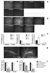
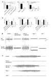
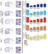
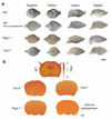
The mechanisms involved in the pathogenesis of epilepsy, a chronic neurological disorder that affects approximately one percent of the world population, are not well understood. Using a mouse model of epilepsy, we show that seizures induce elevated expression of vascular cell adhesion molecules and enhanced leukocyte rolling and arrest in brain vessels mediated by the leukocyte mucin P-selectin glycoprotein ligand-1 (PSGL-1, encoded by Selplg) and leukocyte integrins alpha(4)beta(1) and alpha(L)beta(2). Inhibition of leukocyte-vascular interactions, either with blocking antibodies or by genetically interfering with PSGL-1 function in mice, markedly reduced seizures. Treatment with blocking antibodies after acute seizures prevented the development of epilepsy. Neutrophil depletion also inhibited acute seizure induction and chronic spontaneous recurrent seizures. Blood-brain barrier (BBB) leakage, which is known to enhance neuronal excitability, was induced by acute seizure activity but was prevented by blockade of leukocyte-vascular adhesion, suggesting a pathogenetic link between leukocyte-vascular interactions, BBB damage and seizure generation. Consistent with the potential leukocyte involvement in epilepsy in humans, leukocytes were more abundant in brains of individuals with epilepsy than in controls. Our results suggest leukocyte-endothelial interaction as a potential target for the prevention and treatment of epilepsy.
Figures


Comment in
-
Brain inflammation initiates seizures.Nat Med. 2008 Dec;14(12):1309-10. doi: 10.1038/nm1208-1309. Nat Med. 2008. PMID: 19057551 No abstract available.
-
Leukocyte-endothelial adhesion mechanisms in epilepsy: cheers and jeers.Epilepsy Curr. 2009 Jul-Aug;9(4):118-21. doi: 10.1111/j.1535-7511.2009.01312.x. Epilepsy Curr. 2009. PMID: 19693331 Free PMC article. No abstract available.
-
Breaching the barrier and inflaming epilepsy research.Epilepsy Curr. 2009 Sep-Oct;9(5):148-50. doi: 10.1111/j.1535-7511.2009.01324.x. Epilepsy Curr. 2009. PMID: 19826510 Free PMC article. No abstract available.
Similar articles
-
Macrophage migration inhibitory factor increases leukocyte-endothelial interactions in human endothelial cells via promotion of expression of adhesion molecules.J Immunol. 2010 Jul 15;185(2):1238-47. doi: 10.4049/jimmunol.0904104. Epub 2010 Jun 16. J Immunol. 2010. PMID: 20554956
-
L- and P-selectins collaborate to support leukocyte rolling in vivo when high-affinity P-selectin-P-selectin glycoprotein ligand-1 interaction is inhibited.Am J Pathol. 2005 Mar;166(3):945-52. doi: 10.1016/S0002-9440(10)62314-0. Am J Pathol. 2005. PMID: 15743805 Free PMC article.
-
P-Selectin Glycoprotein Ligand-1 Deficiency Protects Against Aortic Aneurysm Formation Induced by DOCA Plus Salt.Cardiovasc Drugs Ther. 2022 Feb;36(1):31-44. doi: 10.1007/s10557-020-07135-1. Epub 2021 Jan 11. Cardiovasc Drugs Ther. 2022. PMID: 33432452
-
Are you in or out? Leukocyte, ion, and neurotransmitter permeability across the epileptic blood-brain barrier.Epilepsia. 2012 Jun;53 Suppl 1(0 1):26-34. doi: 10.1111/j.1528-1167.2012.03472.x. Epilepsia. 2012. PMID: 22612806 Free PMC article. Review.
-
Therapeutic strategies in autoimmune diseases by interfering with leukocyte endothelium interaction.Curr Pharm Des. 2006;12(29):3799-806. doi: 10.2174/138161206778559696. Curr Pharm Des. 2006. PMID: 17073678 Review.
Cited by
-
Neuroinflammation and status epilepticus: a narrative review unraveling a complex interplay.Front Pediatr. 2023 Nov 21;11:1251914. doi: 10.3389/fped.2023.1251914. eCollection 2023. Front Pediatr. 2023. PMID: 38078329 Free PMC article. Review.
-
The correlation of temporal changes of neutrophil-lymphocyte ratio with seizure severity and the following seizure tendency in patients with epilepsy.Front Neurol. 2022 Oct 20;13:964923. doi: 10.3389/fneur.2022.964923. eCollection 2022. Front Neurol. 2022. PMID: 36341114 Free PMC article.
-
Causes of CNS inflammation and potential targets for anticonvulsants.CNS Drugs. 2013 Aug;27(8):611-23. doi: 10.1007/s40263-013-0078-6. CNS Drugs. 2013. PMID: 23771734 Review.
-
Patterns of postictal cerebral perfusion in idiopathic generalized epilepsy: a multi-delay multi-parametric arterial spin labelling perfusion MRI study.Sci Rep. 2016 Jul 4;6:28867. doi: 10.1038/srep28867. Sci Rep. 2016. PMID: 27374369 Free PMC article.
-
Intracellular and circulating neuronal antinuclear antibodies in human epilepsy.Neurobiol Dis. 2013 Nov;59:206-19. doi: 10.1016/j.nbd.2013.07.006. Epub 2013 Jul 21. Neurobiol Dis. 2013. PMID: 23880401 Free PMC article.
References
-
- Strzelczyk A, Reese JP, Dodel R, Hamer HM. Cost of epilepsy: a systematic review. Pharmacoeconomics. 2008;26:463–476. - PubMed
-
- Holmes GL. Seizure-induced neuronal injury: animal data. Neurology. 2002;59:S3–S6. - PubMed
-
- Duncan JS. Seizure-induced neuronal injury: human data. Neurology. 2002;59:S15–S20. - PubMed
-
- Vezzani A, Granata T. Brain inflammation in epilepsy: experimental and clinical evidence. Epilepsia. 2005;46:1724–1743. - PubMed
Publication types
MeSH terms
Substances
Grants and funding
LinkOut - more resources
Full Text Sources
Other Literature Sources
Medical
Molecular Biology Databases

