. 2009 Jan 9;284(2):1155-65.
doi: 10.1074/jbc.M806219200. Epub 2008 Nov 19.

Agonist and antagonist recognition by RIG-I, a cytoplasmic innate immunity receptor

Affiliations

Agonist and antagonist recognition by RIG-I, a cytoplasmic innate immunity receptor

C T Ranjith-Kumar et al. J Biol Chem. .

Abstract

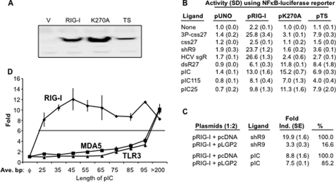
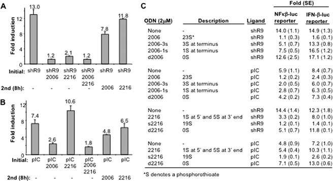
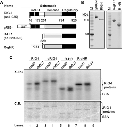
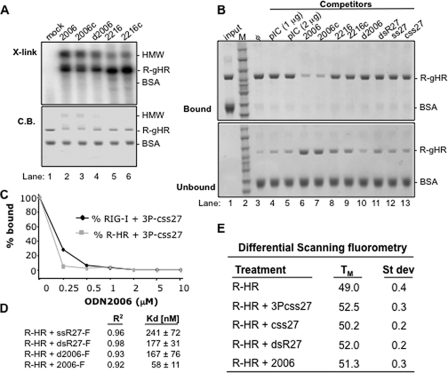
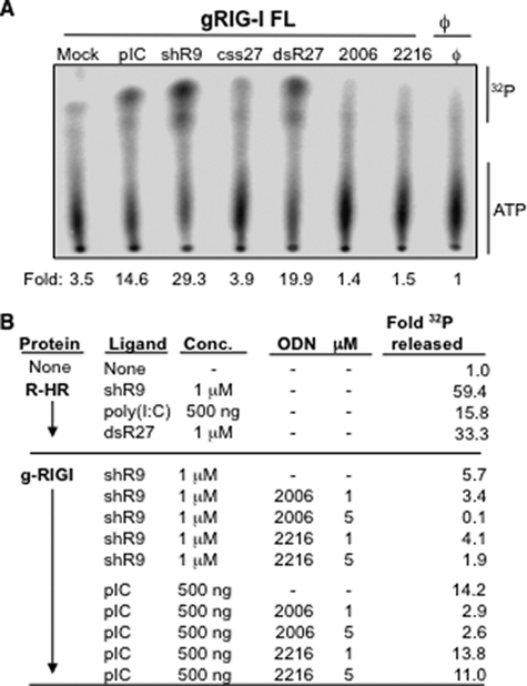
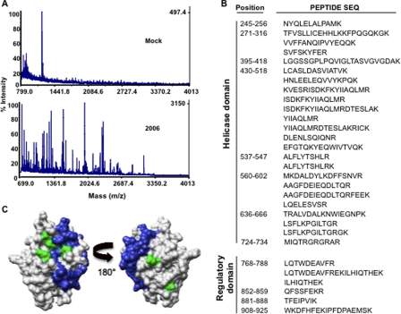
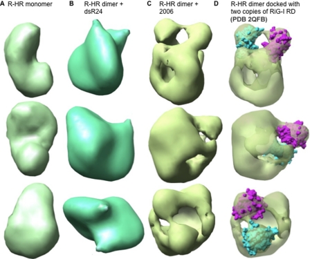
Cytoplasmic RNA receptors are important in the detection of and response to viral infections. We analyzed ligand recognition by the retinoic acid-inducible protein I (RIG-I) protein in biochemical assays and in transiently transfected cells and characterized the requirements for both single- and double-stranded RNA agonists for RIG-I activation of signaling. RIG-I mutants such as K270A and T409A/S411A that were defective in signaling with triphosphorylated single-stranded RNAs were perfectly capable of signaling with dsRNAs. Furthermore, phosphorothioated oligodeoxynucleotides were found to antagonize RIG-I signaling. Both agonists and antagonist bind purified RIG-I protein and a truncated RIG-I protein that lacked the signaling domain. The agonists were necessary to activate RIG-I ATPase activity in vitro, whereas antagonist inhibited ATPase activity. Differential scanning fluorometry showed that RIG-I bound to agonists, and antagonists have different denaturation properties, suggesting a difference in protein conformations. Last, single particle reconstruction was used to generate three-dimensional models of the RIG-I dimers in complex with an agonist and an antagonist. The two complexes exhibited dramatically different structures.


Figures

FIGURE 1.
FIGURE 1.
Single- and double-stranded ligands have different properties for RIG-I signaling. A, Western blot analysis of RIG-I and mutants. RIG-I protein and the two alanine substitution mutants are expressed in transiently transfected 293T cells. The Western blot was probed with a polyclonal antibody against C-terminal region of RIG-I purchased from Santa Cruz Biotechnology, Inc. V, vector. B, the effects of two RIG-I mutations on response to different ligands. Numbers denote -fold induction that is the ratio of the induced versus uninduced samples assayed. The assay was performed using luciferase reporter regulated by a promoter containing NF-κB binding sites. Each number represents the mean of at least three independent assays, and the value of 1 S.E. is shown in parentheses. Throughout this work the following names are used for the various ligands; 3P-css27 is a 27-nt RNA produced by in vitro transcription, and css27 is the same RNA that lacks 5′-triphosphates. ShR9 is a 60-nt hairpin RNA produced by in vitro transcription (see supplemental Fig. 1). HCV sgR is the hepatitis C virus subgenomic replicon RNA transcribed from linearized plasmid pFK/I389neo/NS3-3′/5.1. DsR27 is a double-stranded RNA made by annealing two single-stranded oligonucleotides of 27-nt each; pIC is poly(I:C) purchased from Invitrogen and has a molecular mass that is in excess of 200 bp with very little fragments below this length. pIC115 and pIC25 are poly(I:C)s of 115 and 25 bp, respectively. C, effects of shR9-induced RIG-I activity in the presence of LGP2. The ratio of RIG-I to LGP2 is 1:2. D, the effects of poly(I:C) length on induction of RIG-I activation of an NF-κB reporter in HEK293T cells. The same series of pIC was used to examine MDA5 and TLR3 signaling and yielded different responses. Each point is the graph shows the mean and S.E. of three independent assays.
FIGURE 2.
FIGURE 2.
Modified ssDNAs are potent antagonists for RIG-I signaling. A, effects of modified ODN on RIG-I signaling induced by shR9. This experiment also incorporates a timing of addition, and ODN(s) was either transfected into the cells at the same time as the agonist shR9 or 8 h after shR9 transfection. The mean of the -fold induction is shown as a number above the bars, and the range for one S.E. is shown by the lines above the bar. B, the inhibitory effects of ODNs with poly(I:C) (pIC) as the agonist can be different from that of cells induced by shR9. Note that 2216 can slightly enhance the stimulatory effect of pIC. This result has been reproduced in more than five independent assays. As in panel A, the timing of the introduction of the ligands is shown below the graph. C, the phosphorothioate backbone of the ODNs is a major determinant for inhibitory activity of ODNs. Role of phosphorothioate backbone is analyzed using NF-κB and IFN-β reporters. The number and locations of the phosphorothioates in the ODNs are listed under the descriptions.
FIGURE 3.
FIGURE 3.
Ligand binding by recombinant RIG-I protein and its derivatives. A, schematics of recombinant proteins expressed by baculovirus-infected insect cells. Two constructs expressed are full-length RIG-I and RIG-I with CARD domain deleted (R-HR). These constructs are expressed with and with out the GST tag that could be removed by treatment with thrombin. The site of cleavage is denoted with a black triangle. The constructs with GST are depicted with prefix g. The three domains of the RIG-I protein are shown in different colors, with key residue numbers shown. aa, amino acids. B, SDS-PAGE of the purified recombinant proteins. Each gel image shows the GST-tagged protein and the version in which the GST was removed and then purified away. M, molecular mass standards. C, results of UV cross-linking assay to examine ligand binding by the four versions of RIG-I protein. The top panel contains an image from an autoradiogram depicting the complexes of the RIG-I with the ligands named above the gel image. The bottom image contains the same gel that was stained with Coomassie Blue (C.B.) to reveal the location of RIG-I and its derivatives along with the internal negative control, BSA.
FIGURE 4.
FIGURE 4.
Properties of antagonist binding to RIG-I or its derivatives. A, results from a UV cross-linking assay demonstrating that purified R-gHR can be cross-linked to ODNs. The top panel is an autoradiogram of the cross-linked products in a SDS-PAGE. The bottom panel is the SDS-PAGE stained to reveal the locations of the proteins used in the reaction. BSA is used as an internal negative control in all of the reactions. HMW denotes an oligomeric form of RIG-I that is preferentially detected with strong antagonists. B, results of a pulldown assay examining competition of different ligands with R-gHR. The assay uses biotinylated 2006 bound to streptavidin resin, which can specifically bind R-gHR, but not the internal specificity control, BSA. The binding assay was performed in the presence of various competing ligands shown on the top of the gel. C, a competition assay examining UV cross-linking to a internally labeled 5′-triphosphorylated RNA in the presence of increasing concentrations of antagonist 2006. D, fluorescent anisotropy to determine affinity for ligand. Fluorescein isothiocyanate-labeled ligands were used to measure Kd values using R-HR protein, and the R2 values show the fit of the data to the binding isotherm. E, results of differential scanning fluorometry used to measure the melting point (TM) of RIG-I in the presence of different ligands. Results shown are from three independent experiments.
FIGURE 5.
FIGURE 5.
Effects of agonists and antagonists on RIG-I ATPase activity. A, a demonstration that the ATPase activity of the GST-tagged version of RIG-I is stimulated by RNA agonists and that antagonist ODNs do not stimulate ATPase activity. B, further characterization of ATPase activity. The top portion of the results shows that R-HR retains ligand-induced ATPase activity despite lacking the CARD domain. The bottom portion shows that antagonist oligonucleotide 2006 can inhibit the ATPase activity of RIG-I. 2216 inhibited only shR9-dependent ATPase activity but not poly(I:C)-dependent ATPase activity.
FIGURE 6.
FIGURE 6.
Analysis of the domains in RIG-I that contacts the antagonist 2006. A, a sample of the mass spectra generated by the analysis of the peptides that can be cross-linked to a ligand. The top spectrum shows the background for a mock reaction, whereas the bottom panel shows the spectrum where 2006 was present in the reversible cross-linking reaction. B, a summary of the peptides identified from the reactions with 2006. C, location of the peptides found in the RIG-I regulatory domain that were cross-linked to 2006. The green colors show the location of residues His-830, Ile-875, and Lys-888 that were previously identified to interact with dsRNA by Cui et al. (19).
FIGURE 7.
FIGURE 7.
Reconstruction of monomers of the R-HR domain of RIG-I and R-HR in complex with the agonist dsR24 or the antagonist 2006. The R-HR molecule reconstructed was a monomer, whereas the ligand-bound forms were clearly dimers. A, R-HR monomer. B, R-HR dimer in complex with dsR24. C, R-HR dimer in complex with 2006. D, same as C but with crystal structure of RIG-I regulatory domains docked. Two molecules of RIG-I regulatory domain (shown in cyan and purple) are docked on each R-HR molecule of the dimer. More data for the building of these molecular models are shown in supplemental Fig. 4.

Similar articles

Cited by

References

    1. Akira, S., Uematsu, S., and Takeuchi, O. (2006) Cell 124 783-801 - PubMed
    1. Gaspari, A. A. (2006) J. Am. Acad. Dermatol. 54 Suppl. 2, 67-80 - PubMed
    1. Kobayashi, T., Takaesu, G., and Yoshimura, A. (2006) Nat. Immunol. 7 123-124 - PubMed
    1. Underhill, D. M. (2004) Curr. Opin. Immunol. 16 483-487 - PubMed
    1. Yoneyama, M., Kikuchi, M., Natsukawa, T., Shinobu, N., Imaizumi, T., Miyagishi, M., Taira, K., Akira, S., and Fujita, T. (2004) Nat. Immunol. 5 730-737 - PubMed

Publication types

MeSH terms