Enhanced expression of Janus kinase-signal transducer and activator of transcription pathway members in human diabetic nephropathy
- PMID: 19017763
- PMCID: PMC2628622
- DOI: 10.2337/db08-1328
Enhanced expression of Janus kinase-signal transducer and activator of transcription pathway members in human diabetic nephropathy
Abstract
Objective: Glomerular mesangial expansion and podocyte loss are important early features of diabetic nephropathy, whereas tubulointerstitial injury and fibrosis are critical for progression of diabetic nephropathy to kidney failure. Therefore, we analyzed the expression of genes in glomeruli and tubulointerstitium in kidney biopsies from diabetic nephropathy patients to identify pathways that may be activated in humans but not in murine models of diabetic nephropathy that fail to progress to glomerulosclerosis, tubulointerstitial fibrosis, and kidney failure.
Research design and methods: Kidney biopsies were obtained from 74 patients (control subjects, early and progressive type 2 diabetic nephropathy). Glomerular and tubulointerstitial mRNAs were microarrayed, followed by bioinformatics analyses. Gene expression changes were confirmed by real-time RT-PCR and immunohistological staining. Samples from db/db C57BLKS and streptozotocin-induced DBA/2J mice, commonly studied murine models of diabetic nephropathy, were analyzed.
Results: In human glomeruli and tubulointerstitial samples, the Janus kinase (Jak)-signal transducer and activator of transcription (Stat) pathway was highly and significantly regulated. Jak-1, -2, and -3 as well as Stat-1 and -3 were expressed at higher levels in patients with diabetic nephropathy than in control subjects. The estimated glomerular filtration rate significantly correlated with tubulointerstitial Jak-1, -2, and -3 and Stat-1 expression (R(2) = 0.30-0.44). Immunohistochemistry found strong Jak-2 staining in glomerular and tubulointerstitial compartments in diabetic nephropathy compared with control subjects. In contrast, there was little or no increase in expression of Jak/Stat genes in the db/db C57BLKS or diabetic DBA/2J mice.
Conclusions: These data suggest a direct relationship between tubulointerstitial Jak/Stat expression and progression of kidney failure in patients with type 2 diabetic nephropathy and distinguish progressive human diabetic nephropathy from nonprogressive murine diabetic nephropathy.
Figures


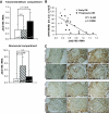
 , early diabetic nephropathy; ▪, progressive diabetic nephropathy. B: Correlation of tubulointerstitial Jak-2 mRNA expression (as measured by real-time RT-PCR) with eGFR in patients with early (×, n = 11) and progressive (•, n = 12) diabetic nephropathy. C: Representative Jak-2 immunohistographs of kidney biopsies from patients with no kidney disease (Control), progressive diabetic nephropathy (Prog. DN), hypertensive nephropathy (HTN), IgA nephropathy (IgAN), or lupus nephritis (LN). Jak-2 expression was substantially and significantly increased in the proximal tubular cells and glomeruli of the patients with diabetic nephropathy compared with the control and not in other progressive kidney diseases. (Please see


Similar articles
-
JAK-STAT Activity in Peripheral Blood Cells and Kidney Tissue in IgA Nephropathy.Clin J Am Soc Nephrol. 2020 Jul 1;15(7):973-982. doi: 10.2215/CJN.11010919. Epub 2020 Apr 30. Clin J Am Soc Nephrol. 2020. PMID: 32354727 Free PMC article.
-
Effect of liraglutide on the Janus kinase/signal transducer and transcription activator (JAK/STAT) pathway in diabetic kidney disease in db/db mice and in cultured endothelial cells.J Diabetes. 2019 Aug;11(8):656-664. doi: 10.1111/1753-0407.12891. Epub 2019 Feb 28. J Diabetes. 2019. PMID: 30575282
-
JAK-STAT signaling is activated in the kidney and peripheral blood cells of patients with focal segmental glomerulosclerosis.Kidney Int. 2018 Oct;94(4):795-808. doi: 10.1016/j.kint.2018.05.022. Epub 2018 Aug 6. Kidney Int. 2018. PMID: 30093081 Free PMC article.
-
JAK inhibition in the treatment of diabetic kidney disease.Diabetologia. 2016 Aug;59(8):1624-7. doi: 10.1007/s00125-016-4021-5. Epub 2016 Jun 22. Diabetologia. 2016. PMID: 27333885 Free PMC article. Review.
-
JAK inhibition and progressive kidney disease.Curr Opin Nephrol Hypertens. 2015 Jan;24(1):88-95. doi: 10.1097/MNH.0000000000000079. Curr Opin Nephrol Hypertens. 2015. PMID: 25415616 Free PMC article. Review.
Cited by
-
Allicin Ameliorated High-glucose Peritoneal Dialysis Solution-induced Peritoneal Fibrosis in Rats via the JAK2/STAT3 Signaling Pathway.Cell Biochem Biophys. 2024 Oct 25. doi: 10.1007/s12013-024-01593-2. Online ahead of print. Cell Biochem Biophys. 2024. PMID: 39448419
-
Role and Mechanisms of Tyro3 in Podocyte Biology and Glomerular Disease.Kidney Dis (Basel). 2024 Jul 24;10(5):398-406. doi: 10.1159/000540452. eCollection 2024 Oct. Kidney Dis (Basel). 2024. PMID: 39430290 Free PMC article. Review.
-
STAT3 Protein-Protein Interaction Analysis Finds P300 as a Regulator of STAT3 and Histone 3 Lysine 27 Acetylation in Pericytes.Biomedicines. 2024 Sep 14;12(9):2102. doi: 10.3390/biomedicines12092102. Biomedicines. 2024. PMID: 39335615 Free PMC article.
-
Alantolactone alleviates epithelial-mesenchymal transition by regulating the TGF-β/STAT3 signaling pathway in renal fibrosis.Heliyon. 2024 Aug 13;10(16):e36253. doi: 10.1016/j.heliyon.2024.e36253. eCollection 2024 Aug 30. Heliyon. 2024. PMID: 39253189 Free PMC article.
-
M2 macrophage infusion ameliorates diabetic glomerulopathy via the JAK2/STAT3 pathway in db/db mice.Ren Fail. 2024 Dec;46(2):2378210. doi: 10.1080/0886022X.2024.2378210. Epub 2024 Aug 1. Ren Fail. 2024. PMID: 39090966 Free PMC article.
References
-
- Mauer SM, Lane P, Zhu D, Fioretto P, Steffes MW: Renal structure and function in insulin-dependent diabetes mellitus in man. J Hypertens Suppl 10: S17–20, 1992 - PubMed
-
- Chavers BM, Bilous RW, Ellis EN, Steffes MW, Mauer SM: Glomerular lesions and urinary albumin excretion in type I diabetes without overt proteinuria. N Engl J Med 320: 966–970, 1989 - PubMed
-
- Nangaku M: Mechanisms of tubulointerstitial injury in the kidney: final common pathways to end-stage renal failure. J Int Med 43: 9–17, 2004 - PubMed
-
- Bader R, Bader H, Grund KE, Mackensen-Haen S, Christ H, Bohle A: Structure and function of the kidney in diabetic glomerulosclerosis: correlations between morphological and functional parameters. Pathol Res Pract 167: 204–216, 1980 - PubMed
Publication types
MeSH terms
Substances
Grants and funding
LinkOut - more resources
Full Text Sources
Other Literature Sources
Medical
Research Materials
Miscellaneous

