. 2009 Feb 15;18(4):607-20.
doi: 10.1093/hmg/ddn386. Epub 2008 Nov 13.

Disruption of nesprin-1 produces an Emery Dreifuss muscular dystrophy-like phenotype in mice

Affiliations

Disruption of nesprin-1 produces an Emery Dreifuss muscular dystrophy-like phenotype in mice

Megan J Puckelwartz et al. Hum Mol Genet. .

Abstract

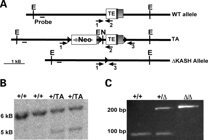
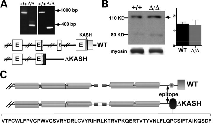
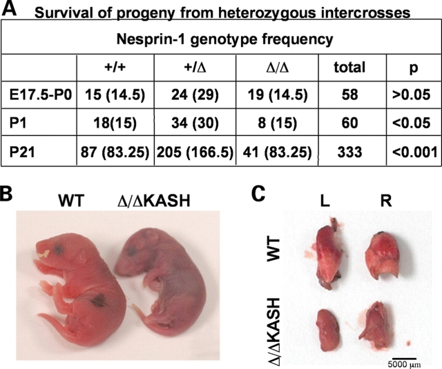
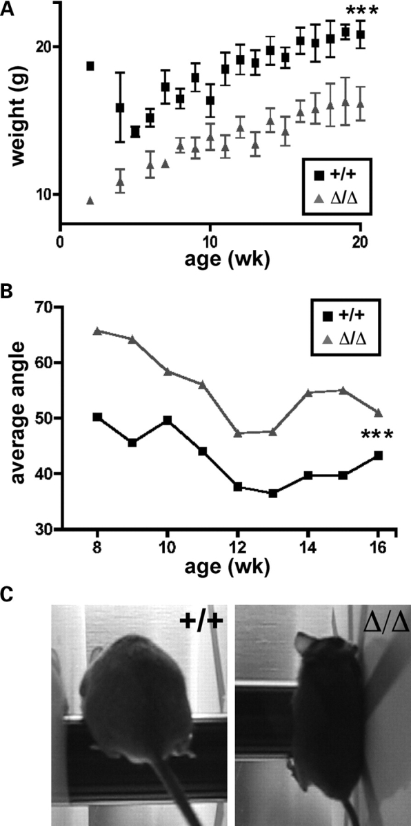
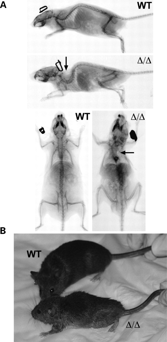
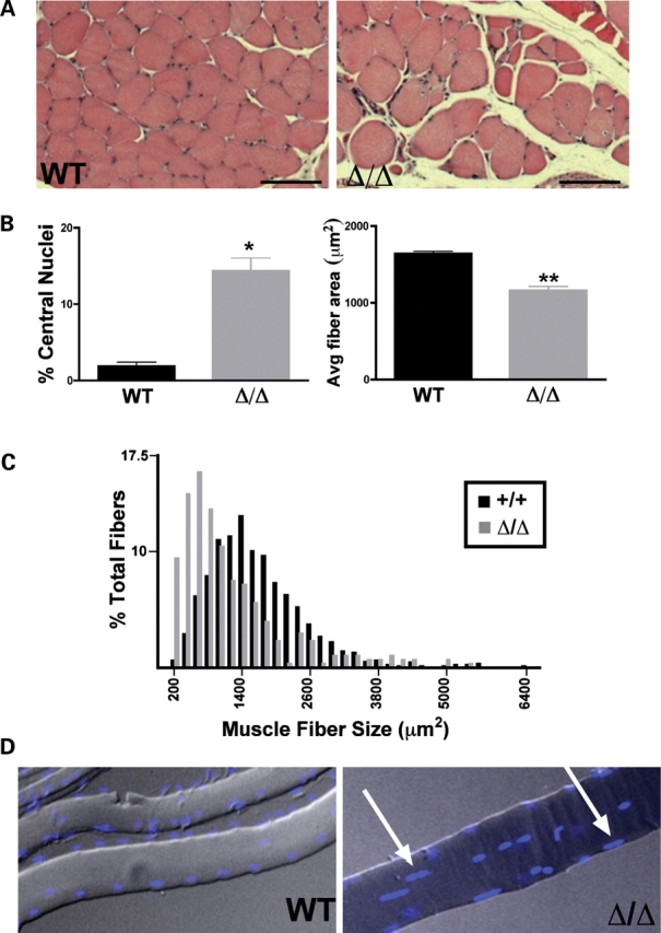
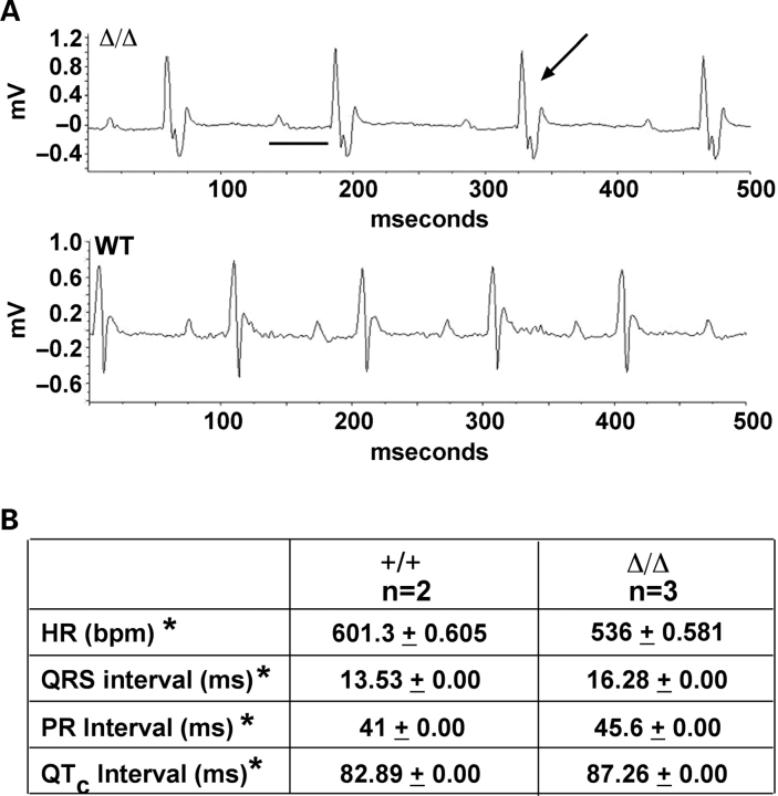
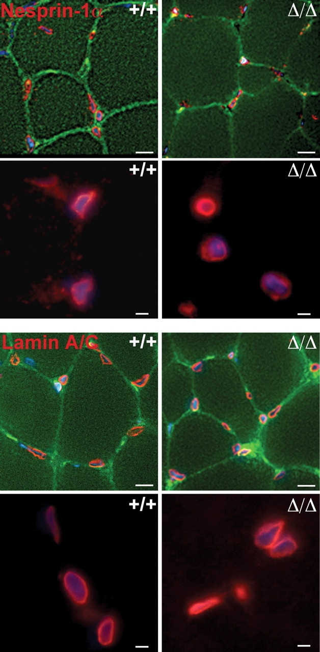
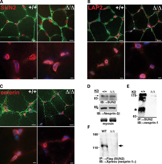
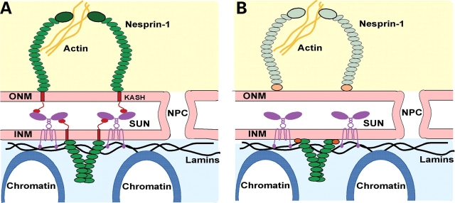
Mutations in the gene encoding the inner nuclear membrane proteins lamins A and C produce cardiac and skeletal muscle dysfunction referred to as Emery Dreifuss muscular dystrophy. Lamins A and C participate in the LINC complex that, along with the nesprin and SUN proteins, LInk the Nucleoskeleton with the Cytoskeleton. Nesprins 1 and 2 are giant spectrin-repeat containing proteins that have large and small forms. The nesprins contain a transmembrane anchor that tethers to the nuclear membrane followed by a short domain that resides within the lumen between the inner and outer nuclear membrane. Nesprin's luminal domain binds directly to SUN proteins. We generated mice where the C-terminus of nesprin-1 was deleted. This strategy produced a protein lacking the transmembrane and luminal domains that together are referred to as the KASH domain. Mice homozygous for this mutation exhibit lethality with approximately half dying at or near birth from respiratory failure. Surviving mice display hindlimb weakness and an abnormal gait. With increasing age, kyphoscoliosis, muscle pathology and cardiac conduction defects develop. The protein components of the LINC complex, including mutant nesprin-1alpha, lamin A/C and SUN2, are localized at the nuclear membrane in this model. However, the LINC components do not normally associate since coimmunoprecipitation experiments with SUN2 and nesprin reveal that mutant nesprin-1 protein no longer interacts with SUN2. These findings demonstrate the role of the LINC complex, and nesprin-1, in neuromuscular and cardiac disease.


Figures

Figure 1.
Figure 1.
Gene targeting of nesprin-1 KASH domain. (A) The targeting allele (TA) included the KASH domain-containing terminal exon (TE, KASH domain=dark gray box, 3′-UTR=light gray box) surrounded by loxP sites and an upstream neomycin cassette. Cre-recombinase was used to remove the neomycin cassette and TE. (B) Southern blot demonstrating homologous recombination of the targeted allele. EcoRI digested embryonic stem cell DNA was blotted and hybridized with the probe shown in (A). (C) PCR genotyping of tail DNA was performed with primers 1, 2 and 3, indicated as arrows in (A), to confirm deletion of the terminal exon. Primer 1, located upstream of the terminal exon and primer 2, located within the terminal exon, yield a 90 bp band in wild-type mice. Primer 3 is located outside of the terminal exon and in conjunction with primer 1 will produce a 254 bp product in Δ/ΔKASH mice.
Figure 2.
Figure 2.
Deletion of the nesprin-1 KASH domain leads to translation of a truncated protein, ΔKASH. (A) 3′RACE was performed on both wild-type and Δ/ΔKASH cDNA. Wild-type cDNA produced the expected 972 bp band, Δ/ΔKASH cDNA produces a 359 bp band corresponding to loss of the last two exons and addition of 183 bp. The image on the left represents the first round of 3′RACE and the gel on the right is the second round from the nested reaction. Below that is a schematic of the genomic region indicating transcribed exons in WT and Δ/ΔKASH mice. In WT mice, the terminal exon contains the KASH domain (dark gray) and the 3′-UTR (light gray). The antibody epitope is indicated by the stippling. In Δ/ΔKASH mice the terminal exon is deleted and readthrough occurs resulting in an mRNA lacking the two terminal exons and containing an additional 183 bp (black). (B) Immunoblotting from skeletal muscle isolated from wild-type (+/+) littermate and Δ/ΔKASH mice express a nesprin-1α isoform that migrates at 120 kDa, indicated by the arrow. The myosin band is shown as a loading control. Densitometry was performed comparing both wild-type and Δ/ΔKASH immunoblotting bands to loading controls. The ratio between signal and control for six wild-type and seven Δ/ΔKASH bands were compared and are shown in the graph provided. There is no significant difference between wild-type and Δ/ΔKASH protein levels. (C) Protein schematic of normal nesprin-1α and ΔKASH. Nesprin-1α normally contains five spectrin repeats (gray bars) and a C-terminal KASH domain (gray box). The ΔKASH protein is also predicted to contain five spectrin repeats but is missing 100 amino acids encoded by the last two exons and instead has 61 amino acids at its C-terminus. These additional 61 residues have no predicted relationship to any known protein (black oval) and do not encode a transmembrane domain. The novel amino acids encoded by the ΔKASH allele are shown at the bottom.
Figure 3.
Figure 3.
Respiratory failure in Δ/ΔKASH neonatal mice. (A) Survival of progeny from heterozygous intercrossed mice. Mice were genotyped between embryonic days 17.5 (E17.5) and birth (P0), at post-natal day 1 (P1) and at 21 days of life (P21). The number of mice observed is indicated for each genotype and the expected is shown in parentheses. Less than half the expected number (49%) of Δ/ΔKASH mice were seen at P21, but they were present at late stage development indicating perinatal lethality. (B) The Δ/ΔKASH mouse appears smaller and cyanotic. (C) Lungs were removed from WT and Δ/ΔKASH P0 mice immediately after sacrifice. The Δ/ΔKASH lungs are not expanded consistent with respiratory failure at birth. L and R indicate left and right lungs, respectively.
Figure 4.
Figure 4.
Female Δ/ΔKASH mice are small and uncoordinated. (A) Whole body mass of WT (+/+) littermates (black squares) and Δ/ΔKASH (gray triangles) female mice differ significantly P < 0.0001, where Δ/ΔKASH mice are much smaller (n = 7 wild-type, n = 8 Δ/ΔKASH). (B) Female Δ/ΔKASH mice do not maintain horizontal position on the rotarod bar as indicated by increased average running angle when compared with age matched controls, (n = 3 female mice per genotype, age 8–16 weeks), P < 0.0003, as illustrated in (C).
Figure 5.
Figure 5.
Δ/ΔKASH mice exhibit kyphoscoliosis. (A) Lateral and posterior–anterior view X-rays of 9–11-month-old WT littermate and Δ/ΔKASH male mice. The Δ/ΔKASH mouse exhibits severe kyphoscoliosis as indicated by arrows. (B) Photographs of mice from (A), note the small size and scruffy coat of the Δ/ΔKASH mouse indicating poor grooming capabilities.
Figure 6.
Figure 6.
Δ/ΔKASH mice have muscle pathology. (A) Cross-section of soleus muscle from 9- to 13-month-old wild-type and Δ/ΔKASH mice. The scale bar represents 100 µm. (B) Graphs indicating that Δ/ΔKASH mice have greater numbers of central nuclei and decreased fiber size in soleus muscle compared with wild-type (n = 4, two male, two female Δ/ΔKASH; n = 3, two male, one female wild-type). (C) Histogram of muscle fiber area from wild-type and Δ/ΔKASH mice indicating that Δ/ΔKASH mice have an increase in the number of small myofibers. (D) Tibialis anterior muscle fibers from wild-type and Δ/ΔKASH skeletal muscle were teased apart and stained for nuclei. Wild-type myofibers have nuclei arranged in long rows, while Δ/ΔKASH myofiber nuclei are clumped (arrows).
Figure 7.
Figure 7.
ECG analysis Δ/ΔKASH mice. (A) Ambulatory ECG tracing from male 9–12-month-old WT and Δ/ΔKASH mice. Δ/ΔKASH have a prolonged PR interval (black line) and a widened QRS (arrow) compared with wild-type indicating conduction system disease (n = 2 male wild-type, n = 3, one female, two male Δ/ΔKASH). (B) Data was compiled from 24 to 30 h of ECG tracing using Physiostat software. Δ/ΔKASH mice have an elongated PR interval and a widened QRS indicative of conduction system defects. HR (bpm), heart rate beats per minute; QTc, corrected QT interval; ms, milliseconds; * indicates P < 0.0001.
Figure 8.
Figure 8.
The KASH domain is not required for nuclear localization of nesprin-1 and lamin A/C. Top panels: Quadriceps muscle from 8- to 12-week-old littermate WT (+/+) and Δ/ΔKASH mice was sectioned and analyzed by immunofluorescence microscopy using antibodies for nesprin-1 (red) and dystrophin (top rows, green). Bottom panels: Lamin A/C also localizes normally at the nuclear membrane (shown in red, dystrophin in green). DAPI shows nuclei (blue). The top rows are shown at 40× magnification to provide muscle fiber context, while the bottom rows are 63× magnification to visualize nuclear rim staining. Scale bar 40×=10 µm, 63×=5 µm.
Figure 9.
Figure 9.
The LINC complex is assembled in Δ/ΔKASH mice. Quadriceps muscle from 8–12-week-old WT (+/+) littermate and Δ/ΔKASH mice was sectioned and analyzed by immunofluorescence microscopy for the localization of the LINC complex proteins, (A) SUN2, (B) LAP2β and (C) emerin (shown in red). Top panels in (A–C) are costained with either anti-dystrophin for the SUN2 and emerin images, or anti-γ-sarcoglycan for the LAP2β images, and shown at 40× magnification to visualize myofibers. Bottom panels in (A–C) are 63× magnification to visualize staining at the nuclear rim. DAPI stains nuclei blue. Scale bar 40×=10 µm, 63×=5 µm. (D) Immunoblotting of skeletal muscle shows that SUN2 and nesprin-2β are expressed equally in wild-type (+/+) and Δ/ΔKASH mice. Myosin is shown as a loading control (left, bottom panel). (E) Coimmunprecipitation from primary myotubes from wild-type and Δ/ΔKASH mice demonstrates that an anti-SUN2 antibody immunoprecipitated wild-type nesprin-1α (*), but not Δ/ΔKASH (right panel). (F) Coimmunoprecipitation from COS7 cells cotransfected with Flag-SUN2 and either Xpress-nesprin-1α (WT) or Xpress-Δ/ΔKASH (Δ/Δ) demonstrates that Xpress-Δ/ΔKASH is not immunoprecipitated by Flag-SUN2 (arrow) indicating that Δ/ΔKASH does cannot interact with SUN2.
Figure 10.
Figure 10.
The LINC complex links the nucleoskeleton with the cytoskeleton. (A) Nesprin-1 isoforms are tethered to both the inner nuclear membrane and outer nuclear membrane through an N-terminal KASH domain. In the nucleus, nesprin-1 binds lamin A/C filaments that in turn bind other proteins and chromatin. Nesprin-1 interacts with SUN proteins in the lumen of the nuclear envelope. These interactions create a bridge between the nucleus and cytoplasm that may play a role in transmitting signals from the cell surface to the nucleus to affect gene expression. (B) The LINC complex in Δ/ΔKASH mice assembles but does not function normally leading to EDMD. ΔKASH protein (shown with orange circle representing alternate C-terminus) cannot interact with the SUN proteins, and therefore the bridge between the cytoplasm and the nucleus is broken leading to neuromuscular disease. The outer nuclear membrane isoforms of nesprin-1 may still be present at the nuclear membrane but do not participate in the LINC complex because no KASH domain is present.

Similar articles

Cited by

References

    1. Favreau C., Dubosclard E., Ostlund C., Vigouroux C., Capeau J., Wehnert M., Higuet D., Worman H.J., Courvalin J.C., Buendia B. Expression of lamin A mutated in the carboxyl-terminal tail generates an aberrant nuclear phenotype similar to that observed in cells from patients with Dunnigan-type partial lipodystrophy and Emery-Dreifuss muscular dystrophy. Exp. Cell Res. 2003;282:14–23. - PubMed
    1. Bonne G., Mercuri E., Muchir A., Urtizberea A., Becane H.M., Recan D., Merlini L., Wehnert M., Boor R., Reuner U., et al. Clinical and molecular genetic spectrum of autosomal dominant Emery-Dreifuss muscular dystrophy due to mutations of the lamin A/C gene. Ann. Neurol. 2000;48:170–180. - PubMed
    1. Wang Y., Herron A.J., Worman H.J. Pathology and nuclear abnormalities in hearts of transgenic mice expressing M371K lamin A encoded by an LMNA mutation causing Emery-Dreifuss muscular dystrophy. Hum. Mol. Genet. 2006;15:2479–2489. - PubMed
    1. Sullivan T., Escalante-Alcalde D., Bhatt H., Anver M., Bhat N., Nagashima K., Stewart C.L., Burke B. Loss of A-type lamin expression compromises nuclear envelope integrity leading to muscular dystrophy. J. Cell Biol. 1999;147:913–920. - PMC - PubMed
    1. Arimura T., Helbling-Leclerc A., Massart C., Varnous S., Niel F., Lacene E., Fromes Y., Toussaint M., Mura A.M., Keller D.I., et al. Mouse model carrying H222P-Lmna mutation develops muscular dystrophy and dilated cardiomyopathy similar to human striated muscle laminopathies. Hum. Mol. Genet. 2005;14:155–169. - PubMed

Publication types

MeSH terms