

Trehalose 6,6'-dimycolate on the surface of Mycobacterium tuberculosis modulates surface marker expression for antigen presentation and costimulation in murine macrophages
- PMID: 19007905
- PMCID: PMC2680729
- DOI: 10.1016/j.micinf.2008.10.006
Trehalose 6,6'-dimycolate on the surface of Mycobacterium tuberculosis modulates surface marker expression for antigen presentation and costimulation in murine macrophages
Abstract
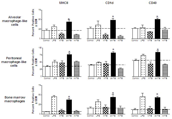
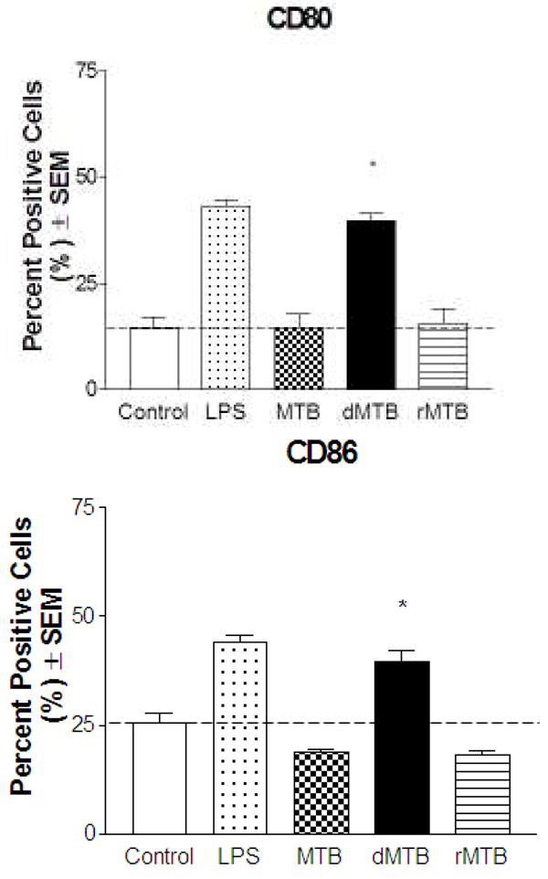
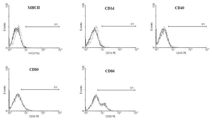
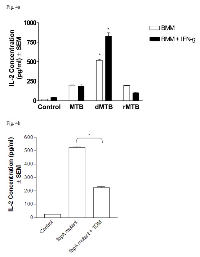
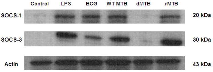
Trehalose 6,6'-dimycolate (TDM) is the most abundant lipid extracted from Mycobacterium tuberculosis (MTB). TDM promotes MTB survival by decreasing phagosomal acidification and phagolysosomal fusion in macrophages. Delipidation of MTB using petroleum ether removes TDM and decreases MTB survival within host cells. TDM reconstituted onto MTB restores its virulent wild-type characteristics. We investigated the role of TDM in regulating surface marker expression in MTB-infected macrophages. Macrophages were infected with wild-type, delipidated, and TDM-reconstituted MTB for 24h and measured for changes in surface marker expression. TDM on MTB was found to specifically target MHCII, CD1d, CD40, CD80 and CD86. Both wild-type and TDM-reconstituted MTB suppressed or induced no change in expression of these surface markers, whereas delipidated MTB increased expression of the same markers. MTB-infected macrophages were also overlaid with MHCII-restricted T cell hybridomas which recognize Antigen 85B. Macrophages infected by wild-type and TDM-reconstituted MTB did not present antigen as well as delipidated MTB-infected macrophages. The evidence shown furthers supports the notion that TDM present on MTB promotes its survival and persistence in host macrophages.
Figures

Similar articles
-
Influence of trehalose 6,6'-dimycolate (TDM) during mycobacterial infection of bone marrow macrophages.Microbiology (Reading). 2002 Jul;148(Pt 7):1991-1998. doi: 10.1099/00221287-148-7-1991. Microbiology (Reading). 2002. PMID: 12101287
-
A mycobacteriophage-derived trehalose-6,6'-dimycolate-binding peptide containing both antimycobacterial and anti-inflammatory abilities.FASEB J. 2013 Aug;27(8):3067-77. doi: 10.1096/fj.13-227454. Epub 2013 Apr 19. FASEB J. 2013. PMID: 23603838
-
Prospective Subunit Nanovaccine against Mycobacterium tuberculosis Infection─Cubosome Lipid Nanocarriers of Cord Factor, Trehalose 6,6' Dimycolate.ACS Appl Mater Interfaces. 2023 Jun 14;15(23):27670-27686. doi: 10.1021/acsami.3c04063. Epub 2023 Jun 1. ACS Appl Mater Interfaces. 2023. PMID: 37262346
-
Cord factor as an invisibility cloak? A hypothesis for asymptomatic TB persistence.Tuberculosis (Edinb). 2016 Dec;101S:S2-S8. doi: 10.1016/j.tube.2016.09.023. Epub 2016 Sep 28. Tuberculosis (Edinb). 2016. PMID: 27743706 Review.
-
Multiple roles of cord factor in the pathogenesis of primary, secondary, and cavitary tuberculosis, including a revised description of the pathology of secondary disease.Ann Clin Lab Sci. 2006 Autumn;36(4):371-86. Ann Clin Lab Sci. 2006. PMID: 17127724 Review.
Cited by
-
Understanding delayed T-cell priming, lung recruitment, and airway luminal T-cell responses in host defense against pulmonary tuberculosis.Clin Dev Immunol. 2012;2012:628293. doi: 10.1155/2012/628293. Epub 2012 Apr 1. Clin Dev Immunol. 2012. PMID: 22545059 Free PMC article. Review.
-
Manipulation of costimulatory molecules by intracellular pathogens: veni, vidi, vici!!PLoS Pathog. 2012;8(6):e1002676. doi: 10.1371/journal.ppat.1002676. Epub 2012 Jun 14. PLoS Pathog. 2012. PMID: 22719245 Free PMC article. Review.
-
Insights into the molecular determinants involved in Mycobacterium tuberculosis persistence and their therapeutic implications.Virulence. 2021 Dec;12(1):2721-2749. doi: 10.1080/21505594.2021.1990660. Virulence. 2021. PMID: 34637683 Free PMC article. Review.
-
Lactoferrin modulation of mycobacterial cord factor trehalose 6-6'-dimycolate induced granulomatous response.Transl Res. 2010 Oct;156(4):207-15. doi: 10.1016/j.trsl.2010.06.001. Epub 2010 Jun 30. Transl Res. 2010. PMID: 20875896 Free PMC article.
-
Recombinant Bacille Calmette-Guérin coexpressing Ag85B-IFN-γ enhances the cell-mediated immunity in C57BL/6 mice.Exp Ther Med. 2017 May;13(5):2339-2347. doi: 10.3892/etm.2017.4273. Epub 2017 Mar 28. Exp Ther Med. 2017. PMID: 28565847 Free PMC article.
References
-
- Bloch H, Sorkin E, Erlenmeyer H. A toxic lipid component of the tubercle bacillus (cord factor). I. Isolation from petroleum ether extracts of young bacterial cultures. Am Rev Tuberc. 1953;67:629–643. - PubMed
-
- Flynn JL, Chan J. Immune evasion by Mycobacterium tuberculosis: living with the enemy. Curr Opin Immunol. 2003;15:450–455. - PubMed
-
- Hestvik AL, Hmama Z, Av-Gay Y. Mycobacterial manipulation of the host cell. FEMS Microbiol Rev. 2005;29:1041–1050. - PubMed
-
- Indrigo J, Hunter RL, Jr, Actor JK. Influence of trehalose 6,6′-dimycolate TDM during mycobacterial infection of bone marrow macrophages. Microbiology. 2002;148:1991–1998. - PubMed
Publication types
MeSH terms
Substances
Grants and funding
LinkOut - more resources
Full Text Sources
Research Materials
