. 2009 Apr;118(2):209-22.
doi: 10.1007/s00412-008-0189-x. Epub 2008 Nov 13.

ATRX marks the inactive X chromosome (Xi) in somatic cells and during imprinted X chromosome inactivation in trophoblast stem cells

Affiliations

ATRX marks the inactive X chromosome (Xi) in somatic cells and during imprinted X chromosome inactivation in trophoblast stem cells

Claudia Baumann et al. Chromosoma. 2009 Apr.

Abstract

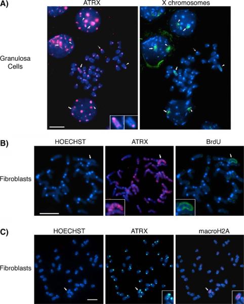
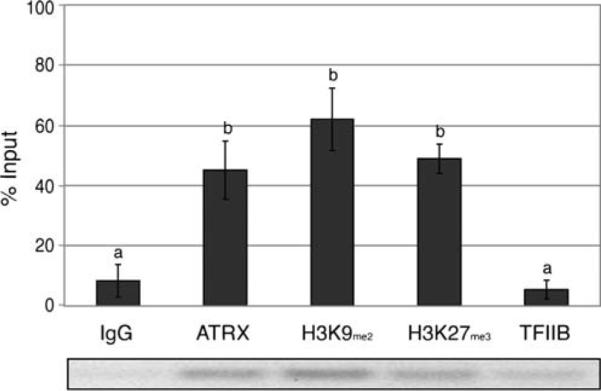
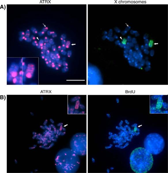
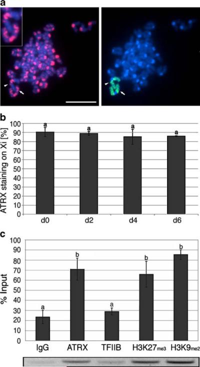
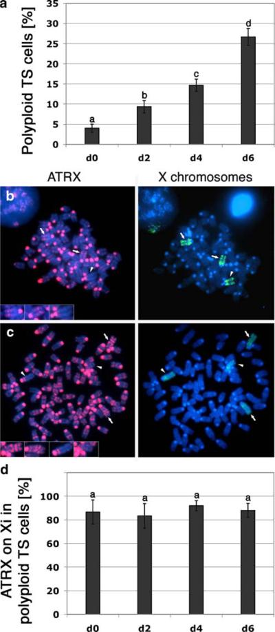
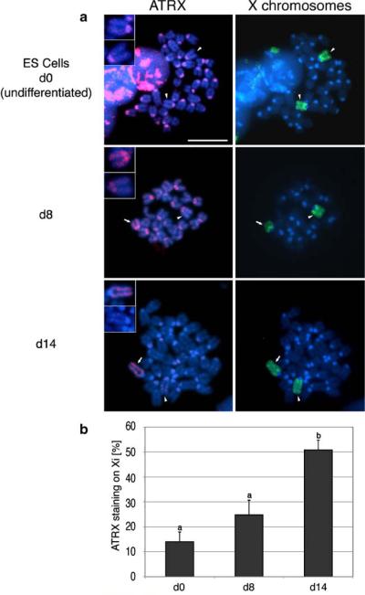
Mammalian X chromosome inactivation (XCI) is an essential mechanism to compensate for dosage imbalances between male and female embryos. Although the molecular pathways are not fully understood, heterochromatinization of the Xi requires the coordinate recruitment of multiple epigenetic marks. Using fluorescence in situ hybridization analysis combined with immunocytochemistry, we demonstrate that the chromatin remodeling protein ATRX decorates the chromatids of a single, late replicating X chromosome in female somatic cells and co-localizes with the bona fide marker of the Xi, macroH2A1.2. Chromatin immunoprecipitation using somatic, embryonic stem (ES) cells and trophoblast stem (TS) cells as model for random and imprinted XCI, respectively, revealed that, in somatic and TS cells, ATRX exhibits a specific association with sequences located within the previously described H3K9me2-hotspot, a region 5' to the X inactive-specific transcript (Xist) locus. While no ATRX-Xi interaction was detectable in undifferentiated ES cells, an enrichment of ATRX was observed after 8 days of differentiation, indicating that ATRX associates with the Xi following the onset of random XCI, consistent with a potential role in maintenance of XCI. These results have important implications regarding a previously described escape from imprinted XCI in ATRX-deficient mice as well as cases of skewed XCI in patients with ATRX syndrome.


Figures

Fig. 1
Fig. 1
Chromosomal localization patterns of ATRX in somatic cells. a Combined fluorescent immunochemistry for ATRX protein (red) and DNA-FISH (green) to determine the position of the X chromosomes on metaphase spreads from a mouse granulosa cell. DNA was visualized by counterstaining with Hoechst 33258. While some autosomes exhibit only a faint banding pattern, a striking accumulation of ATRX on the sister chromatids of one of the two X chromosomes is apparent (bold arrow, inset). The ATRX pattern on the other homolog is indistinguishable from autosomes (arrow head, inset). ATRX protein is found at pericentromeric heterochromatin domains (thin arrow) in autosomes and the sex chromosomes. Notably, ATRX associations are also detectable at the subnuclear domain occupied by one of the two X chromosomes in interphase nuclei (bold arrows, and Supplemental Figure S2-B for nonbiased co-localization analysis). b Analysis of replication timing by BrdU incorporation provides evidence for an association of ATRX protein with the Xi in embryonic fibroblasts. The late replicating inactive X chromosome is clearly labeled with BrdU (green) within the metaphase spread (arrow, inset) and is the only chromosome presenting ATRX protein accumulation (red) along its chromatids (arrow, inset). c Co-immunolocalization of ATRX protein (green) and macroH2A (red), a marker of the inactive X chromosome (Xi), on paraformaldehyde-fixed spreads showing ATRX protein at pericentromeric heterochromatin (inset) and at the chromatids of one chromosome (arrow), which is identified as the Xi by macroH2A co-localization (red, inset). Scale bar=10 μm
Fig. 2
Fig. 2
The ATRX protein is preferentially enriched at DNA sequences 5′ of the Xist locus in female embryonic fibroblasts. Chromatin immunoprecipitation (ChIP) assays of X chromosome-specific sequences were performed on primary mouse embryonic fibroblasts using specific antibodies against ATRX, H3K27me3, and H3K9me2 with antibodies against TFIIB and IgG as controls. Amplification of precipitated genomic DNA with an anti-ATRX antibody revealed a significant enrichment of X chromosome-specific DNA fragments corresponding with the constitutive H3K9me2-hotspot located 5′ of the Xist compared to the negative control (IgG), indicating a specific molecular association with the Xi. A representative gel image is shown. Error bars represent the STD of three independent experiments and different superscripts indicate significant differences (p<0.05)
Fig. 3
Fig. 3
ATRX marks the inactive X chromosome in trophoblast stem cells. a Indirect immunofluorescence detection of ATRX and DNA-FISH analysis for the X chromosomes on metaphase spreads of trophoblast stem cells. ATRX (red)is present at pericentric heterochromatin domains (thin arrow) in both autosomes and the sex chromosomes (arrowhead and bold arrow). Notably, ATRX exhibits a preferential accumulation at the chromatids of one of the two X chromosomes (see bold arrow and right inset), while the other X chromosome reveals pericentric staining only (arrowhead, left inset). b Asynchronous replication timing, determined by BrdU incorporation (green), identifies the inactive X chromosome (bold arrow and inset) and exhibits a preferential enrichment for ATRX (red)in undifferentiated TS cells (arrow). Scale bars=10 μm
Fig. 4
Fig. 4
ATRX is a stable marker of the inactive X chromosome throughout TS cell differentiation. a Analysis of ATRX immunolocalization patterns (red) and subsequent DNA-FISH analysis (green)of the X chromosomes revealed a stable association of the protein with the Xi (arrow, insets) throughout TS cell differentiation as depicted for day 4 of differentiation. The active X chromosome remains indistinguishable from the autosomes (arrowhead, insets). Scale bars represent 10 μm. b Quantification of ATRX associations with the Xi in TS cell metaphase spreads. Data indicate the mean percentage of diploid metaphase spreads with ATRX association at a single X chromosome at different days (d0–d6) following spontaneous differentiation. Error bars represent the STD of three independent experiments, and different superscripts indicate significant differences (p< 0.05). c ChIP assays were performed on trophoblast stem cells using specific antibodies against ATRX, H3K27me3, and H3K9me2. Precipitated genomic DNA was amplified, and a significant enrichment of DNA fragments encoding the constitutive H3K9me2-hotspot located 5′ of the Xist locus for ATRX and the histone modifications compared to the controls (IgG and TFIIB) was detected. A representative gel image is shown below the graph. Error bars represent the STD of three independent experiments, and different superscripts indicate significant differences (p<0.05)
Fig. 5
Fig. 5
ATRX marks the inactive X chromosomes in polyploid trophoblast giant cells. a The incidence of polyploid metaphases depending on the stage of differentiation was quantified and is displayed as the mean percentage of three independent experiments. Different superscripts indicate significant differences (p<0.05). Error bars represent the STD. b In hyperdiploid metaphases, ATRX marks two of the three X chromosomes (red, bold arrows, insets) in agreement with the rule of one active X chromosome per diploid chromosome set. c Polyploid trophoblast giant cell metaphase presenting ATRX staining on two Xi (red, bold arrows, insets), while there is no such association detectable at the two Xa (arrowheads, insets). d Proportion of trophoblast giant cells showing ATRX protein localization to the inactive X chromosomes in polyploid metaphases in three independent experiments. Different superscripts indicate significant differences (p<0.05). Error bars represent the STD
Fig. 6
Fig. 6
Association of ATRX with the inactive X chromosome is a late event during ES cell differentiation. a In undifferentiated (PGK12.1) ES cell metaphases, ATRX (red) localizes to pericentromeric heterochromatin in all chromosomes. Both X chromosomes (upper panel; arrow and arrowhead) are indistinguishable in regard to ATRX labeling. The association of ATRX with Xi is initially detected following 8 days of spontaneous differentiation (middle panel; arrow and inset) and subsequently increases after 14 days of differentiation (lower panel, arrow, insets). b Quantitative analysis of ATRX associations with the Xi during ES cell differentiation. Data indicate the mean percentage of metaphases with preferential ATRX staining on a single X chromosome in undifferentiated, d8 and d14 differentiated cells. Error bars represent the STD of three independent experiments, and different superscripts indicate significant differences (p<0.05)
Fig. 7
Fig. 7
ATRX is enriched at chromosomal sequences corresponding to the H3K9me2-hotspot on the Xi following spontaneous differentiation of ES cells. Chromatin immunoprecipitation (ChIP) analysis of Xi chromosome-specific sequences on undifferentiated ES cells (d0, white bars) and throughout ES cell differentiation (d5, gray bars) and (d8, black bars) using specific antibodies against ATRX, H3K27me3, and H3K9me2 (antibodies against TFIIB and IgG served as controls). A significant enrichment of DNA fragments encoding sequences within the constitutive H3K9me2-hotspot on the Xi was only observed for samples immunoprecipitated with anti-ATRX antibodies after 8 days of differentiation, while the histone modification H3K9me2 was detectable at all time points investigated. Error bars represent the STD of three independent experiments, and different superscripts indicate significant differences (p<0.05). Representative gel images for each stage of differentiation are displayed below the graph

Similar articles

Cited by

References

    1. Abbondanzo S, Gadi I, Stewart C. Methods Enzymology. vol. 225. Academic Press; San Diego: 1993. Derivation of embryonic stem cell lines; pp. 803–823. - PubMed
    1. Bannister AJ, Zegerman P, Partridge JF, Miska EA, Thomas JO, Allshire RC, Kouzarides T. Selective recognition of methylated lysine 9 on histone H3 by the HP1 chromo domain. Nature. 2001;410:120–124. - PubMed
    1. Baumann C, Schmidtmann A, Muegge K, De La Fuente R. Association of ATRX with pericentric heterochromatin and the Y chromosome of neonatal mouse spermatogonia. BMC Mol Biol. 2008;9:29. - PMC - PubMed
    1. Berube NG, Smeenk CA, Picketts DJ. Cell cycle-dependent phosphorylation of the ATRX protein correlates with changes in nuclear matrix and chromatin association. Hum Mol Genet. 2000;9:539–547. - PubMed
    1. Boggs BA, Cheung P, Heard E, Spector DL, Chinault AC, Allis CD. Differentially methylated forms of histone H3 show unique association patterns with inactive human X chromosomes. Nat Genet. 2002;30:73–76. - PubMed

Publication types

MeSH terms