. 2009 Jan;83(2):993-1008.
doi: 10.1128/JVI.01647-08. Epub 2008 Nov 12.

Structural and functional studies of the promoter element for dengue virus RNA replication

Affiliations

Structural and functional studies of the promoter element for dengue virus RNA replication

María F Lodeiro et al. J Virol. 2009 Jan.

Abstract

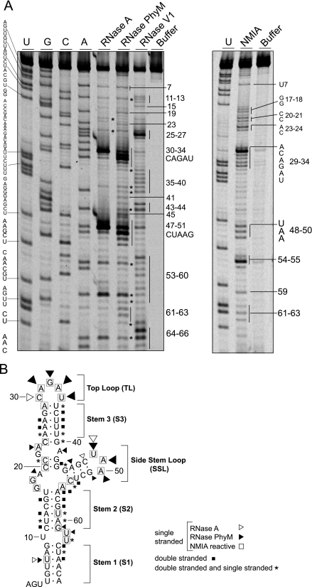
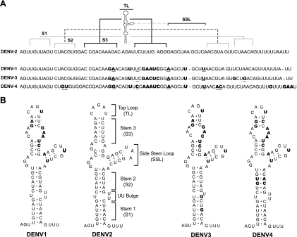
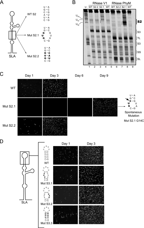
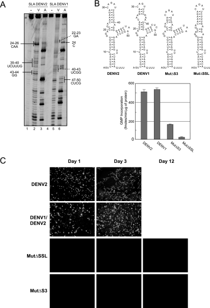
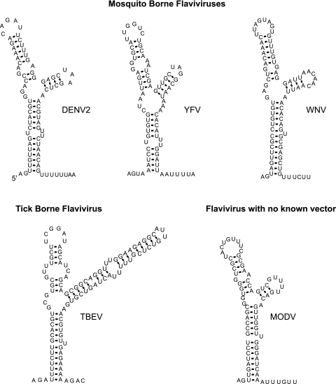
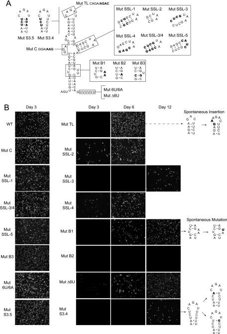
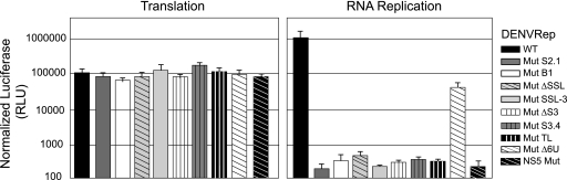
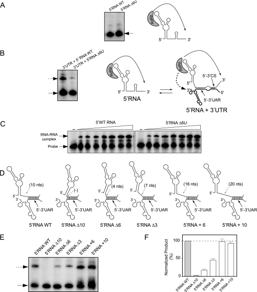
The 5' untranslated region (5'UTR) of the dengue virus (DENV) genome contains two defined elements essential for viral replication. At the 5' end, a large stem-loop (SLA) structure functions as the promoter for viral polymerase activity. Next to the SLA, there is a short stem-loop that contains a cyclization sequence known as the 5' upstream AUG region (5'UAR). Here, we analyzed the secondary structure of the SLA in solution and the structural requirements of this element for viral replication. Using infectious DENV clones, viral replicons, and in vitro polymerase assays, we defined two helical regions, a side stem-loop, a top loop, and a U bulge within SLA as crucial elements for viral replication. The determinants for SLA-polymerase recognition were found to be common in different DENV serotypes. In addition, structural elements within the SLA required for DENV RNA replication were also conserved among different mosquito- and tick-borne flavivirus genomes, suggesting possible common strategies for polymerase-promoter recognition in flaviviruses. Furthermore, a conserved oligo(U) track present downstream of the SLA was found to modulate RNA synthesis in transfected cells. In vitro polymerase assays indicated that a sequence of at least 10 residues following the SLA, upstream of the 5'UAR, was necessary for efficient RNA synthesis using the viral 3'UTR as template.


Figures

FIG. 1.
FIG. 1.
Enzymatic and chemical probing of the 5′-terminal stem-loop of the DENV genome. (A) An RNA corresponding to the first 160 nucleotides of the viral genome was subjected to RNase A, RNase PhyM, RNase V1, or NMIA treatment, and the cleaved or modified RNAs were analyzed by primer extension. The products of primer extension were analyzed with a sequencing gel along with a control sample without treatment and a sequencing ladder. The nucleotide sequence is indicated on the left, and specific cleavages are indicated on the right by numbers. (B) Optimal MFOLD-predicted RNA secondary structure model of SLA that includes a summary of detected enzymatic and chemical modifications. Nucleotides labeled with asterisks were observed to be sensitive to both single- and double-stranded specific RNases.
FIG. 2.
FIG. 2.
Comparison of the nucleotide sequences and secondary structures of the SLA structures of different DENV serotypes. (A) Alignment of the consensus sequences of the first 70 nucleotides of DENV1, DENV2, DENV3, and DENV4. The regions corresponding to the predicted secondary structures S1, S2, S3, TL, and SSL of DENV2 are indicated on the top. Nucleotide variation between DENV2 and the other three serotypes is indicated in bold. Underlined nucleotides indicate covariations. (B) Comparative RNA secondary structure analysis predicted for the SLA structures of the four DENV serotypes. The nucleotide differences between DENV2 and the other three serotypes are indicated in bold.
FIG. 3.
FIG. 3.
Role of helical regions S2 and S3 in viral replication. (A) Schematic representation of specific mutations within S2 of SLA. The sequences of the WT and the mutants Mut S2.1 and Mut S2.2 are shown, and the nucleotide changes are indicated in bold. (B) Enzymatic probing of WT and Mut S2.1 and Mut S2.2 mutant RNAs. The regions corresponding to the predicted secondary structures S2, S3, TL, SL, and SS are indicated on the right. (C) Expression of DENV proteins in BHK cells transfected with WT and mutated viral RNAs was monitored by IF at 1, 3, 6, and 9 days posttransfection. The spontaneous mutation of a recovered virus obtained after transfection of the Mut S2.1 RNA is indicated with an arrow. (D) Viral replication tolerates variations within the helical S3 region. (Left) Schematic representation of SLA showing specific nucleotides modified in Mut S3.1, Mut S3.2, and Mut S3.3 disrupting or reconstituting the helical region. (Right) Expression of DENV proteins in BHK cells transfected with WT or mutated RNAs. Viral replication was monitored by IF at 1 and 3 days after transfection.
FIG. 4.
FIG. 4.
Functional in vivo and in vitro compatibility of SLAs from different DENV serotypes. (A) Enzymatic probing of the first 70 nucleotides of DENV1 and DENV2. The RNAs were subjected to RNase V1 (V) or RNase A (A) digestion, and the cleaved RNAs were analyzed by primer extension. Differences within the S3 and SSL regions of SLAs from DENV1 and DENV2 are indicated. (B) In vitro activity of viral RdRp for WT and mutated RNA templates with 160 nucleotides. (Top) Sequences of SLAs of the RNAs used as templates for RNA synthesis. (Bottom) Viral polymerase activity, expressed in fmol [α-32P]GMP incorporated into acid-insoluble RNA per minute and per μg of protein, for each RNA template. The reaction was carried out as described in Materials and Methods. Error bars indicate the standard deviations of results from three experiments. (C) Expression of DENV proteins in BHK cells transfected with RNA of WT DENV2, a chimeric DENV carrying the SLA from DENV1 (DENV1/DENV2), and mutants with deletions of the SSL or S3 (Mut ΔSSL or Mut ΔS3, respectively) was determined. Viral replication was monitored by IF for up to 12 days posttransfection.
FIG. 5.
FIG. 5.
Predicted 5′-end stem-loop structures of flaviviruses. The optimal MFOLD-predicted RNA secondary-structure models for mosquito-borne and tick-borne flaviviruses and a flavivirus with no known vector are shown. Results are shown for WNV M12294, yellow fever virus (YFV) NC-002031, tick-borne encephalitis virus (TBEV) U27495, and Modoc virus (MODV) AJ242984.
FIG. 6.
FIG. 6.
Functions of structural elements of SLA common in flavivirus genomes. (A) Schematic representation of specific mutations within SLA. The modified nucleotides are indicated for each case: in Mut TL, the sequence of the loop CAGA was replaced by AGAC; in Mut B1, the sequence of the UU bulge was replaced by AA; in Mut B2, the UU bulge was replaced by a single A residue; in Mut B3, a UU mismatch was replaced by a CG base pair; in Mut C, the sequence of the GGA bulge was replaced by AAG; in Mut SSL-1, the stem of the SSL was stabilized by 5 GC base pairs; in Mut SSL-2, the stem of the SSL includes a 2-base-pair deletion; in Mut SSL-3, the 4 nucleotides GAGC on one side of the stem of the SSL were replaced by CUCG to disrupt the stem; in Mut SSL-4, the 4 nucleotides CUCG on the other side of the SSL were replaced by GAGC to disrupt the stem; in Mut SSL-3/4, the nucleotides modified in Mut SSL-3 and Mut SSL-4 were introduced together to reconstitute the stem; in Mut SSL-5, the sequence of the SL UAA was replaced by CAUC; in Mut S3.4, the upper half of S3 was disrupted; in Mut S3.5, the predicted base pairs in S3 were reconstituted; in Mut Δ6U, the 6 U residues downstream of SLA were deleted; and in Mut 6U/6A, the oligo(U) track was replaced by an oligo(A) sequence. (B) Expression of DENV proteins in BHK cells transfected with WT and SLA-mutated RNAs described for panel A. Viral replication was monitored by an IF assay as a function of time after RNA transfection using specific anti-DENV antibodies. Viruses recovered in the supernatants from transfected cells were used to purify the RNA for sequencing analysis. In the right panel, the spontaneous mutations are shown in bold.
FIG. 7.
FIG. 7.
Mutations within SLA impair RNA synthesis without affecting translation of the input RNA. Shown are translation and RNA replication of WT and mutant DENV replicons in BHK cells. Normalized luciferase levels (in relative luciferase units [RLU]) are shown using a logarithmic scale at 10 h after transfection to estimate translation of input RNA and at 3 days after transfection to evaluate RNA replication. Error bars indicate the standard deviations of results from three independent transfections.
FIG. 8.
FIG. 8.
Role of the region between SLA and the 5′UAR in viral RNA synthesis. (A) In vitro RdRp activity of the recombinant NS5 for the first 160 nucleotides of the DENV genome WT or the mutant Δ6U as a template (5′RNA WT or 5′RNA Δ6U, respectively, as indicated above the gel). The radiolabeled RNA products were analyzed with denaturing 5% polyacrylamide gels. The arrow indicates the mobility of the product. On the right, a schematic representation of the initiation process is shown. (B) The efficiency of trans initiation polymerase activity depends on the oligo(U) track. The 5′RNA WT or the 5′RNA Δ6U mutant (160 nucleotides) was incubated with the WT 3′UTR molecule (458 nucleotides) and the viral polymerase. The radiolabeled products were analyzed with denaturing 5% polyacrylamide gels, as previously described (2, 11). The arrows indicate the mobilities of the two products. On the right, a schematic representation of the interaction of the RNA molecules representing the 5′ and 3′ ends of DENV RNA and the viral polymerase initiating RNA synthesis at the 3′ end of the template is shown. The hybridized 5′-3′UAR and 5′-3′CS are also shown. (C) RNA mobility shift assays showing the interaction between the 3′SL WT probe and the 5′RNA WT or 5′RNA Δ6U, as indicated at the top. The mobilities of the 3′SL probe and the RNA-RNA complex are indicated on the left. (D) Schematic representation of the hybridized 5′- and 3′-terminal regions of the viral genome for the WT and the mutants with deletions or insertions in the SLA-5′UAR spacer region: 5′RNAΔ10, 5′RNAΔ6, 5′RNAΔ3, 5′RNA+6, and 5′RNA+10. (E) Efficiency of trans initiation polymerase activity for different 5′RNA molecules. The 5′RNA molecules described for panel D were used for the trans initiation assay. The products were analyzed as described above. (F) The two radiolabeled products obtained in panel E were quantified, and the products obtained by trans initiation are expressed relative to the amount of product obtained by cis initiation for each mutant and referred to as percentages relative to WT levels [(3′RNA value/5′RNA value) × (5′RNA WT value/3′RNA WT value) × 100]. Error bars indicate the standard deviations of results from three independent experiments.

Similar articles

Cited by

References

    1. Alvarez, D. E., A. L. De Lella Ezcurra, S. Fucito, and A. V. Gamarnik. 2005. Role of RNA structures present at the 3′UTR of dengue virus on translation, RNA synthesis, and viral replication. Virology 339200-212. - PubMed
    1. Alvarez, D. E., C. V. Filomatori, and A. V. Gamarnik. 2008. Functional analysis of dengue virus cyclization sequences located at the 5′ and 3′UTRs. Virology 375223-235. - PubMed
    1. Alvarez, D. E., M. F. Lodeiro, S. J. Luduena, L. I. Pietrasanta, and A. V. Gamarnik. 2005. Long-range RNA-RNA interactions circularize the dengue virus genome. J. Virol. 796631-6643. - PMC - PubMed
    1. Blaney, J. E., Jr., A. P. Durbin, B. R. Murphy, and S. S. Whitehead. 2006. Development of a live attenuated dengue virus vaccine using reverse genetics. Viral Immunol. 1910-32. - PubMed
    1. Blaney, J. E., Jr., N. S. Sathe, L. Goddard, C. T. Hanson, T. A. Romero, K. A. Hanley, B. R. Murphy, and S. S. Whitehead. 2008. Dengue virus type 3 vaccine candidates generated by introduction of deletions in the 3′ untranslated region (3′-UTR) or by exchange of the DENV-3 3′-UTR with that of DENV-4. Vaccine 26817-828. - PMC - PubMed

Publication types

LinkOut - more resources